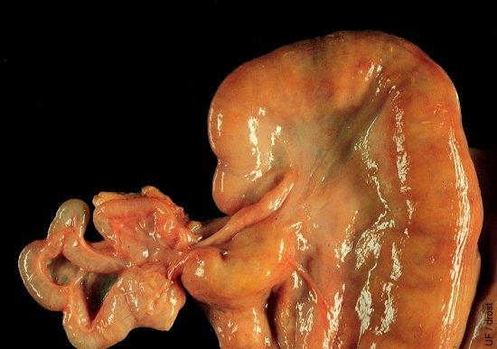
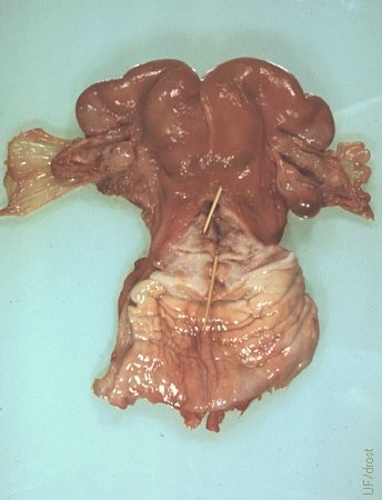
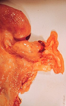
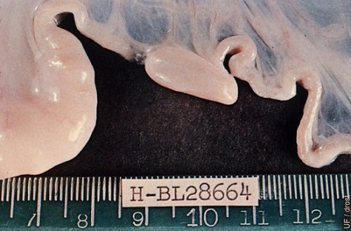
Hydrosalpinx.
The degree of distension varies with the location of the blockage. Minimal distension is difficult to diagnose by palpation per rectum or even by ultrasonography. The effect on fertility is the same.
Drost M (1971)
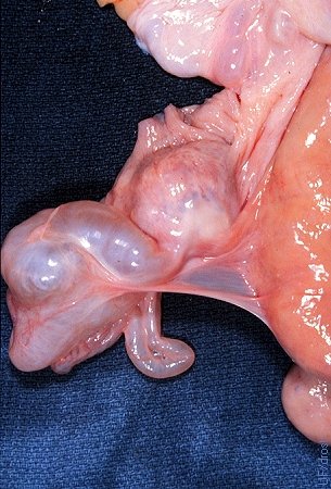
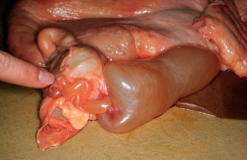
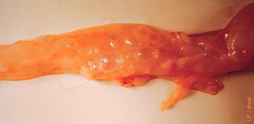
Hydrosalpinx - Close-up.
The left oviduct is tightly distended due to occlusion as the result of adhesions.
Drost M (1971)
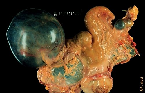
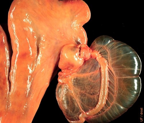
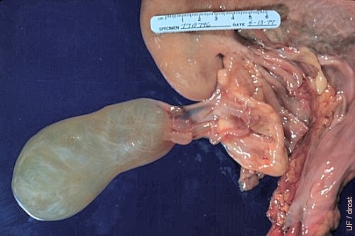
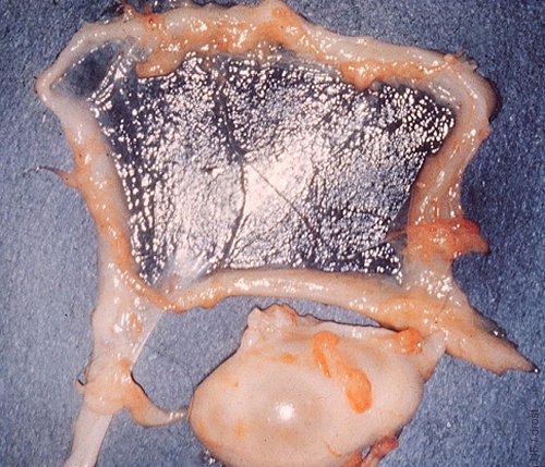
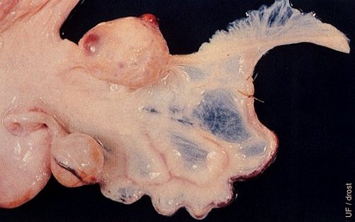
Hydrosalpinx.
The left oviduct is grossly distended with fluid. This structure presents a challenge in differential diagnosis with ovarian cysts. The contralateral ovary displays a corpus luteum which rules out an endocrine problem. The cow is subfertile and can only conceive in the right horn.
Drost M (1971)
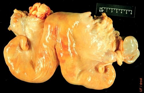
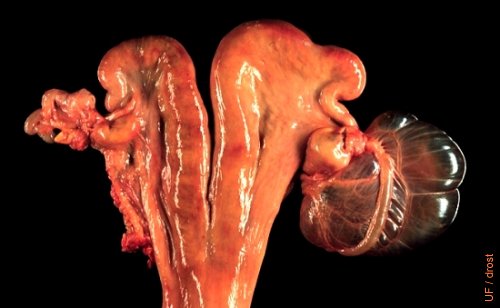
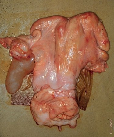
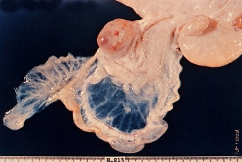
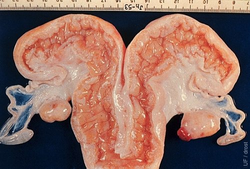
Hydrosalpinx.
The right oviduct was blocked leading to hydrosalpinx.
Drost M (1971)
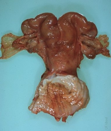
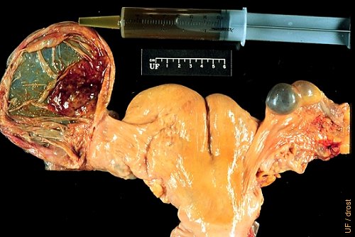
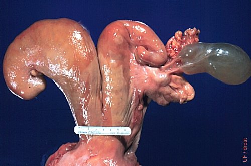
Hydrosalpinx.
Cervical stenosis without occlusion. Owner had difficulty passing the insemination syringe through the internal os of the cervix of this Shorthorn heifer. In addition, the right oviduct was blocked leading to hydrosalpinx. Stenosis was probably due to pipet trauma, and not related to the hydrosalpinx.
Drost M (1971)
Cystic Ovarian Bursa.
Greatly distended cystic ovarian bursa, filled with serosanginous fluid.
Drost M (1974)
Collapsed Cystic Ovarian Bursa.
60 ml of serosanginous fluid were aspirated from the left cystic ovarian bursa. Cystic ovarian bursae are the result of complete adhesions of the fimbriae leading to accumulation of fluid. The right ovary shows a large hemorrhagic follicle and a normal follicle.
Drost M (1974)
Cystic Ovarian Bursa and Hydrosalpinx.
The right ovarian bursa is greatly distended. The left oviduct is blocked and slightly distended. There were bilateral adhesions of the ovarian adnexa.
Drost M (1974)
Close-up of Cystic Ovarian Bursa.
The large fluctuant ovarian bursa can be palpated per rectum.
Drost M (1974)
Elongated Cystic Ovarian Bursa.
The large, elongated cystic ovarian bursa is shown in proportion to the uterus.
Drost M (1974)
Close-up of Elongated Cystic Ovarian Bursa.
Hydrosalpinx invariably occurs in conjunction with a cystic ovarian bursa due to occlusion of the infundibular end of the oviduct.
Drost M (1974)
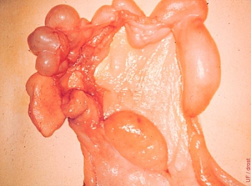
Parovarian Adhesions.
The right ovary is surrounded by adhesions (peri-ovarian) which have also led to the occlusion of the ovarian bursa which appears as a pedunculated fluctuant mass.
Drost M (1974)
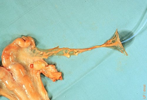
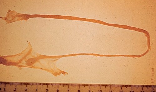
Uterine Tube Dissected Out.
The oviduct has been dissected out to demonstrate its considerable length. A canula has been inserted at the infundibular end. The ampullar section has been straightened out, and the isthmus is still convoluted.
Drost M (1974)
Close-up of Elongated Cystic Ovarian Bursa.
The fluid-filled ovarian bursa has become a pedunculated mass.
Drost M (1974)

Cannulated Uterine Tube.
The oviduct is mock annulated to illustrate the placement of the Silastic cannula in the infundibulum of the oviduct for the surgical recovery of embryos prior to Day 5. Notice the Day 3 to 4 corpus hemorrhagicum.
Drost M (1974)
Oviductal Anomaly.
A rare oviductal anomaly in which the uterine end of the oviduct ends blindly.
Roberts SJ (1973)
Blocked Uterine Tube.
Dramatically distended oviduct due to complete blockage. The ovary is inactive and appears small by comparison.
Drost M (1974)
Cystic Oviductal Diverticula.
Cystic diverticula of the oviduct.
Roberts SJ (1973)
Oviductal Diverticula - Close-up.
Intramucosal cysts or cystic diverticula of the oviduct. The tip of the uterine horn is on the right. These minute cysts result from a salpingitis whereby the epithelium of the free edges of the mucosal folds are denuded and these folds fuse. These cysts apparently do not prevent conception and are only found incidentally on necropsy or in slaughterhouse specimens.
Roberts SJ (1973)
Ovulation Tags.
Ovulation tags are strands of fibrin, remnants of the blood clots associated with ovulation.
Roberts SJ (1973)
Segmental Hydrosalpinx.
Two sacculations are present in the mid-ampullary portion of the oviduct.
Tanabe TY and Almquist GO (1966)
Mesonephric Duct Cyst.
A mesonephric duct cyst is present near the isthmus of the oviduct. The oviduct has been injected with dye for better visualization. The cyst does not occlude the lumen, hence does not adversely affect fertility.
Tanabe TY and Almquist GO (1966)
Bilateral Infundibular Aplasia.
The ovarian bursae are congenitally absent. The ampullae end in a blind fluid-distended pouch, a rare occurrence.
Tanabe TY and Almquist GO (1966)
Segmental Tubal Aplasia.
A 5 mm section of the oviductal isthmus is missing, leading to secondary hydrosalpinx.
Tanabe TY and Almquist GO (1966)