The Visual Guide to
Bovine Reproduction
- Bulls
- Testis and Epididymis
- Accessory Sex Glands
- Scrotum
- Penis
- Prepuce
- Semen
- Breeding Soundness Evaluation
- Breeding Capacity
- Vasectomy
Male Reproductive System: Accessory Sex Glands
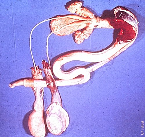
Male Reproductive Tract.
Complete reproductive tract of the bull showing the testes, accessory sex glands and the penis. The tunics of the left testis have been opened to show the epididymis. The fibroelastic penis shows the sigmoid flexure and the retractor penis muscles. Of the accessory sex glands the lobular seminal, vesicular glands are readily visible at the base of the (empty) bladder. The ampullae, the dilated portions of the vasa deferentia are also shown dorsal to the bladder. The prostate gland is not visible. The annular portion is located just posterior to the confluence of the seminal vesicular glands and the disseminate portion lies along the root of the penis. The bulbourethral glands are also not visible they are surrounded by the red bulbourethral muscles at the base of the penis.
Drost M (1980)
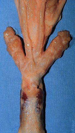
Normal Seminal Vesicular Glands.
The seminal vesicular glands are symmetrical. Also shown are the distended terminal portions of the vasa deferentia, called the ampullae. All four meet at the colliculus seminalis. 18 month old Brahman bull.
Larsen RE (1980)
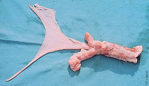
Asymmetrical Seminal Vesicular Glands.
The left seminal vesicular gland is larger than the right, possibly due to early seminal vesiculitis.
Drost M (1980)
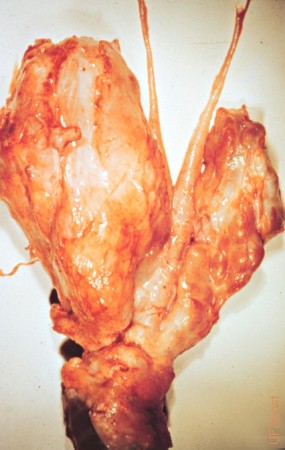
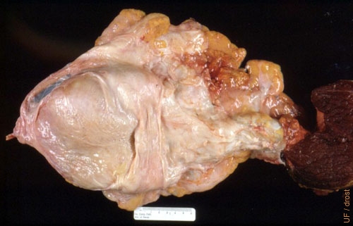
Seminal Vesiculitis.
Both seminal vesicular glands are enlarged, especially the left one. The typical lobulation (vesicular pattern), of the left gland, is no longer apparent and adhesions are present.
Drost M (1980)
Purulent Seminal Vesiculitis 1.
Enlarged pus-filled seminal vesicular glands will feel relatively smooth and fluctuant when palpated per rectum. The most common infectious agent is Arcanobacterium pyogenes.
Drost M (1980)
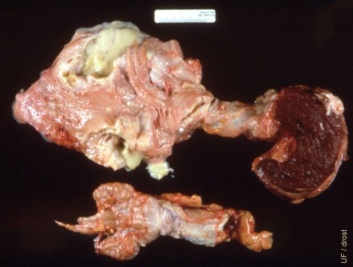
Purulent Seminal Vesiculitis 2.
Bilateral purulent seminal vesiculitis due Arcanobacterium pyogenes. The seminal vesicles are grossly enlarged and lost their vesicular appearance and feel. The latter may be visually appreciated in the smaller normal specimen at the bottom of the image.
Drost M (1980)
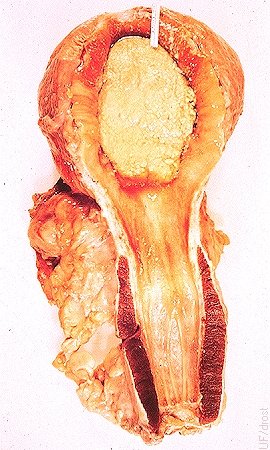
Bladder Paralysis.
The paralyzed bladder contained sand-like crystals. Paralysis occurred after surgical removal of the seminal vesicular glands.
Roberts SJ (1973)
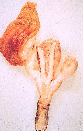
Adhesions to Rectum.
The left seminal vesicular gland is adhered to the rectum. A fistulous tract was present between the seminal vesicular gland and the rectum. Arcanobacterium pyogenes was cultured from the exudate.
Roberts SJ (1973)