The Visual Guide to
Bovine Reproduction
- Bulls
- Testis and Epididymis
- Accessory Sex Glands
- Scrotum
- Penis
- Prepuce
- Semen
- Breeding Soundness Evaluation
- Breeding Capacity
- Vasectomy
Male Reproductive System: Penis
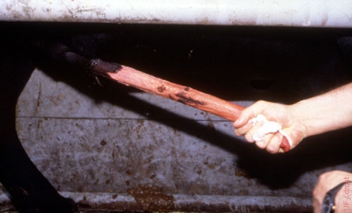
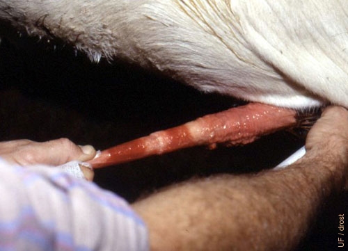
Manual Extension.
The bull can be made to extend his penis with the aid of an electroejaculator, or (with some practice) by manually stimulating the base of the penis per rectum, until the glans penis protrudes from the prepuce. It should then be grasped with the help of a piece of gauze.
Larsen RE (2001)
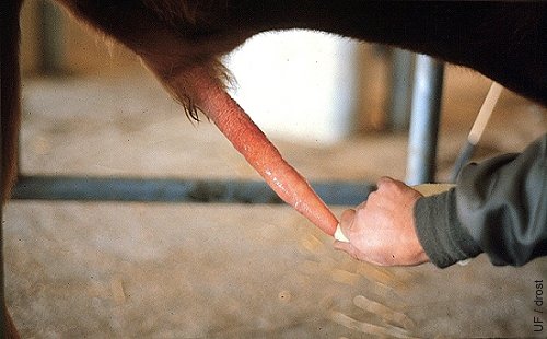
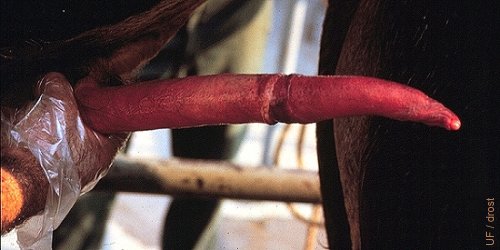
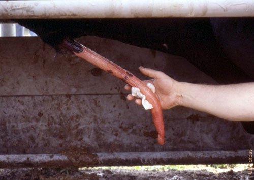
Examination of the Penis.
The penis is manually extended by grasping the tip (glans) with a piece of gauze. It is then examined for the presence of any abnormalities or lesions.
Larsen RE (1982)
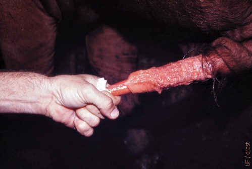
Persistent Frenulum.
Persistent frenulum is the result of the retention of the attachment between visceral and parietal layers of the inner lamina of the prepuce. This condition is believed to be hereditary. It is advisable to ligate the frenulum prior to transsection because the structure frequently contains well developed blood vessels.
Wolfe DF (2005)
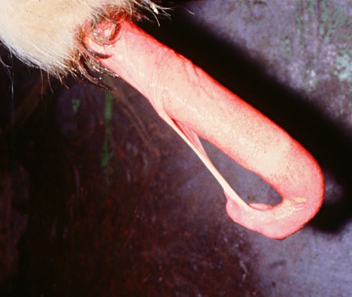
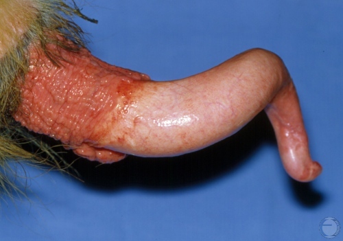
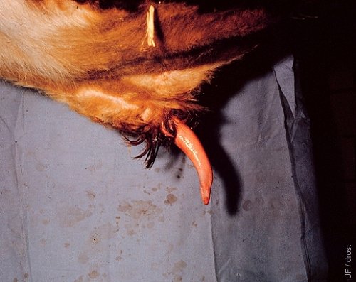
Frenulum.
Occasionally, a persistent frenulum and retention of the attachment between visceral and parietal layers of the inner lamina of the prepuce occurs. The frenulum is a band of tissue that extends from the ventral tip of the glans penis to the prepuce. Separation of the penis from the sheath normally begins at 1 month of age in the bull, and ends with complete separation by 8 months. When the frenulum persists there is usually a blood vessel present in the center of the tissue band comprising the frenulum. Persistence of the frenulum causes curvature of the extended penis.
Larsen RE (2001)
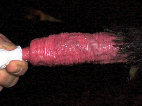
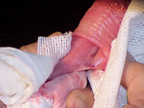
Persistent Frenulum Repair 1.
The frenulum is a band of tissue that extends from the ventral tip of the glans penis to the prepuce. Persistence of the frenulum keep the penis from fully extending and causes curvature of the erected penis.
Shipley C (2006)
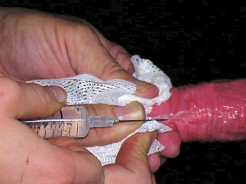
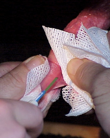
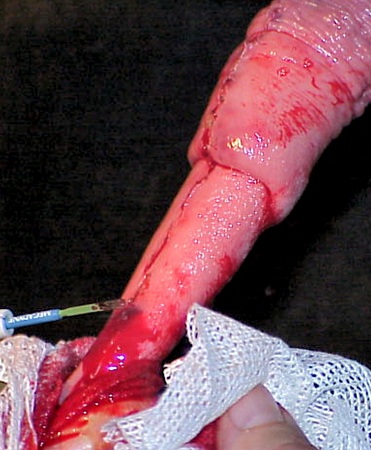
Persistent Frenulum Repair 2.
Injection of local anesthetic agent prior to surgical repair of a persistent frenulum.
Shipley C (2006)
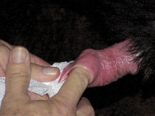
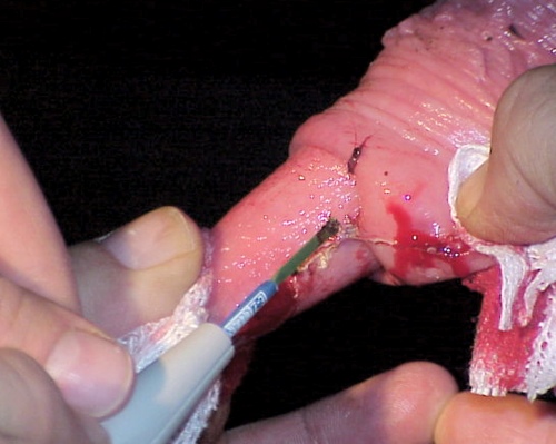
Persistent Frenulum Repair 3.
Exploration of the extent of persistence of the band of tissue that extends from the prepuce to the ventral side of the penis near the glans.
Shipley C (2006)
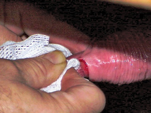
Persistent Frenulum Repair 4.
Transsection of the persistent band.
Shipley C (2006)
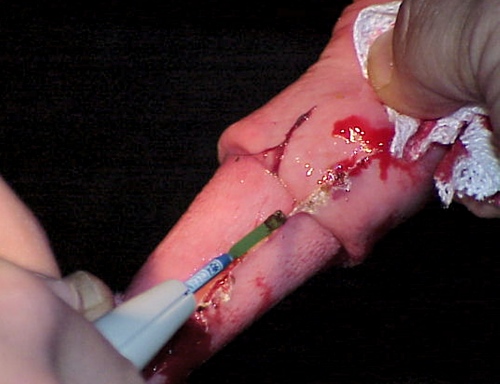
Persistent Frenulum Repair 5.
More persistent adhesion of the preputial lining to the penis.
Shipley C (2006)
Persistent Frenulum Repair 6.
Electrocautery and severing of the vascular portion of the persistent frenulum.
Shipley C (2006)
Persistent Frenulum Repair 7.
Continued electrocautery and severing of the vascular portion of the persistent frenulum.
Shipley C (2006)
Persistent Frenulum Repair 8.
Continued electrocautery and severing of the vascular portion of the persistent frenulum.
Shipley C (2006)
Persistent Frenulum Repair 9.
Final separation of the persistent frenulum. Electrocautery of the last remaining bleeders.
Shipley C (2006)
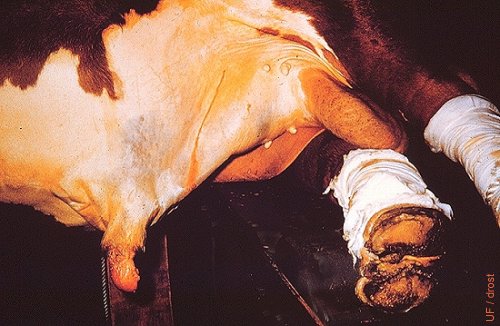
Prepubertal Prepuce.
Forced manual detachment of the prepuce from the free end of the penis during BSE of a bull just going through puberty.
Larsen RE (2001)
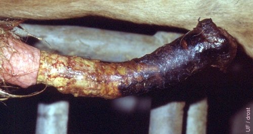
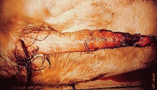
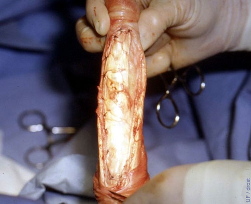
Separation of the Prepuce.
Circumferential separation of the penis and prepuce. Picture taken 2 to 3 weeks after the injury.
Roberts SJ (1973)
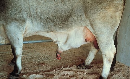
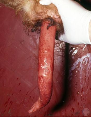
Hematoma of the Penis.
Prolapse of the prepuce due to hematoma of the penis. The diagonal line indicates the surgical approach for removal of the blood clot once it has had time to firm up for 2 to 3 days.
Roberts SJ (1973)
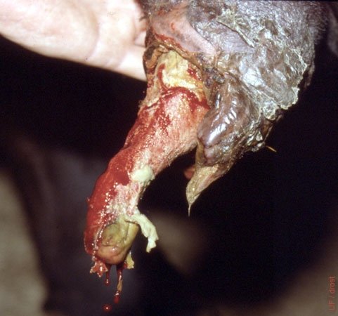
Penile Hematoma.
Rupture of the penis occurred at the points of attachment of the retractor penis muscles. Swelling is due to accumulation of blood. Internal pressure leads to prolapse of the preputial lining. This is also referred to as a broken penis.
Larsen RE (1985)
Penile Hematoma.
Rupture of the penis occurred at the points of attachment of the retractor penis muscles. Swelling is due to accumulation of blood. Internal pressure leads to prolapse of the preputial lining. This is also referred to as a broken penis.
Larsen RE (1985)
Multiple Fibropapillomas.
Pedunculated masses can be removed surgically. Self-immunization via the open wounds can aid in recovery and regression of the papillomatous tissue, much like the effect of an autogenous vaccine prepared from the excised tissues.
Hopper RM (2008)
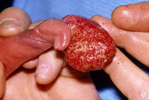
Multiple Large Warts.
Large masses of warts can lead to paraphimosis, trauma and contamination.
Hopper RM (2008)
Fibropapilloma.
Pedunculated masses can be removed surgically. Self-immunization via the open wounds can aid in recovery and regressing of the papillomatous tissue, much like the effect of an autogenous vaccine prepared from the excised tissues.
Roberts SJ (1973)
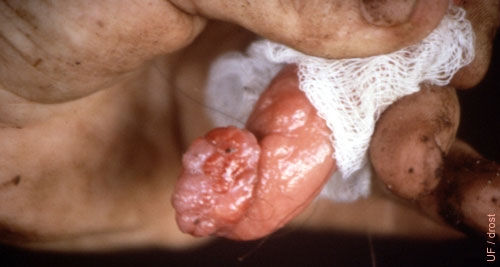
Penile Wart.
This is a singular pendunculated fibropapilloma. It may be successfully removed by excision with or without electrocautery.
Larsen RE (2001)
Wart at Tip of Penis.
Single fairly pedunculated wart at the tip of the glans penis. The mass may be ligated and excised.
Larsen RE (2001)
Single Large Wart.
Single large pedunculated wart at the tip of the glans penis. The mass may be ligated and excised.
Ott R (2013)
Single Large Wart.
Single large pedunculated wart at the tip of the glans penis. The mass may be ligated and excised.
Ott R (2013)
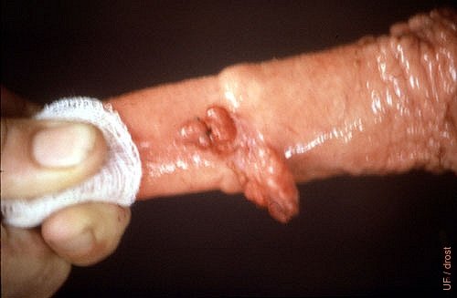
Small Penile Warts.
Several small fibropapillomas are present at the preputial reflection of the penis. They are of no functional significance at this point.
Drost M (1980)
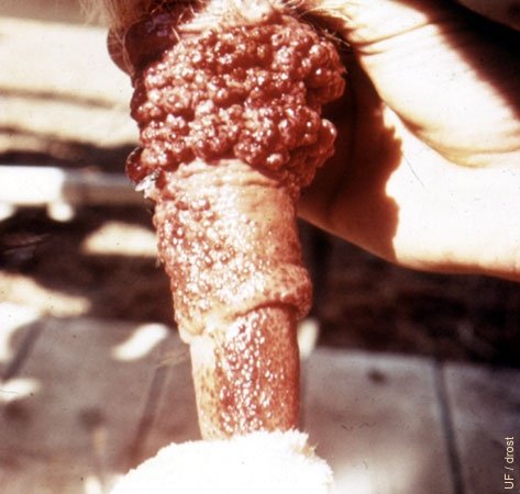
Granular Venereal Disease.
"Granular venereal disease" is an old term denoting the presence of enlarged lymph follicles on the surface of the penis and prepuce, in response to an irritant which may be infectious or chemical.
Roberts SJ (1973)
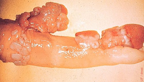
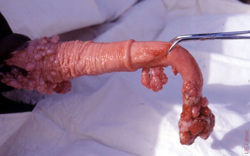
Multiple Fibropapillomata.
Multiple diffusely attached fibropapillomata are more difficult to remove surgically due to problems with hemostasis.
Larsen RE (2001)
Multiple Small Fibropapillomas.
This bull can be treated by ligation and excision of the pendulous tissues. Bleeding may result in autoimmunization against the wart virus. Some of the warts near the tip are traumatized.
Larsen RE (2001)
Diffuse Fibropapillomas.
An autogenous vaccine can be prepared from some of the fibropapillomatous tissue. Excision is problematic due to the diffuse nature and blood supply.
Larsen RE (2001)
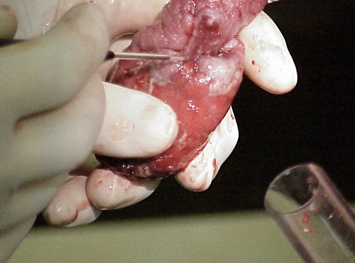
Large Fibropapilloma.
Large fibropapilloma covering the tip of the penis towards the bottom of the image. Normal pink tissue of the penis above.
Shipley C (2006)
Electrocautery.
The electrocautery probe is aimed at the junction between the large fibropapilloma at the tip of the penis and the normal pink tissue of the penis above.
Shipley C (2006)
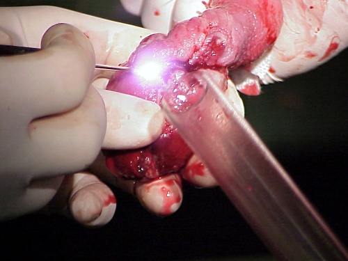
Electrocautery in Progress.
The electrocautery probe is applied to the junction between the large fibropapilloma at the tip of the penis and the normal pink tissue of the penis above. A glass vaginal speculum is used to protect some of the normal tissue of the penis.
Shipley C (2006)
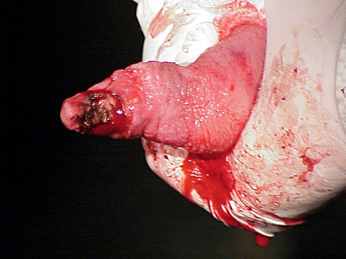
After Electrocautery.
The large consolidated fibropapilloma mass has been removed with electrocautery.
Shipley C (2006)
Laceration of the Penis.
Laceration at the junction of the penis and the preputial reflexion.
Larsen RE (2001)
Laceration of the Penis.
This glans penis was lacerated on a broken glass vial during semen collection with an artificial vagina.
Wright JM (1973)
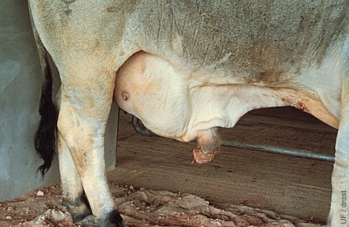
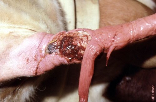
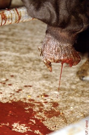
Amputated Preputial Prolapse.
The bull stepped on his own preputial prolapse and traumatically amputated it. Loss of internal lamina of the prepuce resulted in immediate formation of adhesions around the penis.
Larsen RE (2001)
Stepped-on Prolapse.
The bull stepped on his own preputial prolapse and traumatically amputated it. Loss of internal lamina of the prepuce resulted in immediate formation of adhesions around the penis.
Larsen RE (2001)
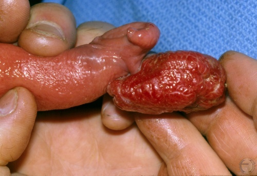
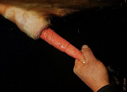
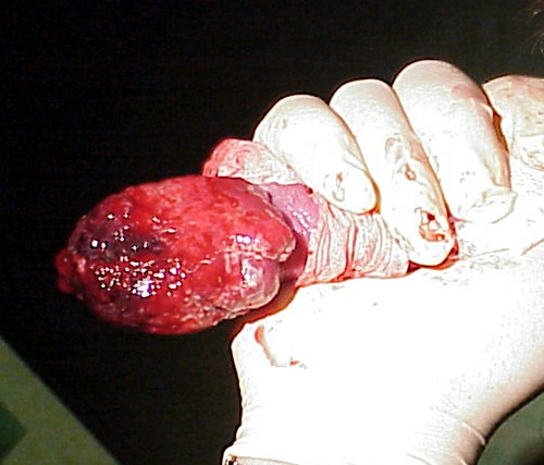
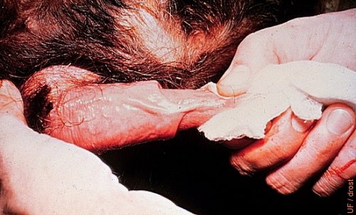
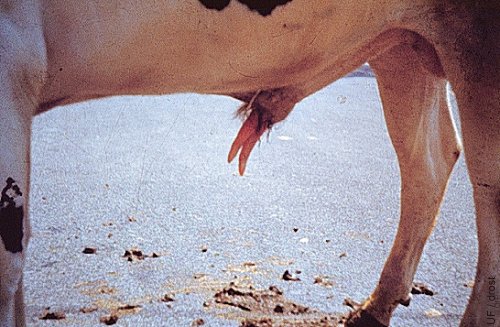
Rare Preputial Injury 1.
Detachment and prolapse of the prepuce with exposed subcutaneous surface of inner lamina hanging from the penis.
Larsen RE (2001)
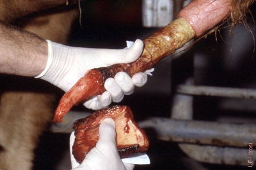
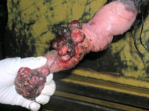
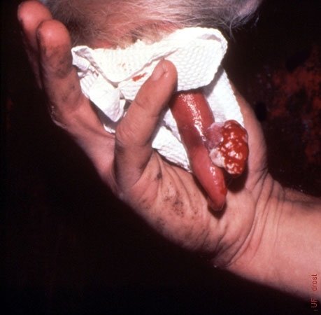
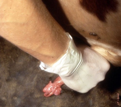
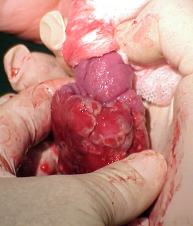
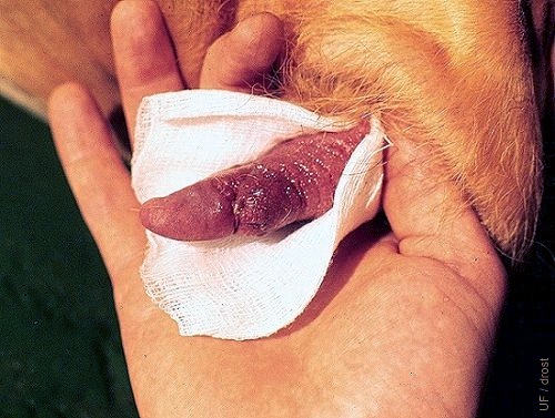
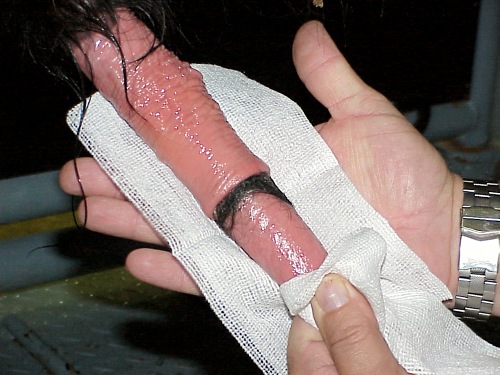
Rare Preputial Injury 2.
Detachment and prolapse of the prepuce with exposed subcutaneous surface of inner lamina hanging from the penis. The prolapsed tissue was surgically removed, and is shown in the lower hand.
Larsen RE (2001)
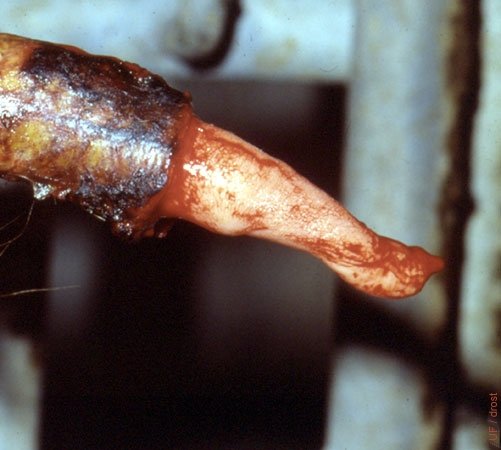
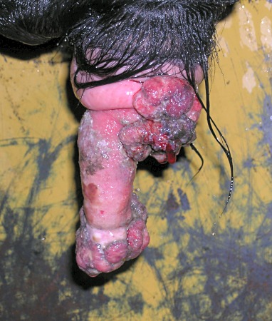
Rare Preputial Injury 3.
Detachment and prolapse of the prepuce with exposed subcutaneous surface of inner lamina hanging from the penis, shown here after removal of the prolapsed tissue. The bull did not return to service.
Larsen RE (2001)
Traumatic Balanitis.
Trauma to the glans penis after paralysis of the penis.
Roberts SJ (1973)
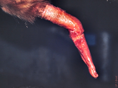
Persistent Frenulum.
Congenital persistent frenulum, usually detected at the time of a Breeding Soundness Evaluation (BSE). The persistent band limits extension of the penis, and may cause deviation of the penis. These bulls should be culled due to the hereditary nature of the condition. The band can be severed and the bull kept for natural service provided the offspring is not kept for breeding purposes. Proximal ligation prior to cutting is recommended because of the common presence of a well developed artery within the band.
Larsen RE (1982)
Diphallus.
Congenital diphallus, a rare duplication of the glans penis in the bull. In the vernacular referred to as "double dick".
Roberts SJ (1973)
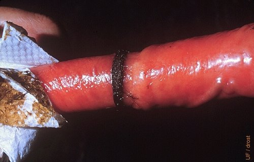
Hair Ring.
Accumulation of hair around the base of the glans may lead to constriction and stenosis. This sometimes occurs in young bulls as a result of homosexual behavior whereby they draw hair into the prepuce.
Drost M (1980)
Hair Ring.
Encircling of the base of the glans by hair may lead to constriction and stenosis. This sometimes occurs in young bulls as a result of homosexual behavior whereby they draw hair into the prepuce.
Shipley C (2006)
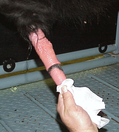
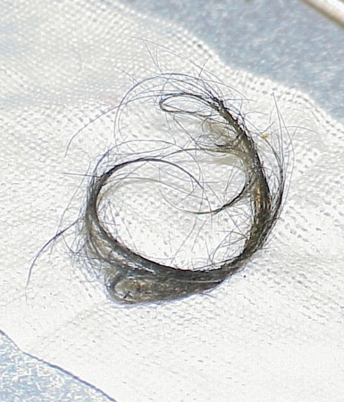
Hair Ring.
Hair trapped by the circular fold of the base of the penis may lead to constriction and stenosis.
Shipley C (2006)
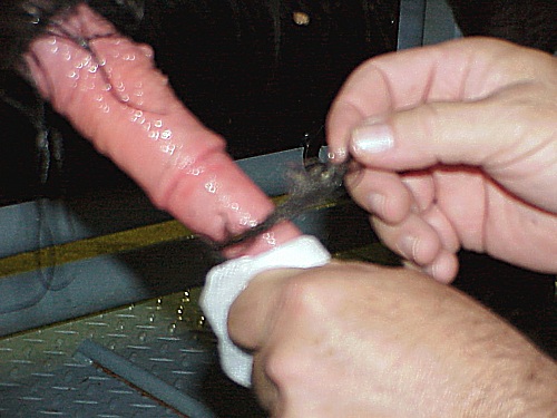
Hair Ring.
Hair trapped by the circular fold of the base of the penis can readily be manually removed.
Shipley C (2006)
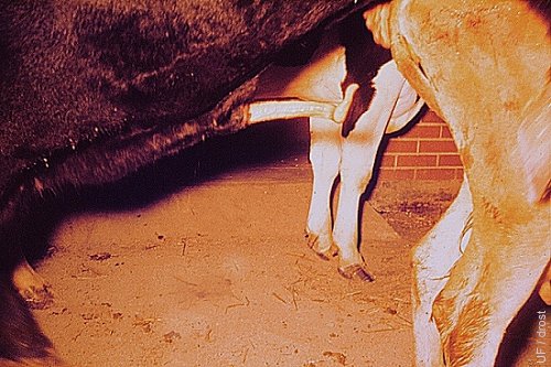
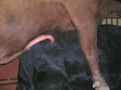
Spiral Deviation.
Spiral deviation of the penis in a 3 year old Angus bull at full erection. When this occurs prior to intromission penetration will be difficult. Studies with a transparent artificial vagina have shown that spiral deviation after intromission occasionally occurs in some bulls at the height of erection.
Roberts SJ (1973)
Spiral Deviation Close Up.
Spiral deviation of the penis at full erection. When this occurs prior to intromission penetration will be difficult.
Ott R (2013)
Spiral Deviation Close Up.
Incipient spiral deviation of the penis prior to full erection.
Ott R (2013)
Penile Deviation.
Deviation of the tip of the penis at full erection. When this occurs prior to intromission, penetration will be difficult. Studies with a transparent artificial vagina have shown that spiral deviation after intromission occasionally occurs in some bulls at the height of erection.
Hopper RM (2008)
Failure of Intromission.
When spiral deviation of the tip of the penis at full erection occurs prior to intromission, the latter will usually fail. This can lead to trauma and hematoma of the penis.
Hopper RM (2008)
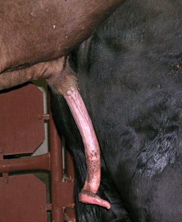
Rainbow Deviation.
Ventral deviation of the penis that prevented intromission in an Angus bull.
Drost M (1987)
Rainbow Deviation.
Ventral or rainbow deviation in a Hereford bull due to weak dorsal ligaments of the penis. Intromission is a problem.
Roberts SJ (1973)
Ventral Deviation.
Weak or injured dorsal ligament of the penis. Scar tissue was present along the dorsal aspect of the penis.
Larsen RE (2001)
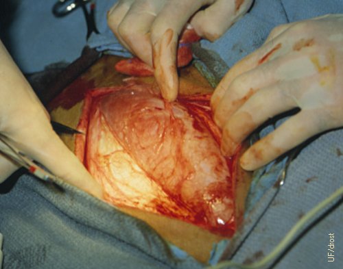
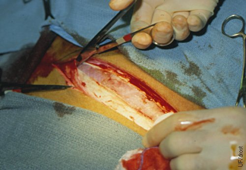
Deviation Correction - Exposure of the Vastus Lateralis.
General anesthesia is required for this procedure. A strip of fascia is removed from the vastus lateralis muscle dorsal to the patella in the direction of the tuber coxae. A 3 cm wide and 12 cm long strip of fascia is removed from the deep tougher layer covering the vastus lateralis muscle.
Larsen RE (1985)
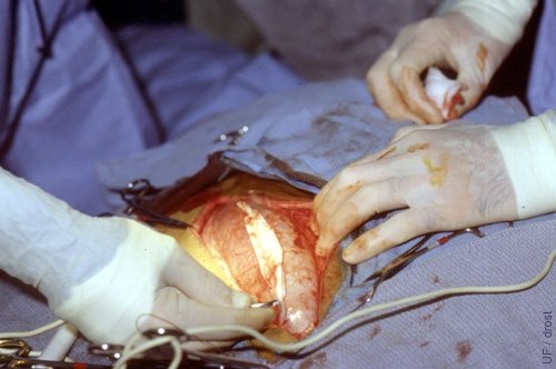
Penis Deviation Correction - Incision of the Fascia Lata.
General anesthesia is required for this procedure. A strip of fascia is removed from the vastus lateralis muscle dorsal to the patella in the direction of the tuber coxae. A 3 cm wide and 12 cm long strip of fascia is removed from the deep tougher layer covering the vastus lateralis muscle.
Larsen RE (1985)
Penis Deviation Correction - Incision of the Fascia Lata.
A strip of fascia is removed from the vastus lateralis muscle dorsal to the patella in the direction of the tuber coxae. A 3 cm wide and 12 cm long strip of fascia is removed from the deep tougher layer covering the vastus lateralis muscle.
Larsen RE (1985)
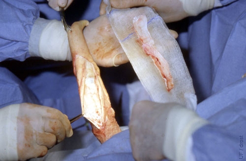
Penis Deviation Correction - Isolated Strip of Fascia.
A 3 cm wide and 12 cm long strip of fascia, removed from the deep tougher layer covering the vastus lateralis muscle, is transferred to the dorsal ligament of the penis.
Larsen RE (1985)
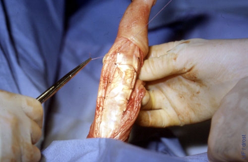
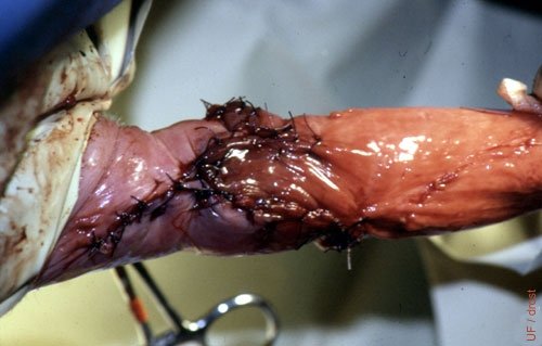
Penis Deviation Correction - Graft in Place.
Fascial strip sutured into place along the dorsal aspect of the penis.
Larsen RE (1985)
Penis Deviation Correction - Graft Sutured in Place.
The fascial graft has been sutured in place.
Larsen RE (1985)
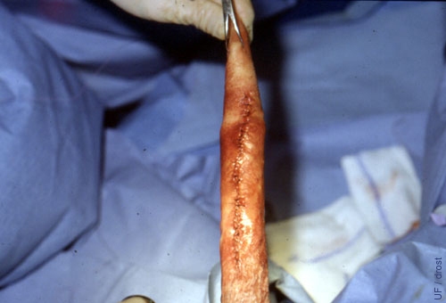
Penis Deviation Correction - Closed Incision Line.
Final closure of the incision line.
Larsen RE (1985)