The Visual Guide to
Bovine Reproduction
Placenta: Abnormal Placenta

Bovomane.
The yellow portion of these expelled fetal membranes is a bovomane which is a rubbery allantoic concretion (calculus) that gels from waste products in the allantoic sac.
Smith MC (2020)
Adventitious Placentation.
The outer surface of the allantoic membranes shows dark red specks which are very small scattered cotyledonary attachments of the allantois to the endometrium.
Smith MC (2020)

Expelled Fetal membranes.
These fetal membranes were spontaneously expelled and still contained a dead fetal twin calf.
Smith MC (2020)

Expelled Fetal membranes.
These fetal membranes were spontaneously expelled and still contained a dead fetal twin calf. Notice the leg of the calf. The entire picture resembles a prolapsed uterus.
Smith MC (2020)
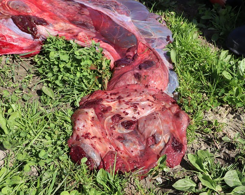
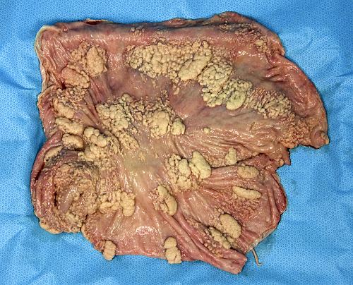
Severe Adventitious Placentation.
Multiple random islets of cotyledonary tissue surround a normal cotyledon. Adventitious placentation can lead to placental dysfunction and result in hydrallantois.
Roberts SJ (1973)
Early Adventitious Placentation.
Small adventitious specks can be seen next to normal cotyledons.
Drost M (1978)
Adventitious Placentation.
The maternal caruncle is surrounded by small scattered areas of adventitious caruncular tissue. The caruncle shown is not entirely intact.
Drost M (1978)
Adventitious Caruncular Tissue.
Third trimester abnormal placentation. Adventitious placentation. This generally leads to hydrops allantois.
Drost M (2009)
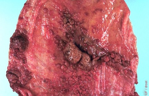
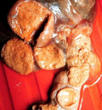
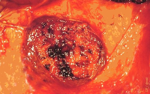
Transsected Giant Caruncle.
Term placenta showing an abnormally large transected caruncle after the delivery of a normal. Calf was by a 2-year old Holstein heifer.
DeHoogh W (2011)
Spurious Giant Caruncle.
Term placenta showing an abnormally large caruncle after delivery of a normal live calf by a 2-year old Holstein heifer. Possible coalescence of several caruncles.
DeHoogh W (2011)
Retained Fetal Membranes.
The fetal membranes are normally expelled within 2 to 6 hours after calving. If they are not delivered by 12 hours they are considered retained.
Drost M (1978)
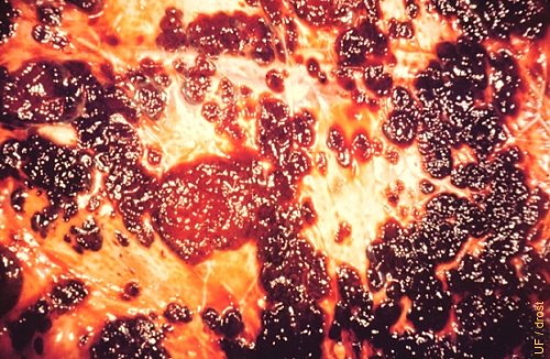
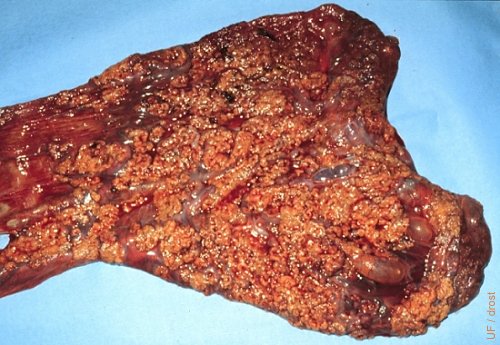
Necrotic Mycotic Placentitis.
The gross appearance of this fungal placentitis resembles that of a placenta infected with brucellosis or campylobacteriosis. Abortion occurred around 6 months of gestation.
Roberts SJ (1973)
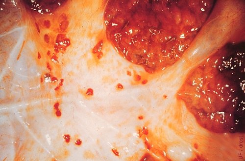
Necrotic Adventitious Placentitis.
Necrotic adventitious caruncular tissue in a heifer. The caruncular tissue had lost its blood supply upon early termination of the pregnancy.
Haibel GK (1983)
Placental Anomaly.
The anterior 60 cm of the gravid horn was devoid of placentomes. The fetal membranes of the right horn contained 60 cotyledons (12 to 14 mm in diameter), while the nongravid horn contained 16 cotyledons (4 to 5 mm in diameter).
Roberts SJ (1973)
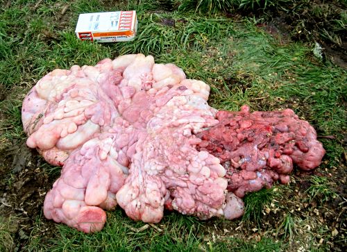
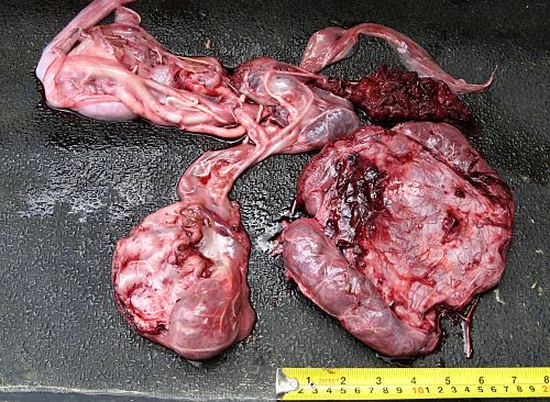
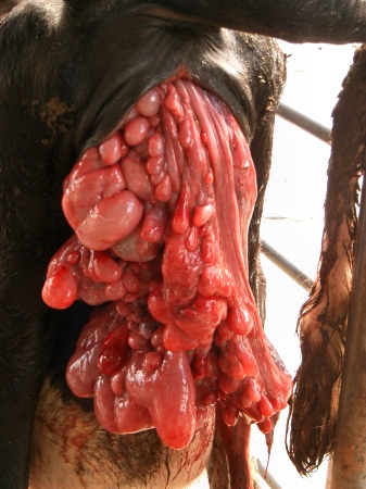
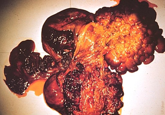
Hydatidiform Mole.
Extensive formation of grape-like clusters of translucent cysts and villi of the placenta expelled along with the fetal membranes of a live calf at term. The length of the box of palpation sleeves at the top of the picture is 25 cm.
Leduc F (2007)
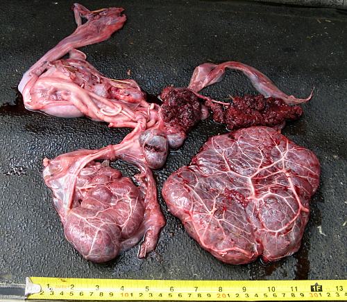
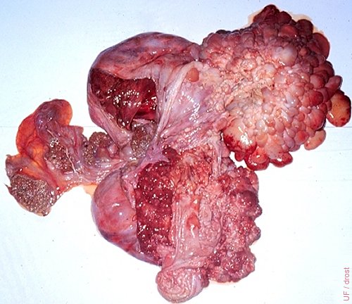
Hydatidiform Mole.
This huge grape-like cluster of placental cysts or villi appeared at the time of calving. A difficult-to-reach live normal calf was subsequently delivered per vaginam; with assistance.
Leduc F (2007)
Cystic Placental Mole.
This is a highly unusual cystic placental mole that followed embryonic death, after which the placenta continued to grow to the size of 3 to 4 month pregnancy; with retention of the corpus luteum. This condition should not be confused with a hydatidiform mole which accompanies a live calf.
Roberts SJ (1973)
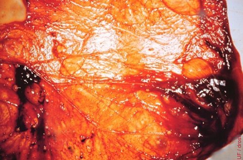
Early Necrosis.
This cotyledon shows early evidence of hemorrhage and necrosis.
Drost M (1978)
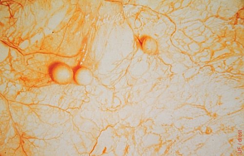
Small Amniotic Cysts.
Small, incidental, epithelial cysts of the amnion. No known clinical significance.
Roberts SJ (1973)
Tumor of the Trophoblast.
Tumor of the trophoblast: a hydatidiform cystic mole.
Roberts SJ (1973)