The Visual Guide to
Bovine Reproduction
Reproductive Technology: In Vitro Embryos
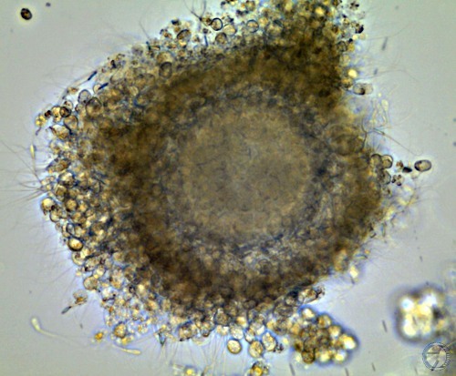
Mature Cumulus Oocyte Complex.
Oocyte complex matured in vitro is surrounded by spermatozoa.
Denicol AC (2013)
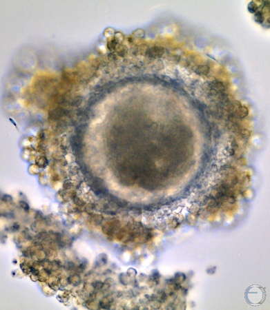
Mature Cumulus Oocyte Complex.
In-vitro fertilization in process. Note a spermatozoon on the left. A piece of the cuulus cell mass has separated at the lower left part of the image.
Denicol AC (2013)
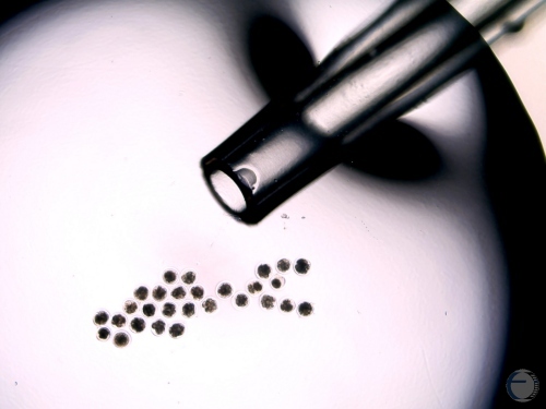
Adding Supplement to Culture Medium.
A pipet is used to add nutrients to the culture medium.
Denicol AC (2013)
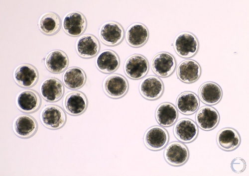
IVF Embryos.
Morulae on Day 5. Several unfertilized, single cell ooctyes are present. The single cell oocyte on the far lower right side shows a polar body.
Denicol AC (2013)
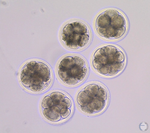
Early IVF Morulae.
Early morulae with identifiable blastomeres, compact morulae, and unfertilized ova.
Denicol AC (2013)
4-cell IVF Embryos.
Day 3 4-cell to 8-cell embryos and several unfertilized ova (UFO).
Denicol AC (2013)
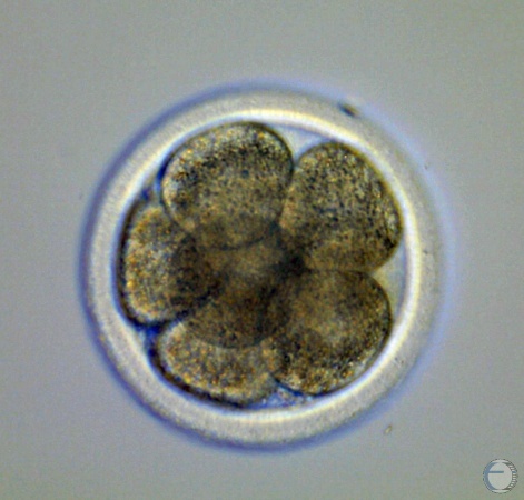
8-cell IVF Embryo.
All 8 blastomeres are of the same size and shape.
Denicol AC (2013)
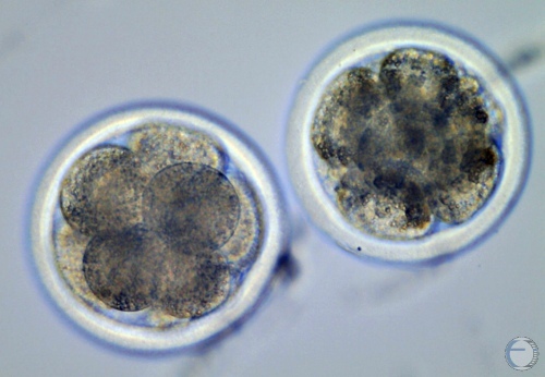
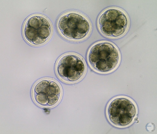
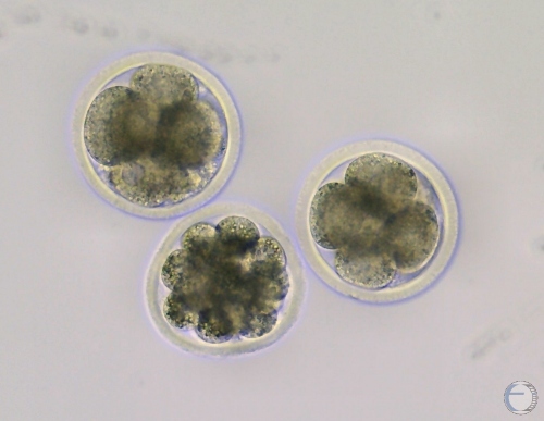
Day 4 to 5 IVF Embryos.
8-cell to 16-cell in-vitro embryos on day 4 to 5 of incubation.
Denicol AC (2013)
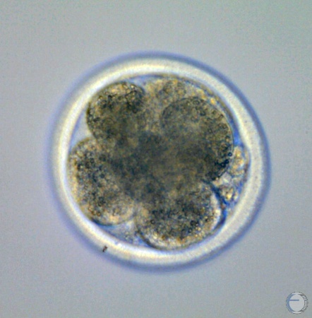
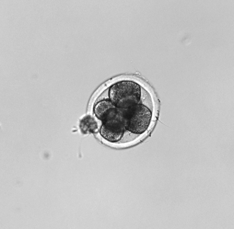
Day 4 to 5 IVF Embryo.
8-cell embryo on day 4 to 5. Note the small polar body at the 4 o'clock position.
Denicol AC (2013)

16-cell IVF Embryo.
16-cell IVF embryo on day 5 after fertilization.
Denicol AC (2013)
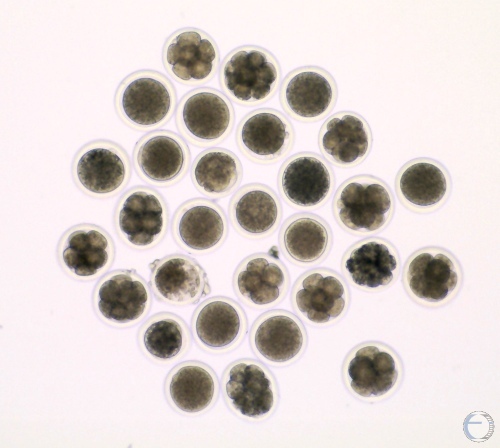
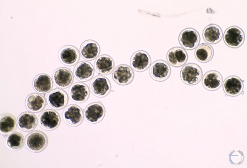
Multiple Day 3 IVF Embryos.
4- to 8-cell embryos, no morulae yet, many UFOs
Denicol AC (2013)
Day 3 IVF Embryos.
Two 4-cell and one 8-cell IVF embryos on Day 3 after fertilization.
Denicol AC (2013)

IVF 2-cell Embryos.
Two-cell embryos 28 to 32 h after in-vitro fertilization [Stage 2]. The embryo on the far right shows signs of degeneration as the cytoplasm is shrinking away from the plasma membrane.
Hansen PJ (2006)
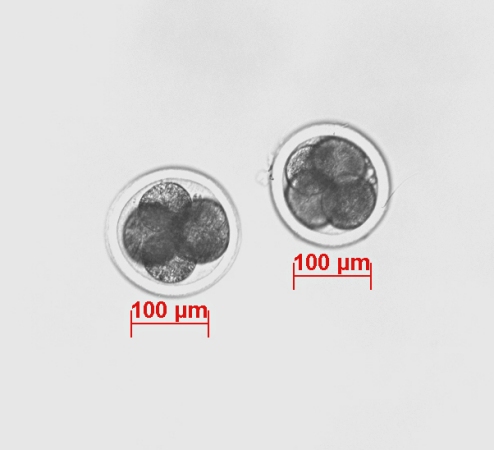
IVF 4-cell Embryos.
Four-cell embryos 40 to 44 h after in-vitro fertilization [Stage 2].
Hansen PJ (2006)
IVF 6-8 cell Embryos.
Six-cell embryo [Stage 2]. A clump of cumulus cells remains adhered to the zona pellucida. There also appear to be a few accessory spermatozoa attached to the zona pellucida.
Hansen PJ (2006)

IVF 8-cell Embryos.
Eight-cell embryos [Stage 2] about 3 days after in-vitro fertilization. The embryo on the right shows signs of pyknosis, which at this point may be an artifact.
Hansen PJ (2006)
IVF 8-cell Embryo.
Eight-cell embryo with a few remaining cumulus cells attached to the zona pellucida [Code 1]. [Code 1: excellent or good; Code 2: fair; Code 3: poor; Code 4: dead or degenerating].
Hansen PJ (2006)

IVF Early Morula.
Early morula [Stage 3, Code 2]. Blastomeres of in-vitro embryos do not compact as tightly as those of in-vivo embryos. [Code 1: excellent or good; Code 2: fair; Code 3: poor; Code 4: dead or degenerating].
Hansen PJ (2006)

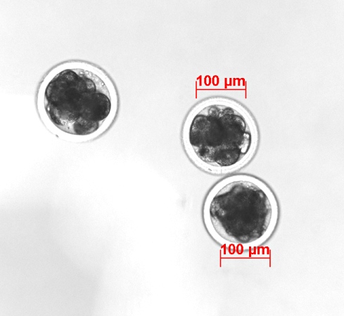
IVF Early Morulae.
Early morulas [Stage 3, Code 1]. IVF embryos tends to be darker than in-vivo embryos. [Code 1: excellent or good; Code 2: fair; Code 3: poor; Code 4: dead or degenerating]
Hansen PJ (2006)
IVF Early Morulae.
Day 5 to 6 after insemination. The embryo at the left is a Code 2. The embryos on the right are Code 1. A more precise evaluation can be made by rolling the embryos under the microscope to gain a three dimensional perspective, and with additional incubation time.
Hansen PJ (2006)
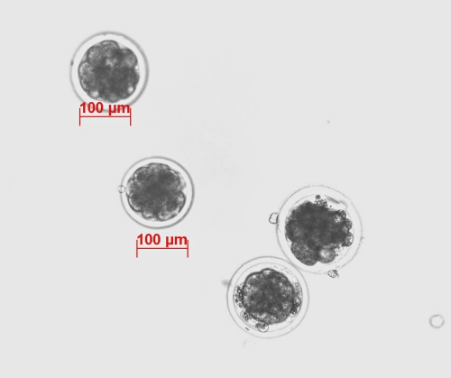
IVF Early Morulae.
Early IVF morulas [Stage 3] Day 5 to 6 after insemination. Top two embryos: Code 1. Bottom left embryo is starting to compact and is a Code 1. The bottom right embryo is a Code 3 (pyknosis and several extruded blastomeres).
Hansen PJ (2006)

IVF Degenerating Early Morulas.
At 8 days after insemination these three embryos resemble early morulas and look degenerated (Code 4). They should have reached the blastocyst stage by day 8.
Hansen PJ (2006)

IVF Morulas.
IVF morulas [Stage 4]. Top left embryo is a Code 1, top right a Code 2. Center left and right embryos are Code 1. Bottom left embryo is a Code 2, bottom right embryo is a Code 1. The latter has a large extruded early blastomere.
Hansen PJ (2006)

IVF Degenerating Morula.
Code 2 morula [Stage 4]. In-vitro fertilized embryos are darker than in vivo embryos. There is a large extruded blastomere at the 5 o'clock position.
Hansen PJ (2006)

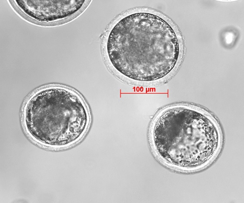
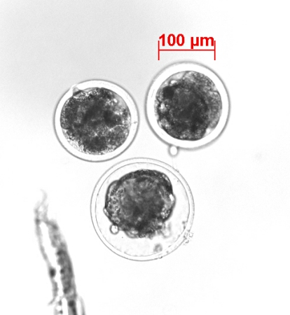
IVF Blastocysts.
The blastocyst stage is normally reached by day 6 to 7 after in-vitro fertilization. Both embryos are Stage 6 and Code 1. The blastocele is starting to noticeably enlarge in the blastocyst on the right. [Code 1: excellent or good; Code 2: fair; Code 3: poor; Code 4: dead or degenerating].
Hansen PJ (2006)
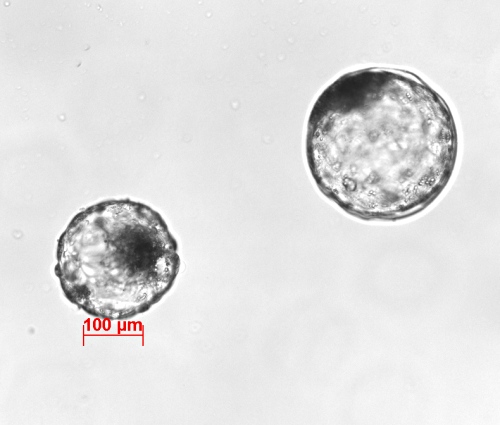
IVF Blastocysts.
Code 1 blastocysts. The top embryo is beginning to expand. The blastocele of the lower right embryo is expanding making the dark inner cell mass more distinct.
Hansen PJ (2006)
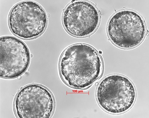
IVF Expanding Blastocysts.
Blastocysts in the process of expanding (Stage 6 to 7). All are Code 1. The thin zona pellucida of the embryo in the center appears compromised and may be at the point of rupture (hatching).
Hansen PJ (2006)

IVF Expanded Blastocysts.
Expanded blastocysts [Stage 7] on the verge of hatching (note the diameter). The smaller embryo on the left has started to collapse within its zona pellucida. All are Code 1.
Hansen PJ (2006)
IVF Collapsed Expanded Blastocyst.
While the thin zona pellucida is still intact, the blastocele of the embryo on the bottom has collapsed (Code 1). The top two embryos are Code 4 because of the stage of development is retarded for their age (day 8). [Code 1: excellent or good; Code 2: fair; Code 3: poor; Code 4: dead or degenerating].
Hansen PJ (2006)

IVF Hatching Blastocyst.
Hatching blastocyst (Stage 8, Code 1). The stretched, thin zona pellucida has ruptured at the bottom of the image. The dark inner cell mass is about to exit first. Note the large diameter.
Hansen PJ (2006)
IVF Hatched Blastocyst.
The smaller embryo is hatched blastocyst [Stage 8]. The blastocele is re-expanding and the embryo re-assumed its spherical shape. The dark inner cell mass is clearly delineated. The other embryo is still surrounded by its very thin zona pellucida.
Hansen PJ (2006)

IVF Hatched Blastocysts.
Recently hatched blastocysts. The top embryo is re-expanding (Stage 9). The bottom embryo is a Stage 8. The embryos are surrounded by trophoblast cells
Hansen PJ (2006)
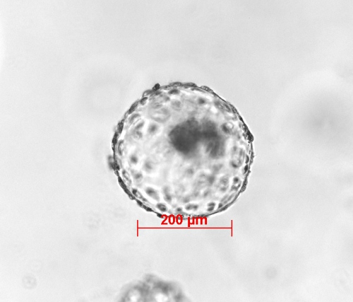
IVF Expanded Hatched Blastocyst.
Nice expanded hatched blastocyst (Stage 9, Code 1). The dark crescent shaped mass is the inner cell mass. The embryo is surrounded by trophoblast cells. Day 8 to 9 after insemination.
Hansen PJ (2006)
IVF Hatched Blastocyst and Zona.
Collapsed hatched blastocyst [Stage 8] with dark inner cell mass and dark peripheral circle of trophoblast cells. The empty zona pellucida is nearby.
Hansen PJ (2006)

Empty Zona Pellucida.
The thin, stretched, ruptured, empty zona pellucida.
Hansen PJ (2006)

IVF Unfertilized Ova.
Picture taken eight days after in vitro insemination. The bottom two oocytes are unfertilized [Stage 1], the top two are fertilized, retarded and degenerated, a 4-cell and 8-cell respectively. Code 4.
Hansen PJ (2006)
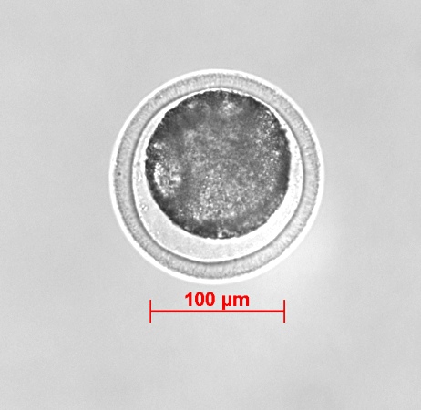
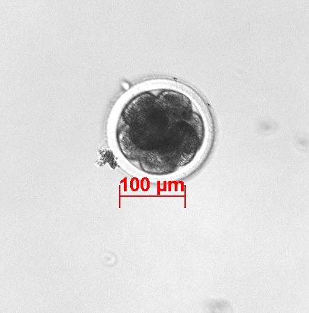
Unfertilized Oocyte.
After several days of incubation there is no evidence of cell division. The contour of the cell mass is smooth in contrast with the slight scalloped surface of a compact morula, which may show the same percentage of perivitelline space. The two can easily be mistaken for one another.
Hansen PJ (2006)
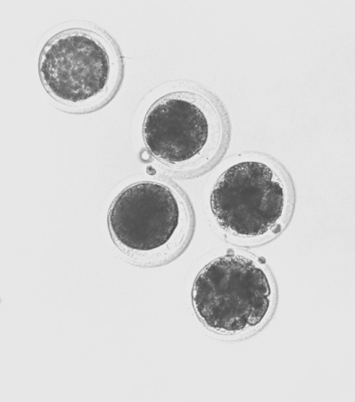
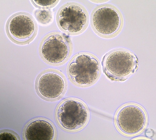
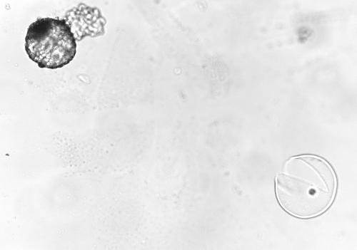
IVF UFOs.
Three days after insemination. The upper left oocyte may have come from an atretic follicle. Its quality is poor based on its granular, light appearance. The center two oocytes appear normal but did not become fertilized. The two embryos on the right are a 4-cell and an 8-cell respectively [Stage 2].
Hansen PJ (2006)