The Visual Guide to
Bovine Reproduction
Ultrasonography: Gender Diagnosis
Fetal Sexing.
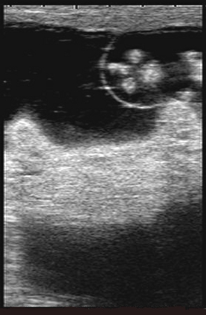
In this oblique view, the tail and female genital tubercle are seen just to the left and right of cross sections of hind limbs. Cross sections of forelimbs and the head are seen to the right.
Colloton J (2006)
Gender Determination.
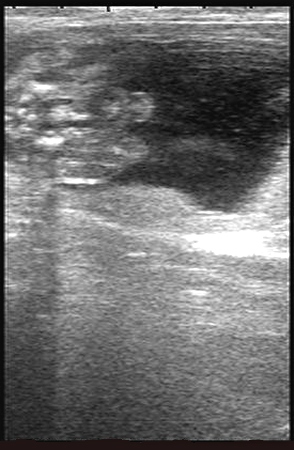
The soft tissue of the scrotum is visible between the thighs. When diagnosing fetal gender, it is not advisable to base the diagnosis on soft tissue structures such as scrotum or teats. The genital tubercle must be identified for the most accurate diagnosis.
Colloton J (2006)
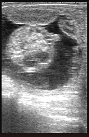
Female Fetus.
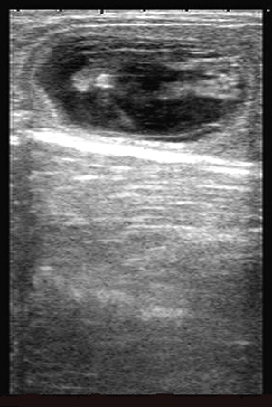
Note the tarsi and cloven hooves to the right. To the left, note the mono-lobed cross section of the tail and the bi-lobed cross section of the female genital tubercle.
Colloton J (2006)
Female Genital Tubercle.
In this frontal view the tail and female genital tubercle are apparent to the left. Note the V-shaped rib cage. The rib cage is a good landmark for cranial / caudal orientation.
Colloton J (2006)
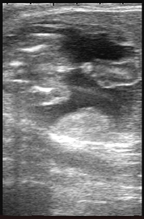
Male Fetus.
In this cross sectional view, the male genital tubercle is seen at 2 o'clock. In a still frame, it is difficult to be sure the protrusion is the genital tubercle and not a portion of the umbilicus. Note the non-echogenic fetal stomach.
Colloton J (2006)
Male Fetus.
The pelvic bones are distinctly seen to the left on this image. The umbilicus and bright male genital tubercle are seen in the center of the image.
Colloton J (2006)
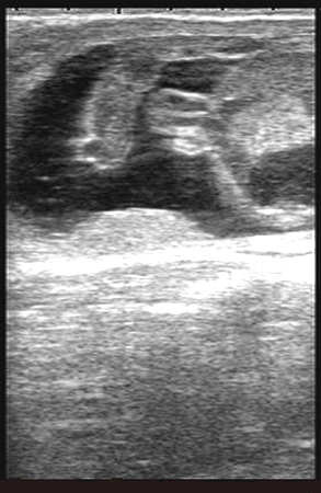
Male Genital Tubercle.
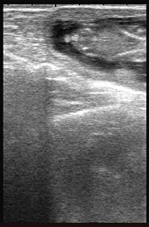
In this cross sectional view of the abdomen the thick umbilicus is seen entering the body. The bright male genital tubercle is visible just below the umbilicus.
Colloton J (2006)