The Visual Guide to
Bovine Reproduction
Teratology: Fetal Monsters
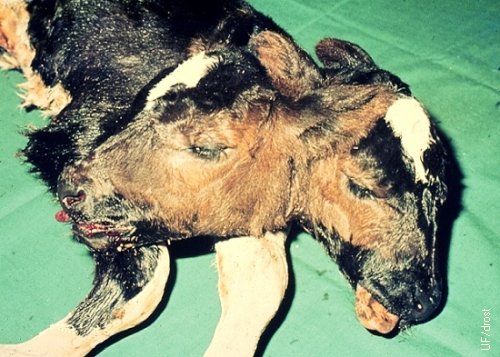
Two-Headed Calf.
This two-headed calf is an incomplete set of twins. This bicephalic calf had one body. The conformation may lead to dystocia. Vaginal delivery is effected by partial fetotomy if the calf is dead, or after severance of the umbilical cord.
Utrecht (1976)
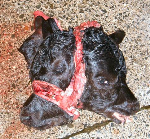
Partial Fetotomy.
The head of this term bicephalic calf was reduced in size by making two cuts with the fetatome, to enable vaginal delivery.
Leduc F (2007)
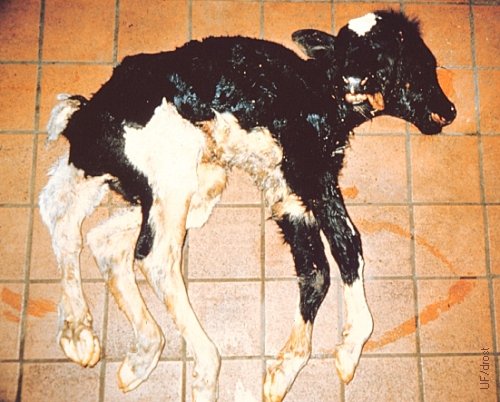
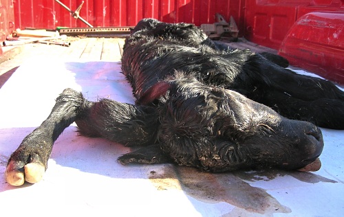
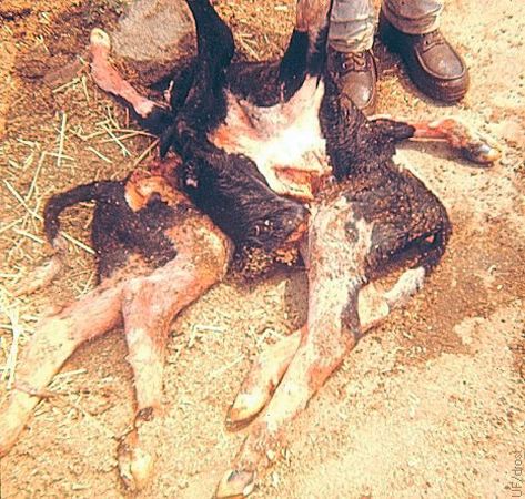
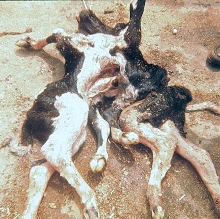
Two-Headed, Five-Legged Calf.
Term fetal monster with two asymmetrical heads and a parasitic (extra) pelvic limb.
Roberts SJ (1973)
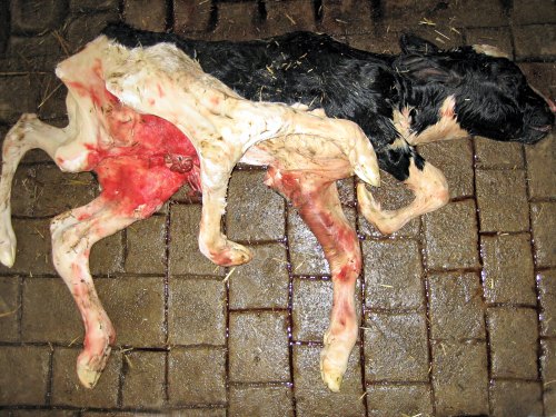
Six-legged Calf.
This six-legged Holstein calf was stillborn at term to a pluriparous cow. The delivery required only minimal assistance. The head, tail and four other limbs appeared normal. The extra set of hindlimbs narrowed as they approached the pelvis and were attached near the head of right femur.
Roodzant JP (2007)
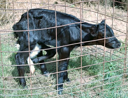
Polymelia - Incomplete Twin.
Six-legged Holstein calf. Aberrant duplication of the development of the hind legs during the early embryonic stage.
Mayo D (2013)
Polymelia - Incomplete Twin.
Right lateral view of a six-legged Holstein calf. Aberrant duplication of the development of the hind legs during the early embryonic stage. Two rear legs and a small part of an extra abdominal wall are shown between the main hind legs.
Mayo D (2013)
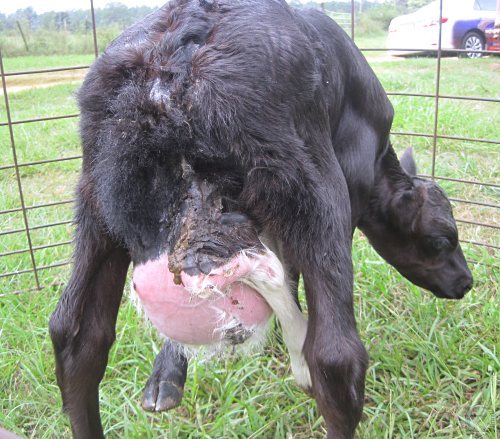
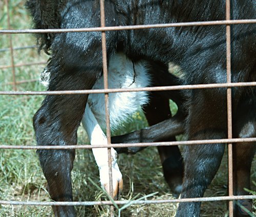
Polymelia - Incomplete Twin.
Rear view of a six-legged Holstein calf. Aberrant partial duplication of the development of the hind legs during the early embryonic stage. Two rear legs and a small part of an extra abdominal wall are shown between the main hind legs.
Mayo D (2013)
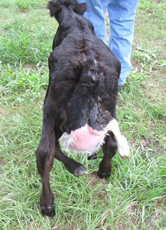
Polymelia - Incomplete Twin.
Rear view of a six-legged Holstein calf. Aberrant partial duplication of the development of the hind legs during the early embryonic stage. Two rear legs and a small part of an extra abdominal wall are shown between the main hind legs.
Mayo D (2013)
Three-legged Angus Calf.
Complete congenital absence of the left front leg.
Dow CA (2009)
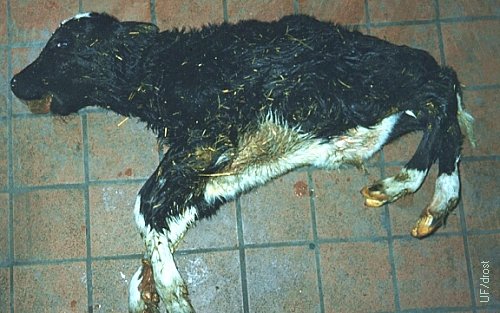
Three-legged Angus Calf.
Complete congenital absence of the left front leg.
Dow CA (2009)
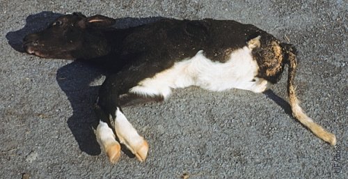
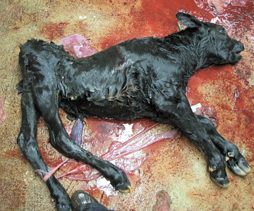
Three-legged Holstein Calf.
Complete congenital absence of the left hind leg.
Varsano S (2012)
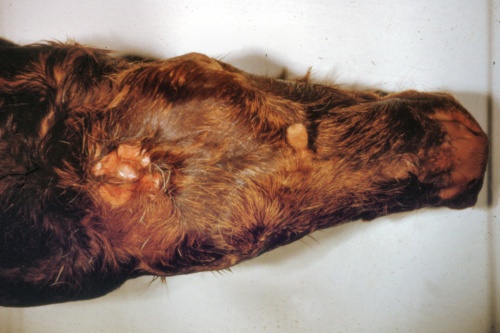
Parasitic Limb.
This Angus calf was born with a parasitic forelimb protruding from the right side of its poll, a rare occurrence. A more common location for a parasitic limb is the area of the whithers.
Miskimins DW (2009)
Parasitic Limb.
This Angus calf was born with a parasitic forelimb protruding from the right side of its poll, a rare occurrence. A more common location for a parasitic limb is the area of the whithers.
Miskimins DW (2009)
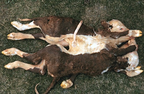
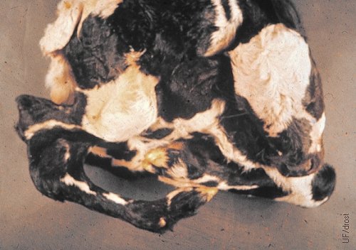
Conjoined Hereford Twins.
Complete Hereford twins conjoined at the chest (thoracopagus).
Drost M (1984)
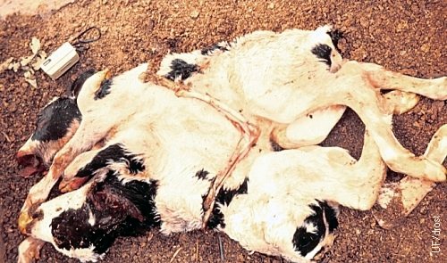
Conjoined Holstein Twins.
Complete Holstein twins conjoined at the chest. One head was severed with the fetatome to enable vaginal delivery.
Drost M (1984)
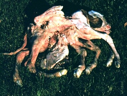
Conjoined Twins.
These conjoined twins were born along with a small normal calf at term. They were dipygus, tetrapus, tetrabrachius, monocephalus, and had 4 kidneys and a hernia.
Roberts SJ (1973)
Conjoined Twins.
Twins conjoined at the chest and with eight legs and one head.
Drost M (1984)
Conjoined Twins.
Twins conjoined at the thorax (thoracopagus) and with eight legs and one head.
Drost M (1984)

Conjoined Twins.
Twins conjoined at the hindquarters with two pelvises and six legs.
Varsano S (2012)
Cyclops.
Cyclops found at the slaughter house. This calf weighed 60 kg, had long hair and did not have a pituitary gland, suggestive of postmaturity as a result of failure by the calf to initiate parturition.
Drost M (1968)
Fetal Giant.
Lateral view of the skull of a postterm (12 months) fetal giant showing a cerebral hernia. While the brain was defective, the pituitary gland was normal in this Guernsey fetus, yet the fetus did not initiate parturition. The hypothalamus may have been deranged.
Roberts SJ (1973)
Fetal Giant.
Dorsal view of the skull of a postterm (12 months) fetal giant showing a cerebral hernia. While the brain was defective the pituitary gland was normal in this Guernsey fetus.
Roberts SJ (1973)
Perosomus Horridus.
Perosomus horridus is a fetal monster with generalized, severe ankylosis and muscle contractures. The spine is short due to double S-shaped lateral twisting of the vertebrae.
Roberts SJ (1973)
Perosomus Elumbis.
Perosomus elumbis; complete lack of development of the hindquarters.
Roberts SJ (1986)
Perosomus Elumbis.
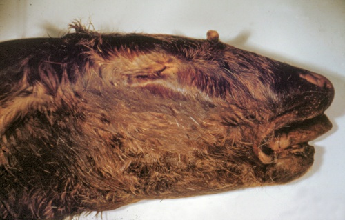
Perosomus elumbis; lack of development of the hindquarters and ankylosis of the hind limbs. The calf also has an abnormally short mandible (brachygnathia; "sow mouth").
Roberts SJ (1973)
Perosomus Elumbis.
This severe congenital defect is characterized by vertebral defects caudal to the thoracic region, and a deformed pelvis. In addition there is ankylosis of the rear limbs.
Willis LA (2007)
Congenital Dropsy.
Congenital dropsy and anasarca in a 5-month old aborted, crossbred fetus. The cow was a Red Holstein, the sire a Hinterwaelder ("Backwoods") bull.
Benz M (2009)
Fetal Anasarca.
Congenital dropsy and anasarca in a 5-month old aborted, crossbred fetus. The cow was a Red Holstein, the sire a Hinterwaelder ("Backwoods") bull.
Benz M (2009)
Abdominal Dropsy.
Congenital dropsy and anasarca in a third trimester Holstein fetus. The distended abdomen had to be lanced to permit vaginal delivery.
Shipley C (2006)
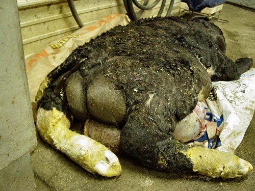
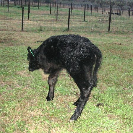
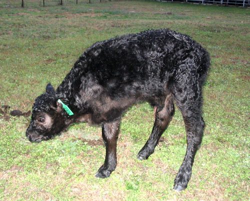
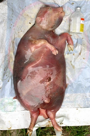
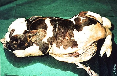
Pulmonary Hypoplasia with Anasarca.
Posterior view of a Maine-Anjou fetus with Pulmonary Hypoplasia with Anasarca (PHA). The huge bilateral swelling is due to fluid, not muscle. The scrotum is the size of a 2-year old bull and is also filled with fluid. The calf was delivered by cesarean section.
Kaiser L (2007)
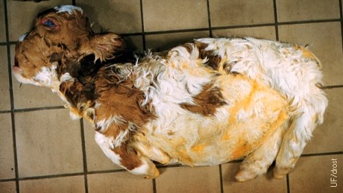
Achondroplasia (Bulldog Calf).
Achondroplastic term Ayrshire calf (bulldog calf). [size of the square tiles is 15 cm].
Roberts SJ (1986)
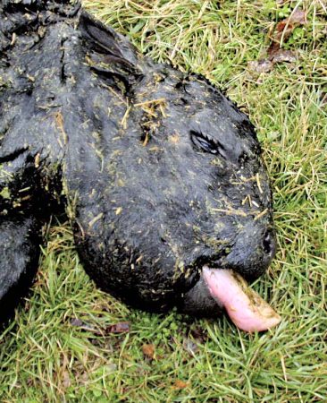
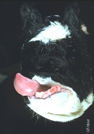
Pulmonary Hypoplasia with Anasarca.
Enlarged edematous head with swollen tongue. The ears are almost invisible and the eyes appear as slits. This newborn calf was sire by a purebred Maine-Anjou, the dam was a registered Chianina.
Kaiser L (2007)
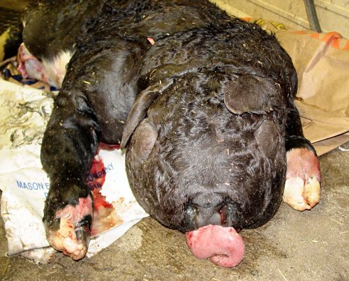
Pulmonary Hypoplasia with Anasarca.
Facial view of the enlarged edematous head with swollen tongue, of a newborn PHA calf. The ears are flat and nearly invisible. The eyes are mere slits.
Kaiser L (2007)
PHA Lung.
Distal trachea and neonatal Pulmonary Hypoplastic Achondroplastic (PHA) lung.
Kaiser L (2007)
Normal Noninflated Neonatal Lung.
Normal noninflated (atelectatic) neonatal lung.
Kaiser L (2007)
Extreme Achondroplasia.
Extreme example of an achondroplastic calf (bulldog calf).
Roberts SJ (1973)
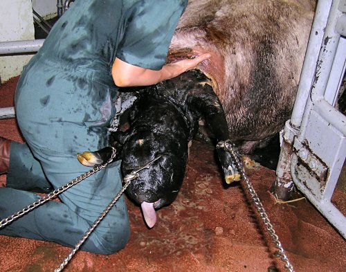
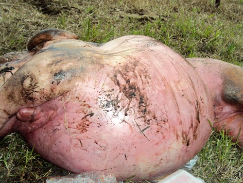
PHA Dystocia.
Assisted delivery of a large edematous fetus afflicted by Pulmonary Hypoplasia and Anasarca.
Varga A (2005)
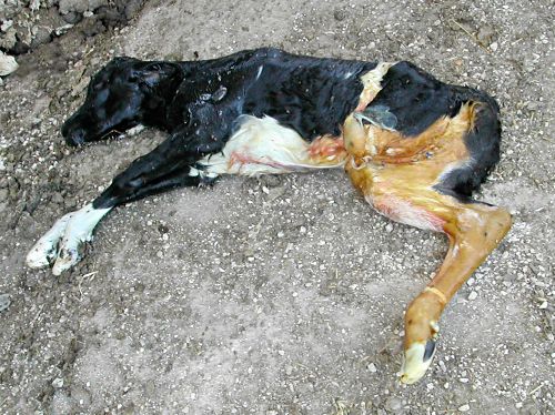
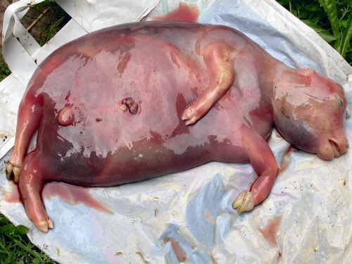
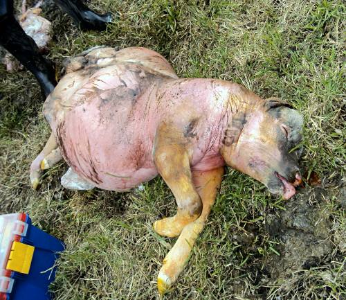
PHA Neonate.
Dorsal view of newborn fetal monster afflicted by Pulmonary Hypoplasia and Anasarca. Generalized edema and massive swelling of the entire body. The scrotum is the size of that of a mature bull, except it is only filled with fluid.
Varga A (2005)
PHA Neonate.
Ventral view of a fresh, dead fetus delivered, with assistance, per vaginam. The umbilical cord is still intact. The scrotum is the size of that of a mature bull, except it is only filled with fluid.
Varga A (2005)
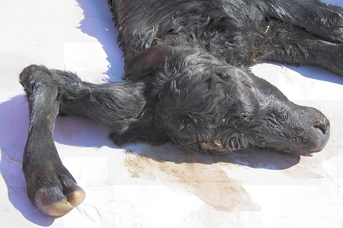
PHA Severe Facial Edema.
Massive swelling of the face of a calf born with PHA. The ears are difficult to identify and the eyes are mere slits.
Varga A (2005)
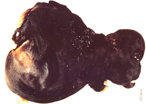
PHA Severe Edema of the Head.
Massive edema and swelling of the head of a calf born with Pulmonary Hypoplasia Anasarca. The ears are difficult to identify and the eyes are mere slits.
Kaiser L (2012)
PHA Severe Edema of the Head.
Massive swelling of the head of a calf born with Pulmonary Hypoplasia Achondroplasia. The ears are difficult to identify and the eyes are mere slits.
Kaiser L (2012)
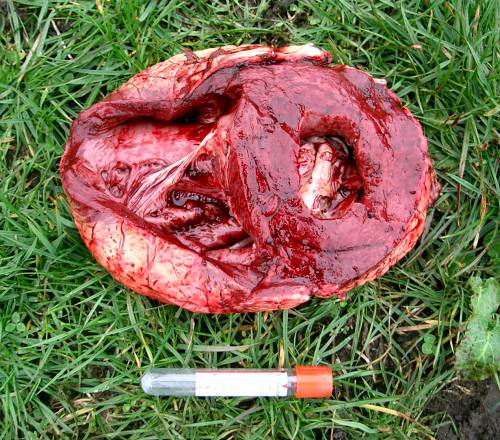
PHA Placenta.
Fresh fetal membranes delivered with the severely edematous fetus. The gross appearance of the membranes was normal.
Varga A (2005)
PHA Necropsy.
Severe mandibular edema and a slightly swollen tongue (brown).
Varga A (2005)
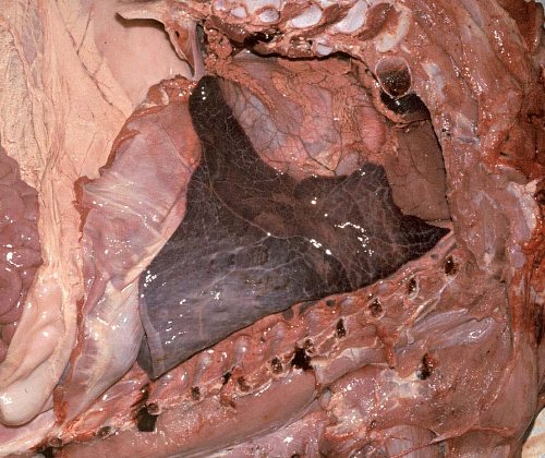
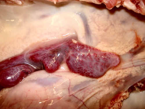
PHA Hypoplastic Lung.
The small dark colored tissue is a hypoplastic atelectatic lung.
Varga A (2005)
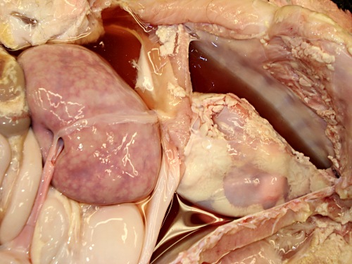
PHA Open Chest and Abdomen.
The thoracic cavity is to the right and contains a small dark lung and the heart. To the left of the diagram is a small pale liver.
Varga A (2005)
PHA Heart.
Heart within the pericardium. The cardiovascular system is normal.
Varga A (2005)
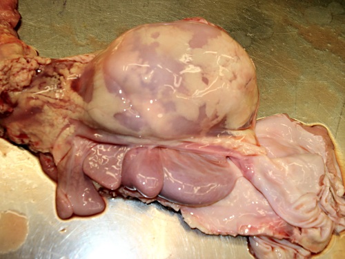
PHA Compartments of the Stomach.
All organs are underdeveloped due to the pressure of the generalized edema.
Varga A (2005)
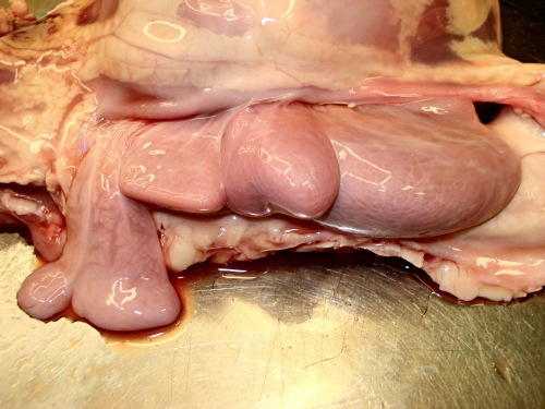
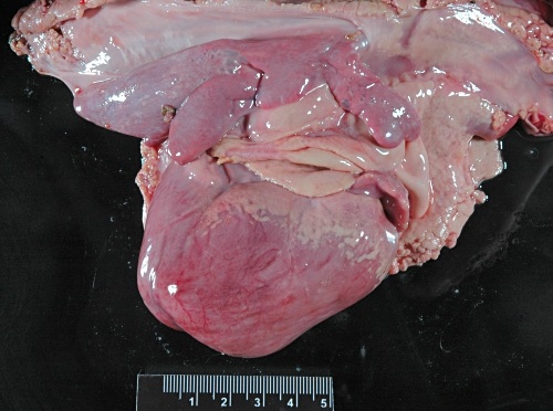
PHA Heart and Lungs.
Heart and hypoplastic lungs. The cardiovascular system is normal.
Varga A (2005)
PHA Close-up of the Lungs.
Close-up of the hypoplastic atelectatic lungs.
Varga A (2005)
PHA Close-up of the Heart.
The pericardium has been opened to show the heart.
Varga A (2005)
PHA Close-up of the Heart.
The pericardium has been removed to show the ventricle and the atrium.
Varga A (2005)

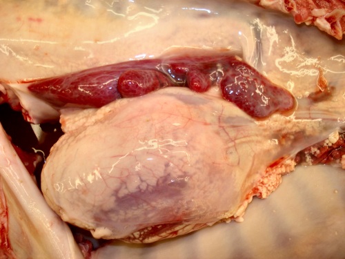
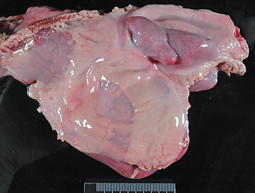
PHA Open Chest.
Exposure of the heart and the small hypoplastic lungs, flanked by the cut ribs.
Varga A (2005)

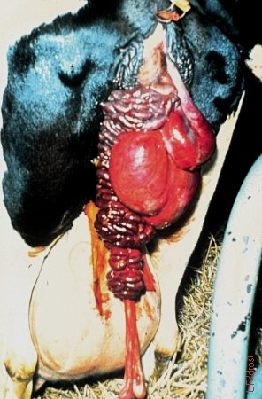
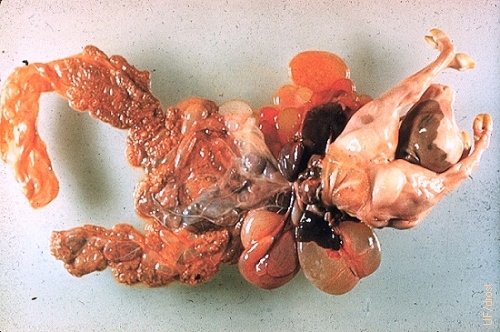
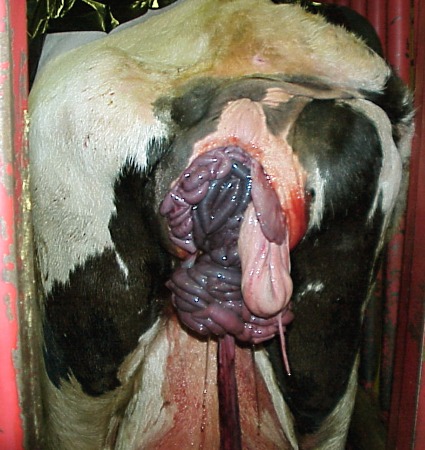
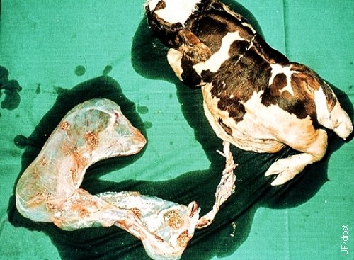
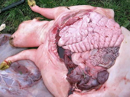
Partial Delivery - Schistosoma Reflexum.
Cow in the process of delivering a calf with Schistosoma reflexum. The extra-abdominal viscera of the fetal monster protrude from the vulva. The gauge of the fetal intestines readily distinguish them from potential maternal viscera. The ankylosed vertebral column frequently makes a vaginal delivery, without the benefit of a partial fetotomy, difficult.
Roberts SJ (1973)
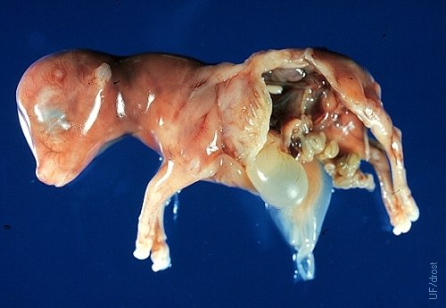
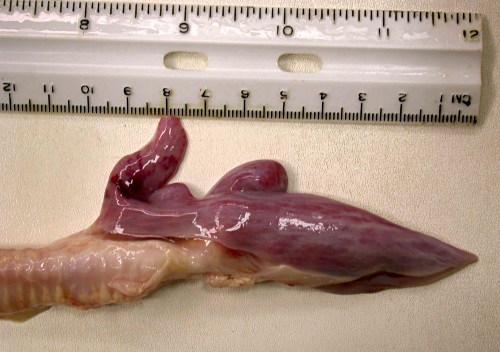
Schistosoma Reflexum - 70-Day Fetus.
A 70-day old fetus with Schistosoma reflexum (incidental finding as a slaughterhouse specimen).
Drost M (1979)
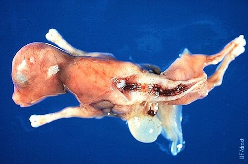
Schistosoma Reflexum - 70-Day Fetus.
A 70-day old fetus with Schistosoma reflexum illustrating the early vertebral defect (incidental finding as a slaughterhouse specimen).
Drost M (1979)
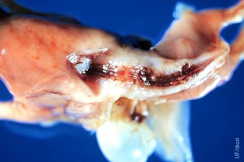
Vertebral Column Schistosoma Reflexum.
Closeup of the defective vertebral column of a 70-day old fetus (incidental finding as a slaughterhouse specimen).
Drost M (1979)
Schistosoma Reflexum - 6-Month Fetus.
Extreme expression of Schistosoma reflexum in a 6-month old fetus.
Drost M (1978)
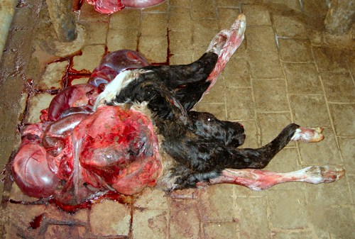
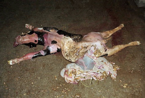
Schistosoma Reflexum - Holstein Neonate.
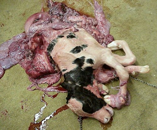
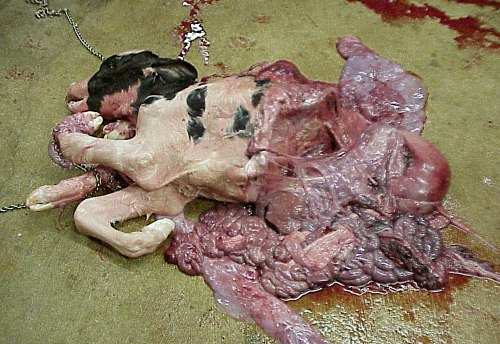
Schistosoma reflexum in a newborn Holstein calf. The vertebral column has doubled back on itself and is ankylosed. Recommended method of delivery is by fetotomy after severing the umbilical cord, if the calf is alive.
Roodzant JP (2007)
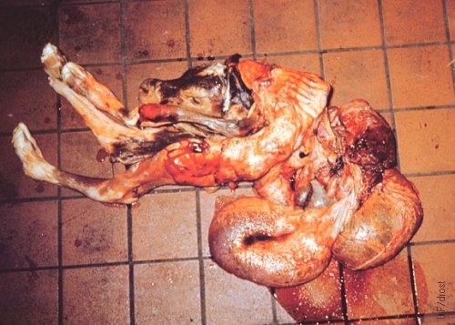
Schistosoma Reflexum - Guernsey Fetus.
Schistosoma reflexum in a newborn Guernsey calf. The vertebral column has doubled back on itself and is ankylosed. Viscera are distended with fluid.
Roberts SJ (1973)
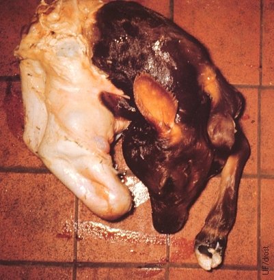
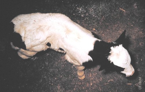
Schistosoma Reflexum - Jersey Neonate.
Schistosoma reflexum in a newborn Jersey calf. The hind legs are contained within a sack of skin.
Roberts SJ (1973)
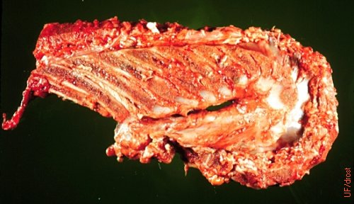
Vertebral Column - Schistosoma Reflexum.
Extreme lordosis, to the point of doubling back upon itself (reflexum) of the vertebral column, is one of the landmarks of this developmental abnormality, as shown in this sagittal section of the vertebral column of an afflicted term fetus.
Drost M (1978)
Schistosomus Reflexus in a Holstein Cow.
Fetal-size intestines presented at the vulva indicate an eviscerating Schistosomus reflexus fetus in the process of delivery.
Shipley C (2006)
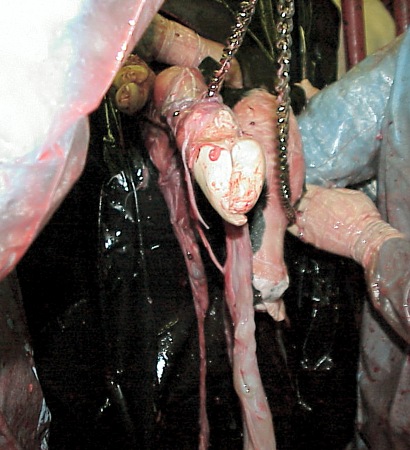
Schistosomus Reflexus in a Holstein Cow.
Presentation of a head and a hindleg during a dystocia in a Holstein cow. Fetal membranes are also presented. The calf turned out to be a Schistosomus reflexus monster.
Shipley C (2006)
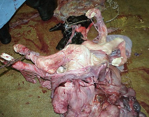
Schistosomus Reflexus in a Holstein Cow.
A term Schistosomus reflexus calf was delivered by traction per vaginam. These fetal monsters are not large, per se, but they are contorted.
Shipley C (2006)
Schistosomus Reflexus in a Holstein Cow.
A term Schistosomus reflexus calf was delivered by traction per vaginam. These fetal monsters are not large, per se, but they are contorted. This calf had a vertebral column that doubled back on itself.
Shipley C (2006)
Schistosomus Reflexus in a Holstein Cow.
A term Schistosomus reflexus calf was delivered by traction per vaginam. These fetal monsters are not large, per se, but they are deformed. This calf presented with a vertebral column that doubled back on itself.
Shipley C (2006)
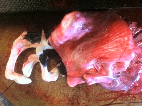
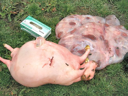
Severe Case of Schistosomus Reflexus.
Term Holstein calf delivered by cesarean section. A severe case of Schistosoma reflexum with complete doubling back of the vertebral column and complete eversion of the abdominal wall. In addition the legs appear to be deformed as well.
Cepero A (2012)
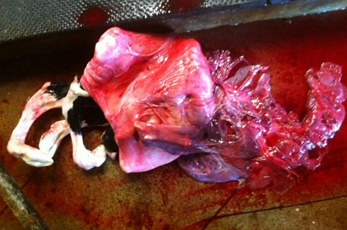
Severe Case of Schistosomus Reflexus.
Term Holstein calf delivered by cesarean section. A severe case of Schistosoma reflexum with complete doubling back of the vertebral column and complete eversion of the abdominal wall. In addition the legs appear to be deformed and contorted. The fetal membranes are still attached on the right.
Cepero A (2012)
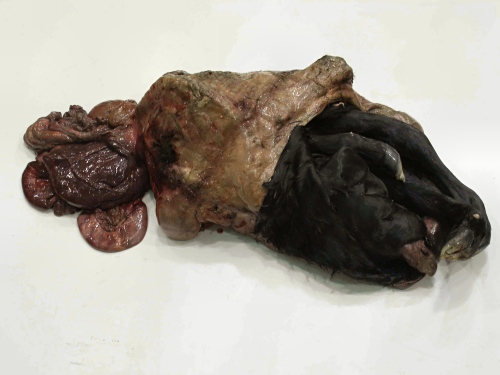
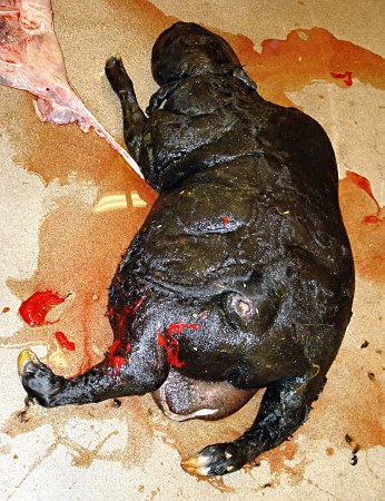
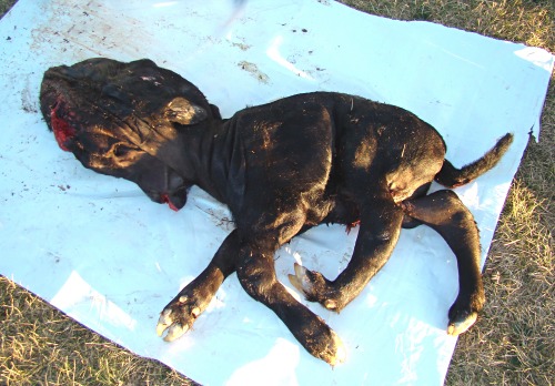
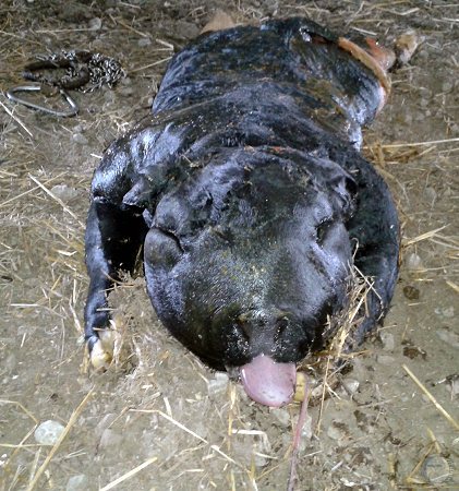
Schistosomus Reflexus / Fetal Death.
Extreme case of Schistosomus reflexus. The fetus died near or intra partum and has started to decompose. The open abdominal wall had everted to enclose the fetus that was doubled back on itself due to reflex of the vertebral column. Some necrotic viscera are present on the left.
Wuenschmann A (2012)
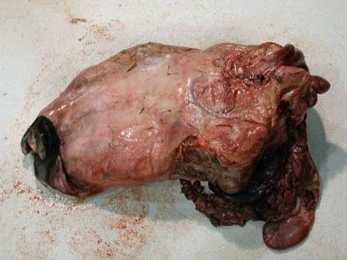
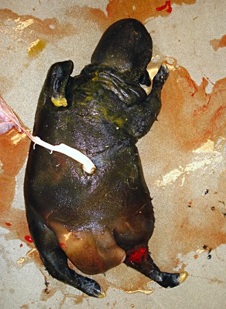
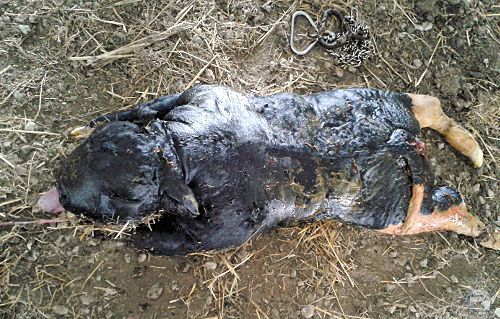
Schistosomus Reflexus / Fetal Death.
Complete eversion of the abdominal wall of the fetus that is doubled back on it self due to the reflexed vertebral column. Some necrotic viscera are present on the right.
Wuenschmann A (2012)
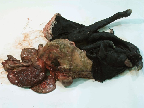
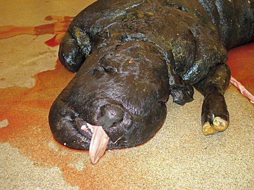
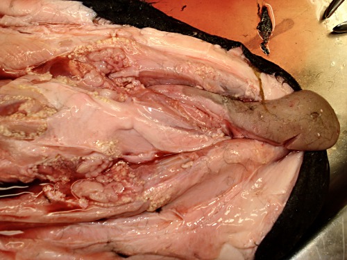
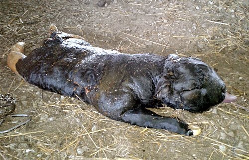
Schistosomus Reflexus / Fetal Death.
The everted abdominal wall has been opened up to show the head on the lower right. The tip of the tongue is sticking out. The fetus is completely turned back on itself and is compacted.
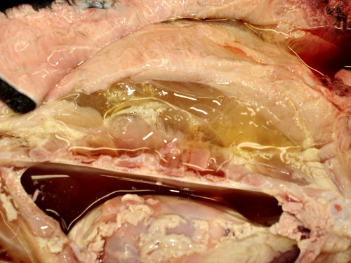
Wuenschmann A (2012)
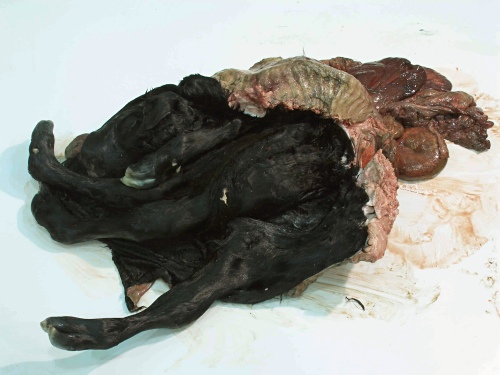
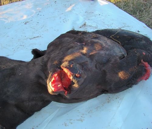
Schistosomus Reflexus / Fetal Death.
The everted abdominal wall has been opened up to show the head on the upper left. The tip of the tongue is sticking out of the right corner of the mouth. The fetus is completely turned back on itself and is compacted.
Wuenschmann A (2012)
Scoliosis.
Pronounced scoliosis in a Holstein calf. The calf also has a wry tail.
Roberts SJ (1973)
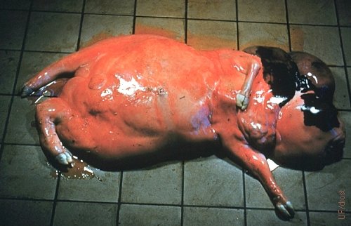
Fetal Anasarca.
Generalized massive edema of all parts of the body (anasarca) of the fetus. As a rule these fetuses are aborted. Most of these fetuses are the result of an autosomal recessive gene.
Kearns BM (2013)
Fetal Anasarca.
Generalized massive edema of all parts of the body (anasarca) of the fetus. As a rule these fetuses are aborted. Most of these fetuses are the result of an autosomal recessive gene.
Kearns BM (2013)
Fetal Anasarca.
Generalized massive edema of all parts of the body (anasarca) of the fetus. As a rule these fetuses are aborted. Most of these fetuses are the result of an autosomal recessive gene.
Kearns BM (2013)
Fetal Anasarca.
Generalized massive edema of all parts of the body (anasarca) of the fetus. As a rule these fetuses are aborted. Most of these fetuses are the result of an autosomal recessive gene.
Roberts SJ (1973)
Fetal Anasarca with Membranes.
Generalized massive edema of all parts of the body (anasarca) of the fetus. As a rule these fetuses are aborted.
Roberts SJ (1973)
Congenital Dropsy.
Fetal anasarca or congenital dropsy in an aborted Ayrshire fetus, due an recessive autosomal gene. [size of the square tile 15 cm].
Roberts SJ (1986)
Fetal Ascitis and Anasarca.
Fetal ascitis and anasarca in an aborted 6 month old fetus, due an recessive autosomal gene. [size of the glove box is 12 x 24 cm]. There is also pronounced edema of the chorioallantois.
Leduc F (2007)
Fetal Edema and Anasarca.
Fetal ascitis and anasarca in an aborted 6 month old fetus, due an recessive autosomal gene. The thymus gland is grossly enlarged.
Leduc F (2007)
Anasarcous Fetal Heart.
Cross section of the heart of a six month old aborted fetus with ascitis and anasarca. The ventricular muscles appear to be hypertrophied.
Leduc F (2007)
Anasarcous 7 Month Fetus.
Male anasarcous Simmental fetus delivered by cesarean section at ~ 7 months of gestation.
Ramírez O (2012)
Anasarcous 7 Month Fetus.
Male anasarcous Simmental fetus with ascitis delivered by cesarean section at ~ 7 months of gestation.
Ramírez O (2012)
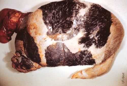
Globosus Amorphus (Classic).
Globosus amorphus (shapeless mass) is an incomplete twin with a vascular connection to the placenta of its twin. All three primary germ layers are present (ectoderm, mesoderm and endoderm).
Roberts SJ (1986)
Globosus Amorphus (Typical).
Globosus amorphus (shapeless mass) is an incomplete twin with a vascular connection to the placenta of its twin.
Drost M (1982)
Globosus Amorphus Opened Up.
Globosus amorphus (shapeless mass) is an incomplete twin with a vascular connection to the placenta of its twin.
Drost M (1982)
Globosus Amorphus - Small Necrotic.
Globosus amorphus (shapeless mass) is an incomplete twin with a vascular connection to the placenta of its twin. Strangulation of the stalk has led to necrosis of the mass in this specimen.
Drost M (1982)
Globosus Amorphus - Small, Opened Up.
Globosus amorphus (shapeless mass) is an incomplete twin with a vascular connection to the placenta of its twin. Strangulation of the stalk has led to necrosis of the mass.
Drost M (1982)
Atypical Globosus Amorphus.
Globosus amorphus (shapeless mass) is an incomplete twin with a vascular connection to the placenta of its twin. This specimen is atypical because of the partial development of the mandible and the soft palate.
Roberts SJ (1973)
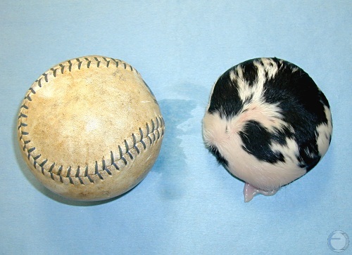
Fetal Globosus Amorphus - 8 mos.
Globosus amorphus was an incomplete twin with a vascular connection to the placenta of its twin at 8 months of gestation. A surprise, soft ball sized slaughter house specimen, was accompanied by a normal Holstein twin fetus.
Drost M (2013)
Fetal Globosus Amorphus - 8 mos.
Globosus amorphus. An incidental finding of a soft ball sized slaughter house specimen that was connected to the placental circulation of a normal Holstein 8 month old fetal twin fetus.
Drost M (2013)
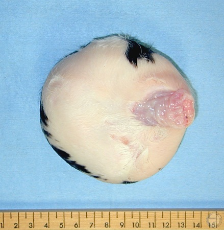
Fetal Globosus Amorphus - 8 mos.
Ventral aspect of a globosus amorphus. An incidental finding of a soft ball sized slaughter house specimen connected to the placental circulation of a normal Holstein 8 month old fetal twin fetus. Note the stump of the umbilical cord on the right.
Drost M (2013)
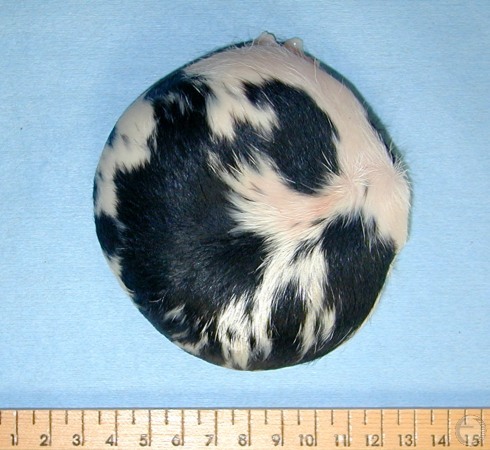
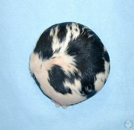
Fetal Globosus Amorphus - 8 mos.
Globosus amorphus was an incomplete twin with a vascular connection to the placenta of its twin. A surprise finding of a soft ball sized slaughter house specimen with a normal Holstein twin fetus. Covered with short hair like its 8 month old twin.
Drost M (2013)
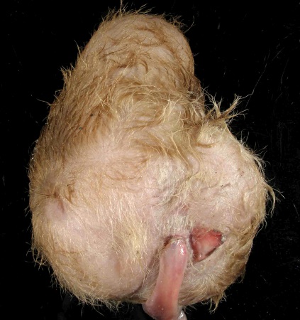
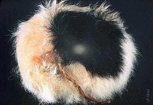
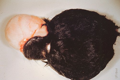
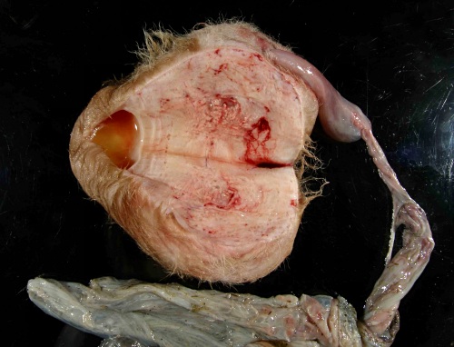
Term Globosus Amorphus.
Globosus amorphus is an incomplete twin with a vascular connection to the placenta of its twin. This specimen is covered with hair which reflects its gestational age.
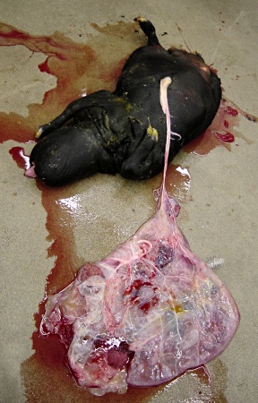
Wuenschmann A (2012)
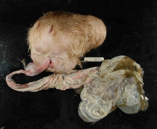
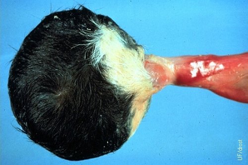
Term Globosus Amorphus.
The amniotic sac is still attached to the Amorphous globosus via its umbilical cord. It shared circulation with its normal twin.
Wuenschmann A (2012)
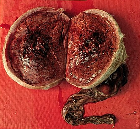
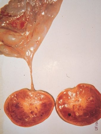
Term Globosus Amorphus.
Cross section of the amorphus globosus. All three primary germ layers (endoderm, mesoderm, ectoderm) are present.
Wuenschmann A (2012)
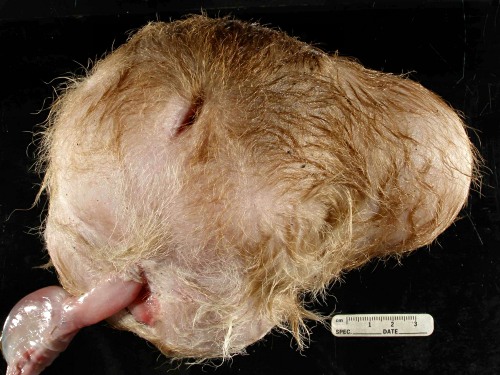
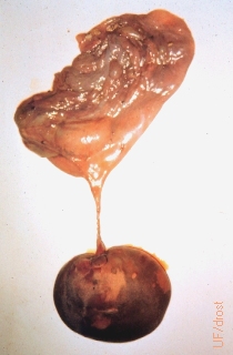
Term Globosus Amorphus.
Complete amorphus globosus. All three primary germ layers (endoderm, mesoderm, ectoderm) are present. The umbilical cord is the vascular connection to the placenta of its twin.
Wuenschmann A (2012)
Holoacardius Amorphus.
Holoacardius amorphus, also called an anideus or an anidean monster (Greek, an = neg., idea = form; no idea!?). An embryonic anideus is a blastoderm in which no embryonic axis develops.
Roberts SJ (1973)