The Visual Guide to
Bovine Reproduction
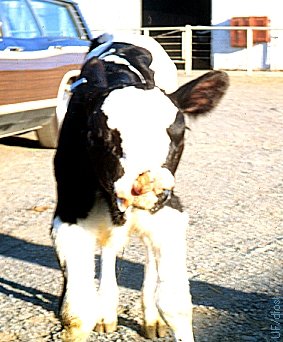
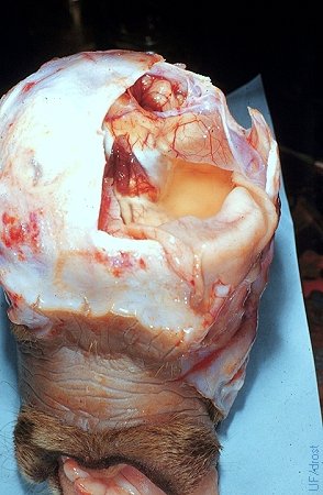
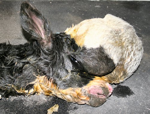
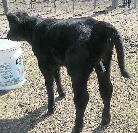
Teratology: Congenital Anomalies
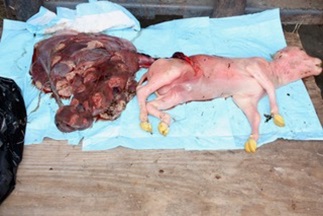
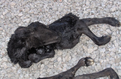
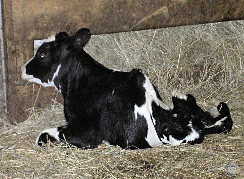
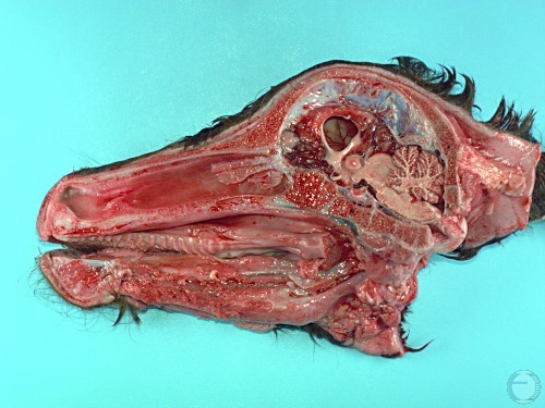
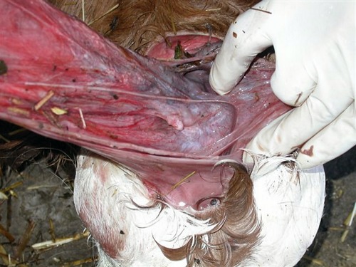
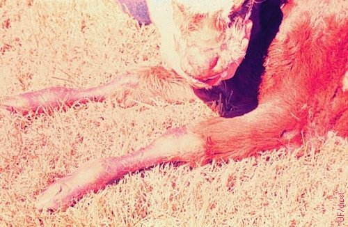
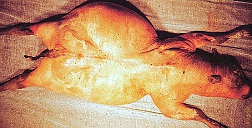
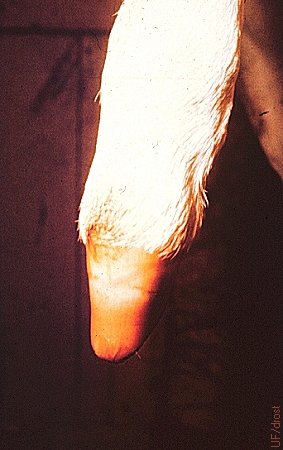
Umbilical torsion in a bovine fetus. Picture 1 of 4.
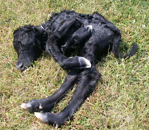
Umbilical torsion in a 177-day-old aborted Red Angus fetus. There were 17 loose counterclockwise twists going from the placenta to the fetus, and tighter twists going clockwise from the fetus to the placenta. The 17 twists were filled with clotted blood. There were two arteries, one vein, and the urachus in the umbilical cord. The vein connected to the liver and the urachus to the bladder. The twist was extremely tight at the fetal abdomen and became more distended and filled with blood clots in the direction of the placenta. A field necropsy was performed, and no other abnormalities were identified.
Kaiser L (2020)
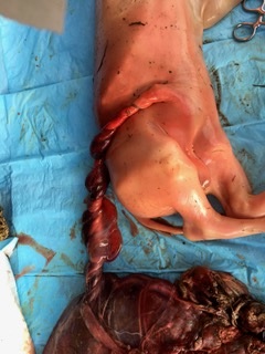
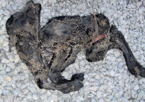
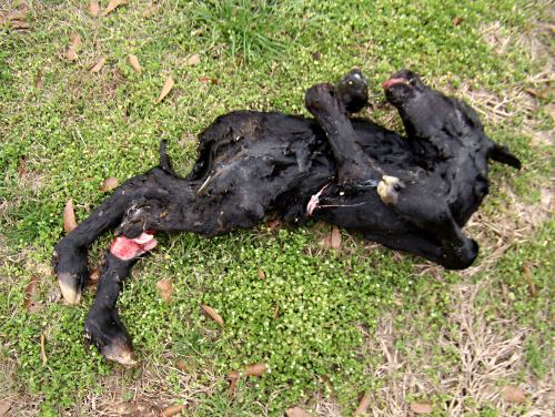
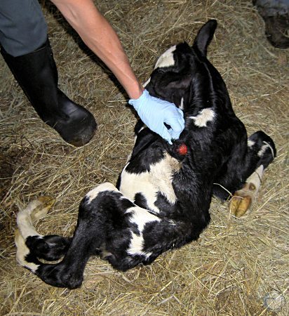
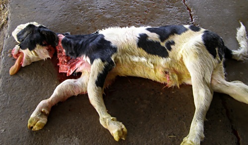
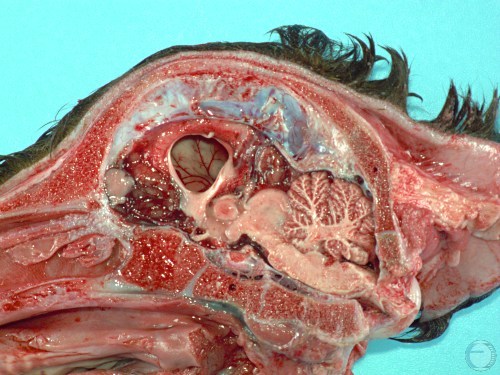
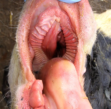
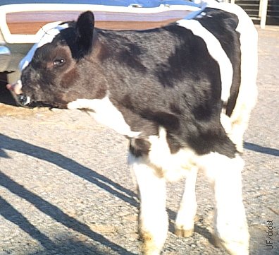
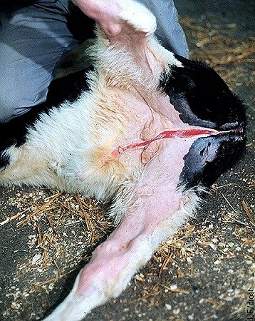
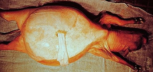
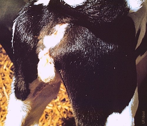
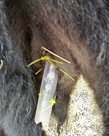
Umbilical torsion in a bovine fetus. Picture 2 of 4.
Close up view of the placenta and the hindquarters of the fetus.
Kaiser L (2020)
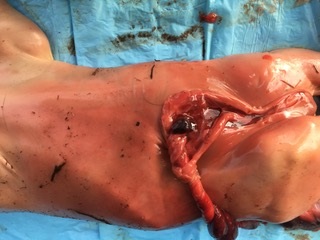
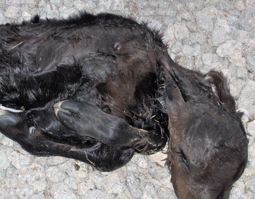
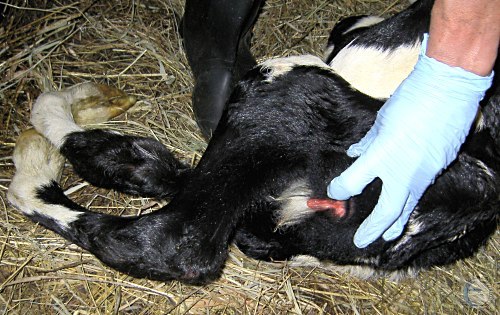
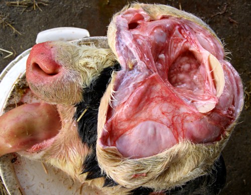
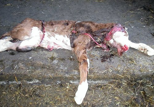
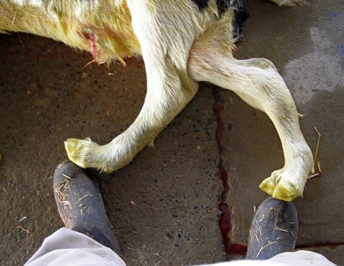
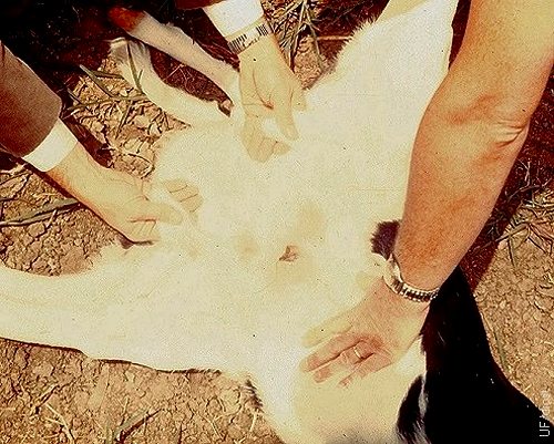
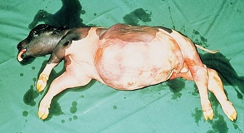
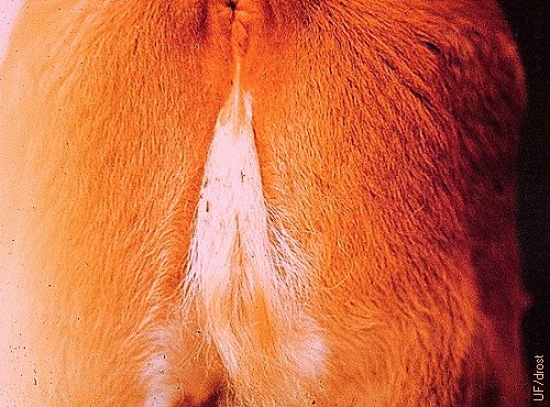
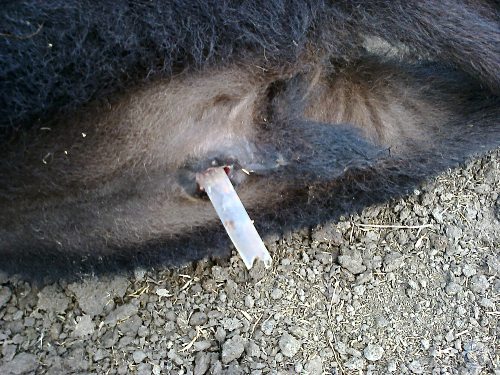
Umbilical torsion in a bovine fetus.
TAbdominal view of the twisted umbilical cord. Anteriorly, the blood vessels lead to the placenta, and posteriorly the urachus to the bladder.
Kaiser L (2020)
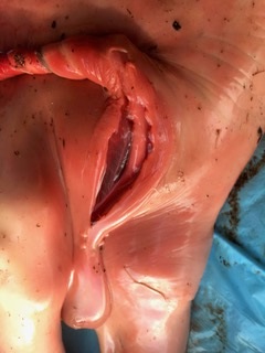
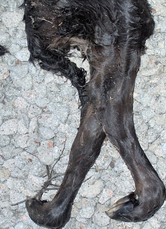
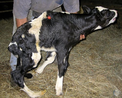
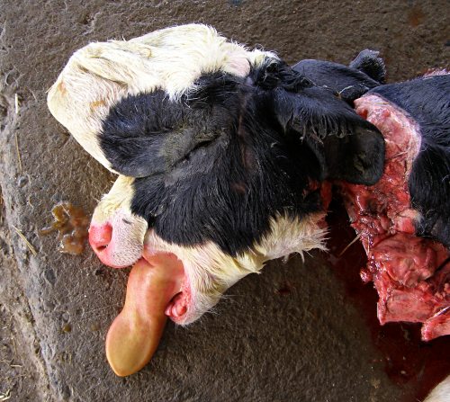
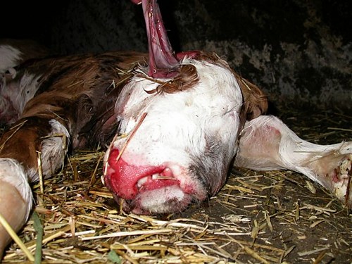
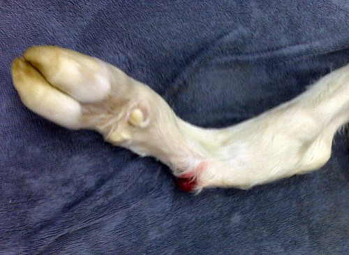
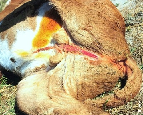
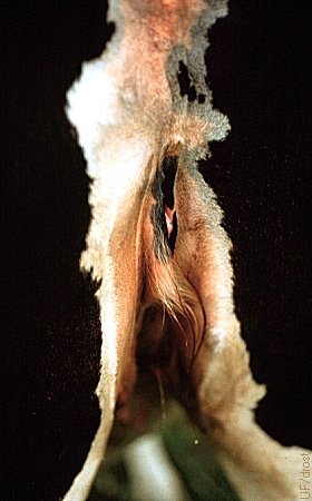
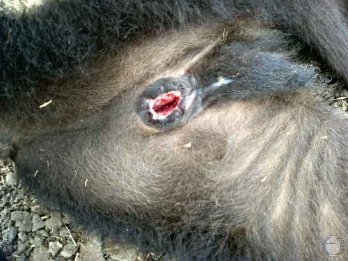
Umbilical torsion in a bovine fetus. Picture 4 of 4.
Close up view of the umbilical cord of a 177-day-old male fetus. Note the scrotum posteriorly.
Kaiser L (2020)
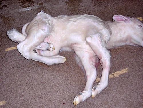
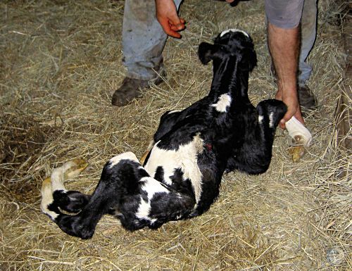
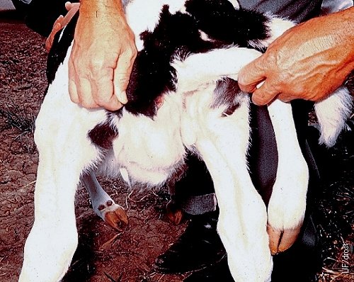
Tibial Hemimelia.
Tibial hemimelia in a Shorthorn calf showing failure of pelvic fusion and an abdominal hernia.
Kaiser L (2008)

Tibial Hemimelia.
Tibial hemimelia in a Shorthorn calf showing failure of pelvic fusion and an abdominal hernia.
Kaiser L (2008)
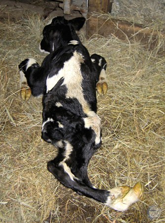
Tibial Hemimelia.
Tibial hemimelia in a Shorthorn calf showing failure of pelvic fusion and an abdominal hernia. Invariably lethal.
Kaiser L (2008)
Tibial Hemimelia.
Tibial hemimelia in a premature calf showing failure of pelvic fusion and distortion of the structure of the hindlegs. Invariably lethal.
Kaiser L (2008)
Tibial Hemimelia.
Tibial hemimelia in a term Shorthorn calf showing failure of pelvic fusion and distortion of the structure of the hindlegs and an abdominal hernia. Invariably lethal.
Kaiser L (2008)
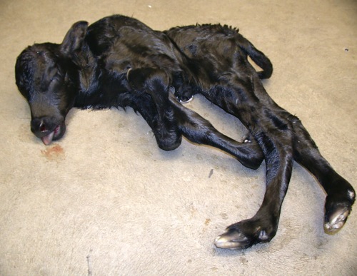
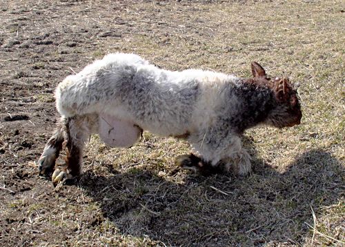
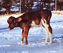
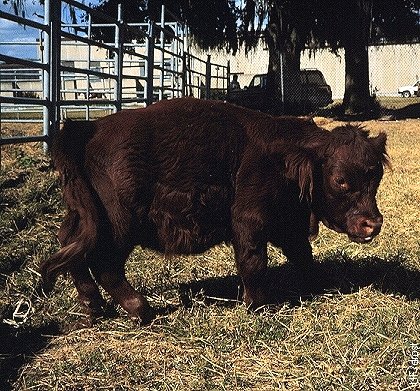
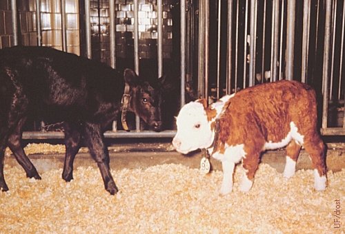
Curly Calf Syndrome.
Arthrogryposis Multiplex Congenita in an Angus calf. The spine is twisted and the calf is small and thin due to inadequate muscular development. A homozygous recessive condition.
Steffen DJ (2008)
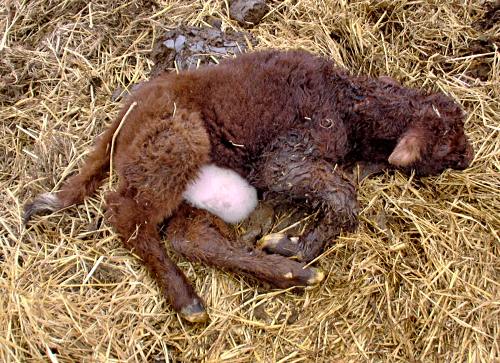
Arthrogryposis Multiplex.
Congenital Arthrogryposis Multiplex in an Angus calf. The spine is twisted and the calf is small and thin due to inadequate muscular development. A homozygous recessive condition.
Steffen DJ (2008)
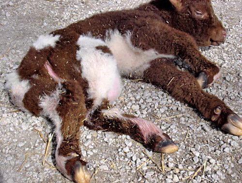
Arthrogryposis Multiplex.
Curly Calf Syndrome in an Angus calf. The spine is twisted and the calf is small and thin due to inadequate muscular development. A homozygous recessive condition.
Steffen DJ (2008)
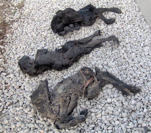
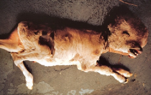
Arthrogryposis Multiplex Congenita.
Arthrogryposis Multiplex Congenita in three Angus calves born at term. The spines are twisted and the calves are small and thin due to inadequate muscular development. A homozygous recessive condition.
Irsik M (2009)
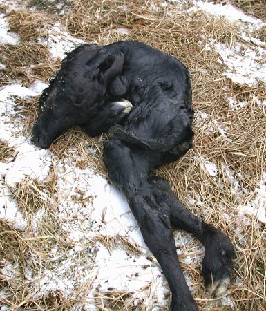
Arthrogryposis Multiplex Congenita.
Arthrogryposis Multiplex Congenita in a term Angus calf. The spine is twisted and the calf is small and thin due to inadequate muscular development. A homozygous recessive condition.
Irsik M (2009)
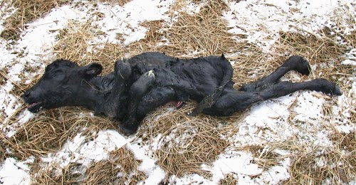
Arthrogryposis Multiplex Congenita.
Curly Calf Syndrome in a term Angus calf. The spine is twisted and the calf is small and thin due to inadequate muscular development. A homozygous recessive condition.
Irsik M (2009)
Arthrogryposis Multiplex Congenita.
Close-up of a term calf with Curly Calf Syndrome in. The spine is twisted and the calf is small and thin due to inadequate muscular development. A homozygous recessive condition.
Irsik M (2009)
Contracted Tendons in an AM Calf.
Contracted tendons in a term calf with Curly Calf Syndrome. The calf is small and thin due to inadequate muscular development.
Irsik M (2009)
Arthrogryposis Multiplex.
Contracted tendons in a term calf with Curly Calf Syndrome. The calf is small and thin due to inadequate muscular development.
Kaiser L (2012)
Arthrogryposis Multiplex.
Twisted spine and contracted tendons in a term Angus calf with Curly Calf Syndrome. The calf is small and thin due to inadequate muscular development.
Kaiser L (2012)
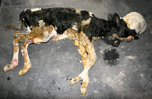
Congenital Arthrogryposis Multiplex.
Twisted spine and contracted tendons of the hindquarters of a term Holstein heifer calf with Curly Calf Syndrome. The posterior half is small and thin due to inadequate muscular development.
Blough E (2013)
Congenital Arthrogryposis Multiplex.
Twisted spine and contracted tendons of the hindquarters of a term Holstein heifer calf with Curly Calf Syndrome. The posterior half is small and thin due to inadequate muscular development.
Blough E (2013)
Congenital Arthrogryposis Multiplex.
Twisted spine and contracted tendons of the hindquarters of a term Holstein heifer calf with Curly Calf Syndrome. The posterior half is small and thin due to inadequate muscular development.
Blough E (2013)
Congenital Arthrogryposis Multiplex.
Twisted spine and contracted tendons of the hindquarters of a term Holstein heifer calf with Curly Calf Syndrome. The posterior half is small and thin due to inadequate muscular development.
Blough E (2013)
Congenital Arthrogryposis Multiplex.
Twisted spine and contracted tendons in a term Holstein heifer calf with Curly Calf Syndrome. Normal vulva.
Blough E (2013)
Congenital Arthrogryposis Multiplex.
Twisted spine and contracted tendons of the hindquarters of a term Holstein heifer calf with Curly Calf Syndrome. The posterior half is small and thin due to inadequate muscular development.
Blough E (2013)
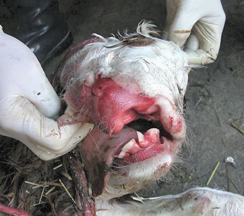
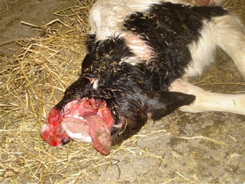
Multiple Anomalies.
Enlargement and deformation of the head necessitated delivery by partial fetotomy. The calf had hydrocephaly, a cleft palate, and abnormal joint formation.
Calderon G (2008)
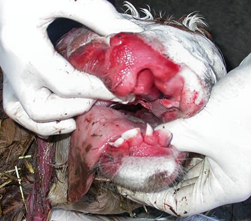
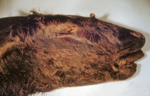
Collapsed Hydrocephalus.
Enlargement and deformation of the head necessitated delivery by partial fetotomy. Grossly swollen tongue.
Calderon G (2008)
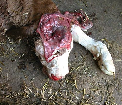
Internal Hydrocephalus.
Internal hydrocephalus is due to excessive accumulation of fluid in the ventricular system of the brain. [External hydrocephalus is rare and due to excessive fluid between the brain and the dura mater].
Calderon G (2008)
Small Internal Hydrocephalus.
Internal hydrocephalus due to excessive accumulation of fluid in the ventricular system of the brain. In addition there is a cleft palate.
Kaiser L (2013)
Small Internal Hydrocephalus
Internal hydrocephalus due to excessive accumulation of fluid in the ventricular system of the brain.
Kaiser L (2013)
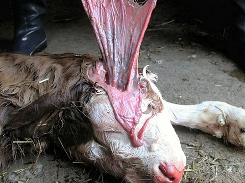
External Hydrocephalus.
External hydrocephalus is rare and due to excessive fluid accumulation between the brain and the dura mater. This term Fleckvieh calf born with a fluid filled external bag that collapsed during the process of delivery. The previous four calves of this pluriparous cow were normal.
Benz M (2009)
External Hydrocephalus.
Close-up of the head of a Fleckvieh term calf born with external hydrocephalus. The thin walled fluid filled sack ruptured during the process of delivery. The calf also had a severe cleft palate (palatoschisis).
Benz M (2009)
External Hydrocephalus.
Close-up of the head of a Fleckvieh term calf born with external hydrocephalus. The calf also had a severe cleft palate (palatoschisis) as featured here.
Benz M (2009)
Extreme Palatoschisis.
Close-up of the head of a Fleckvieh term calf born with a severe cleft palate (palatoschisis). This calf also had an external hydrocephalus.
Benz M (2009)
External Hydrocephalus - Close Up.
Close-up of the head of a Fleckvieh term calf born with an external hydrocephalus. The thin walled sac of the dura mater was not covered by the skin. It ruptured during the process of delivery.
Benz M (2009)
External Hydrocephalus - Close Up.
Close-up of the head of a Fleckvieh term calf born with an external hydrocephalus. The thin wall, of the previously protruding fluid filled sac, is well-vascularized.
Benz M (2009)
External Hydrocephalus - Close Up.
The index finger is inserted in the cavity previously occupied by the fluid filled external hydrocephalic sac. The atlas could be palpated directly.
Benz M (2009)
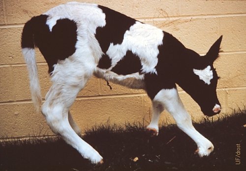
Hindlegs Hyperextension.
Hyperextension due to flaccid tendons and muscles.
Calderon G (2008)
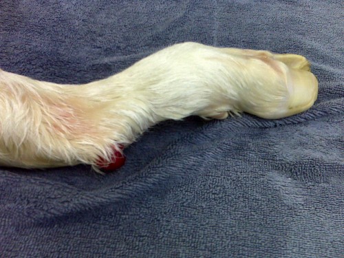
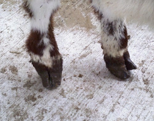
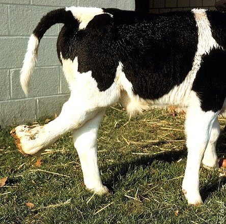
Fetal Fracture.
Healing open, intrauterine, metatarsal fracture just below the hock. Etiology unknown.
Bennett FB (2009)
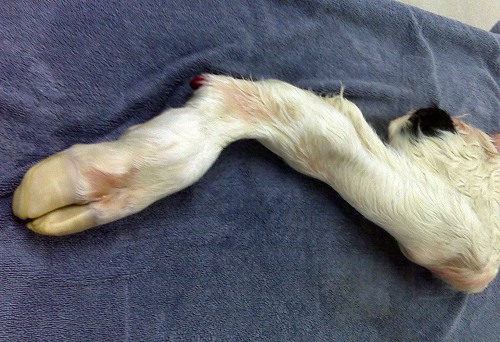
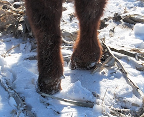
Fetal Fracture.
Healing open, intrauterine, metatarsal fracture with displacement. Etiology unknown. Diagnosed upon delivery at term.
Bennett FB (2009)
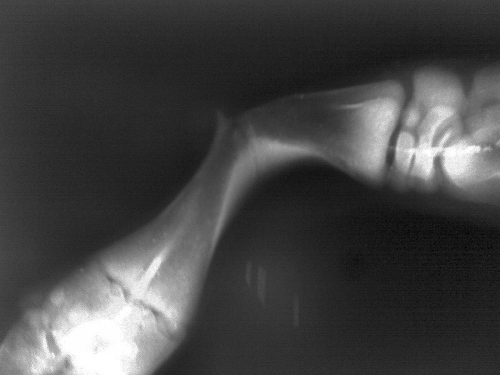
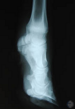
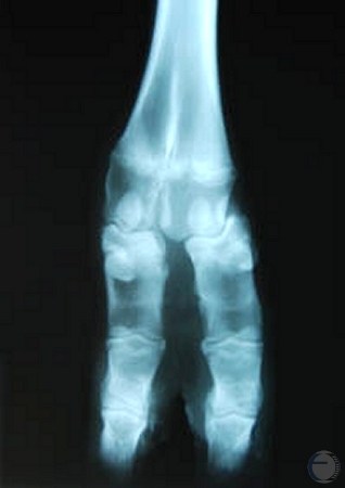
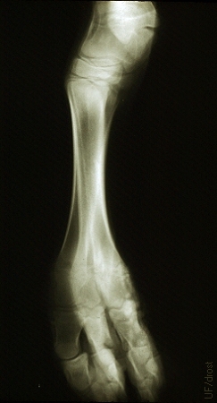
Radiograph of a Fetal Fracture.
Radiograph of a fetal fracture sustained in utero. Diagnosed upon delivery at term.
Bennett FB (2009)
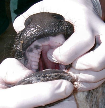
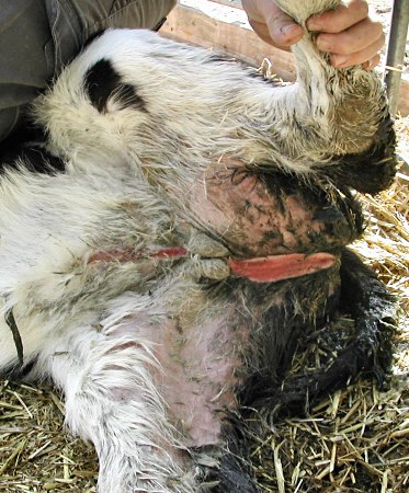
Palatoschisis.
Most calves are not routinely examined for an intact hard palate. The first indication of the presence of a cleft palate is milk coming out of both nostrils after the calf nurses.
Leduc F (2007)
Severe Cleft Palate.
Rostral view of a craniofacial malformation as a result of a severe cleft palate and harelip in a newborn calf.
Braun RK (1971)
Craniofacial Malformation.
Lateral view of a craniofacial malformation as a result of a severe cleft palate in a newborn calf.
Braun RK (1971)
Schistocephalus Bifidus.
Term Holstein calf born with an extreme case of cleft palate (palatoschisis). The upper portion of the face was divided into two equal lobes. Etiology unknown.
Worobey T (2007)
Fetal Giant.
Lateral view of the skull of a postterm (12 months) fetal giant showing a cerebral hernia. While the brain was defective the pituitary gland was normal in this Guernsey fetus, yet the fetus did not initiate parturition. The hypothalamus may have been deranged.
Roberts SJ (1973)
Fetal Giant.
Dorsal view of the skull of a postterm (12 months) fetal giant showing a cerebral hernia. While the brain was defective the pituitary gland was normal in this Guernsey fetus.
Roberts SJ (1973)
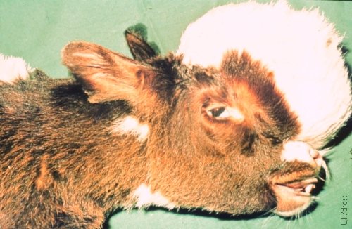
Large Encephalocele.
Encephalocele. When the enlarged cranium does not permit spontaneous vaginal delivery, the fluid should be drained via a stab incision or with a needle. These fetuses do not warrant a cesarean section.
Utrecht (1976)
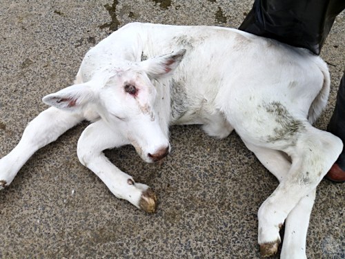
Small Encephalocele.
Small frontal encephalocele. A rare neural tube defect that is characterized by a sac like protrusion of the brain and the membranes through an opening in the skull.
Johnston EH (2013)
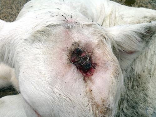
Small Encephalocele.
A rare neural tube defect that is characterized by a sac like protrusion of the brain and the membranes through an opening in the skull. The thin membrane has ruptured. Close up. The hair of the forehead has been clipped.
Johnston EH (2013)
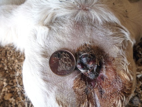
Small Encephalocele.
A rare neural tube defect that is characterized by a sac like protrusion of the brain and the membranes through an opening in the skull. The thin membrane has ruptured. Diameter of the coin is 2.4 cm. The hair of the forehead has been clipped.
Johnston EH (2013)
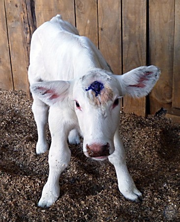
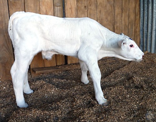
Small Encephalocele.
A rare neural tube defect that is characterized by a sac like protrusion of the brain and the membranes through an opening in the skull. The thin membrane has ruptured. The defect may lead to mental and growth retardation depending on the size of the protrusion.
Johnston EH (2013)
Hydrocephalus in a Limousin Calf.
Hydrocephalus. When the enlarged cranium does not permit spontaneous vaginal delivery, the fluid should be drained via a stab incision or with a needle. These fetuses do not warrant a cesarean section. Doming of the head occurs during non-union of sutures of the temporal, frontal, and parietal bones.
Roberts SJ (1973)
Hydrocephalus Opened Up.
Severe hydrocephalus in a Guernsey calf. Note the membrane-like a calvarium. Hydrocephalus results from an accumulation of excessive fluid in the ventricular system of the brain.
Drost M (1982)
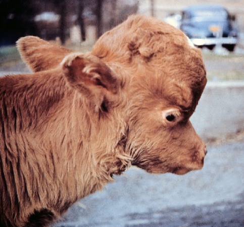
Hydrocephalus in an Ayshire Calf.
In congenital hydrocephalus the obstruction of cerebrospinal fluid is often difficult to demonstrate, but aquaductal stenosis has been postulated. Akabane virus can produce the problem and it is thought that vitamin A deficiency can contribute to the condition.
Roberts SJ (1973)
Hydrocephalus in a Jersey Calf.
Hydrocephalus in a Jersey calf. This is uncommon in calves and can be inherited and congenital. It is often associated with other deformities, e.g. congenital achondroplasia. This calf also did not have a tail, nor an anus, and a had a septal defect of the heart.
Roberts SJ (1973)
Hydrocephalus in a Holstein Calf.
Hydrocephalus in a Holstein calf. This is uncommon in calves and can be inherited. It is neural tube defect characterized by the protrusion of the brain and its surrounding membranes.
Smith MC (2008)
Hydrocephalus in a Holstein Calf.
Excessive accumulation of fluid in the ventricles of the brain. The enlarged head may cause an obstructive dystocia, which may be resolved by (simply) draining the fluid-filled cranium. A cesarean section is not warranted.
Smith MC (2008)
Mild Case of Hydrocephalus.
Excessive accumulation of fluid in the ventricles of the brain. This was likely due to partial obstruction of cerebrospinal fluid.
Murphy AS (2012)
Blind Hydrocephalic Calf.
Mild case of hydrocephalus. There was partial obstruction of cerebrospinal fluid. In this case there is also visual impairment.
Varsano S (2012)
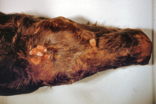
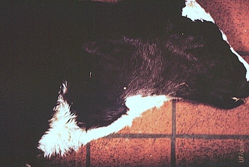
Epitheliogenesis Imperfecta.
Epitheliogenesis imperfecta in an otherwise healthy calf, believed to be due to a single autosomal recessive gene. This defect occurs most commonly on the legs below the knees and hocks, and on the muzzle.
Drost M (1987)
Epitheliogenesis Imperfecta - Close Up.
Epitheliogenesis imperfecta in an otherwise healthy calf, believed to be due to a single autosomal recessive gene. This defect occurs most commonly on the legs below the knees and hocks, and on the muzzle.
Drost M (1987)
Hypospadias in a Holstein Calf.
Hypospadias. The urethral lumen is open along the ventral aspect of the penis in the perineal region. There are two hemiscrota each containing a respective testicle. Extensive urine scalding is evident. This bull calf was afforded palliative treatment with Vaseline.
Drost M (1986)
Hemiscrota.
Hemiscrota. Each testicle is contained in a separate scrotal pouch. This live calf also had a parasitic pelvic limb.
Drost M (1986)
Hypospadias in a Jersey Calf.
Hypospadias in a bull calf. The urethra opens along the ventral aspect of the penis in the perineal area. There are two hemiscrota. Note the absence of the prepuce. Scalding by urine is a problem.
Drost M (1974)
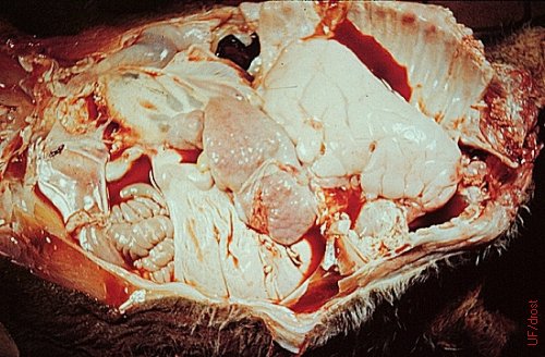
Fetal Ascites.
Ascites in a fetus that was aborted at 5 months and weighed 30 kg (dorsal view). When the distended, fluid-filled abdomen prevents delivery, it may be drained with a pipe forced down the throat.
Drost M (1974)
Hydroperitoneum.
Fetal ascites is a single anomaly. In contrast with this 5 month old, 30 kg aborted fetus, fetuses with ascites are generally carried to term at which time they may cause dystocia. Diagnosis is difficult when the fetus in anterior presentation. When the distended, fluid-filled abdomen prevents delivery, it may be drained with a pipe forced down the throat.
Roberts SJ (1973)
Fetal Ascites.
Fetal ascites. This is a single abnormality and fetuses are usually carried to term at which time they may cause dystocia. The latter can be relieved by drainage of the abdomen, by forcing a pipe down the throat of the fetus. With the fetus in anterior presentation the diagnosis can be difficult to make.
Roberts SJ (1973)
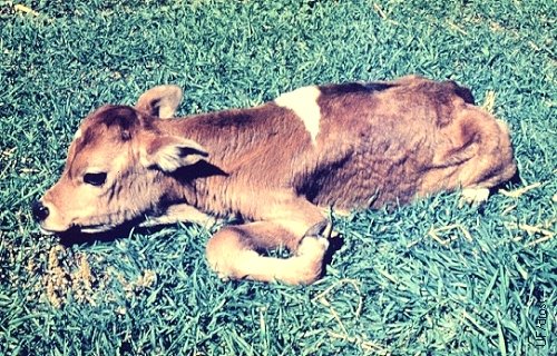
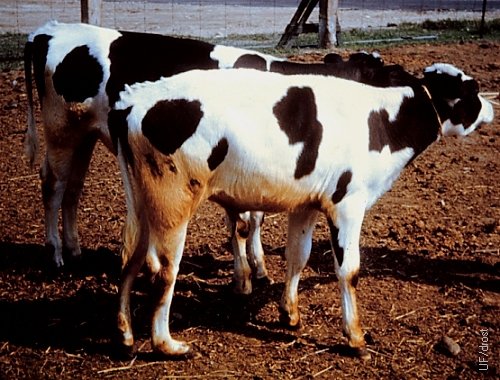
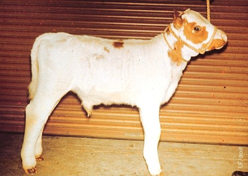
Dwarfism - 8 Months.
Eight month old Santa Gertrudis crossbred dwarf heifer.
Drost M (1978)
Dwarfism - 3 Months.
Three month old dwarf Hereford calf with a normal 3 month old heifer.
Roberts SJ (1973)
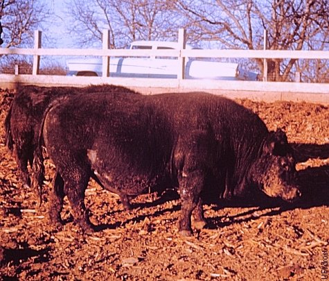
Adult Dwarf.
Mature dwarf Angus bull. This bull was born in an experimental dwarf herd as a result of natural service. Most calves from dwarf dams had to be delivered by cesarean section due fetal-maternal incompatibility.
Drost M (1968)
Ectopic Heart.
Extrathoracic heart (ectopia cordis) in a live newborn Holstein calf. The heart could be seen and be felt to beat.
Roberts SJ (1973)
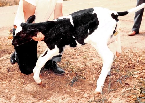
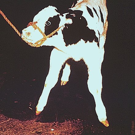
Limber Legs.
Limber legs in a Jersey calf. Apparently inherited as a simple recessive trait. Calves die soon after birth. If viable, they are unable to stand due to generalized muscular atony.
Roberts SJ (1973)
Mild Digital Subluxation.
Mild Digital Subluxation in a term Shorthorn calf showing distortion of the structure of the hindlegs.
Kaiser L (2012)
Mild Digital Subluxation.
Uneven digits. The right digit is slightly bent laterally.
Kaiser L (2013)
Mild Digital Subluxation.
Posterior radiogram of uneven rear digits. Slight lateral deviation of the right carpal bone.
Kaiser L (2013)
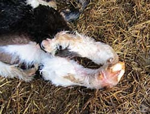
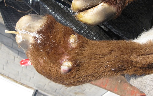
Severe Digital Subluxation.
The calf is on its belly and the left hind leg is abnormally curved at metatarsal phalangeal joints (fetlock).
Kaiser L (2013)
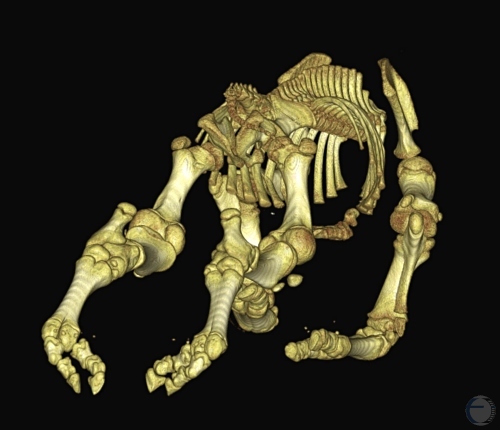
Digital Subluxation - CAT Scan.
Computer recreated image from the CAT (computed tomography or CT) scan (soft tissues have been removed to reveal only the bones). While the bones are abnormally aligned they are normal in size and shape suggesting that the abnormality is in the collateral ligaments.
Kaiser L (2013)
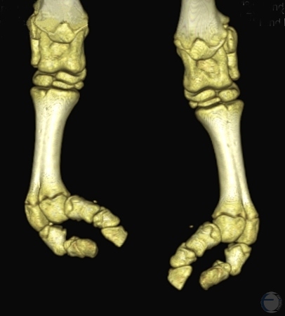
Digital Subluxation - CAT Scan.
Computer recreated image from the CAT (computed tomography or CT) scan. While the bones are abnormally aligned they are normal in size and shape suggesting that the abnormality is in the collateral ligaments.
Kaiser L (2013)
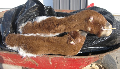
Digital Subluxation.
Digital Subluxation in a term Shorthorn calf showing distortion of the structure of the hindlegs.
Kaiser L (2012)
Digital Subluxation.
Digital Subluxation in a term Shorthorn calf showing distortion of the structure of the hindlegs.
Kaiser L (2012)
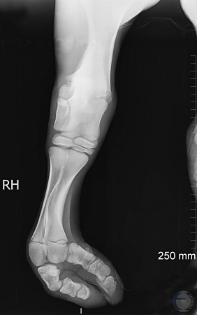
Severe Digital Subluxation - X-ray.
Radiogram of the right hind limb of a calf with severe digital subluxation due to malfunction of the collateral ligament.
Kaiser L (2013)
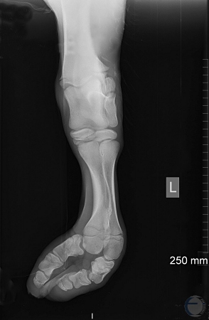
Severe Digital Subluxation - X-ray.
Radiogram of the left hind limb of a calf with severe digital subluxation due to malfunction of the collateral ligament.
Kaiser L (2013)
Parasitic Limb.
Parasitic pelvic limb (notomelus) dangling next to the tail. This calf also has contracted tendons in both thoracic limbs.
Drost M (1979)
Duplication of Bones of Lower Hind Limb.
Duplication of bones in the right hind limb of a calf.
Roberts SJ (1973)
Radiograph of Duplication of Bones.
Radiograph illustrating duplication of bones of the lower hind limb.
Roberts SJ (1973)
Contracted Tendons.
Contracted tendons in the front legs of a calf. These may be associated with uterine crowding when the calf is large or when the amount of amniotic is small (oligamnios).
Drost M (1980)
Spastic Paresis.
Spastic paresis (Elso heel) is characterized by spastic contracture of the gastrocnemius muscle. It is usually unilateral and occurs more commonly in the right rear limb. Spastic paresis is a state of partial paralysis, or incomplete paralysis.
Roberts SJ (1973)
Congenitally Dropsy of the Hind Legs.
Congenitally notched ears and dropsy (stocking) of the hind legs in an Ayrshire calf.
Roberts SJ (1973)
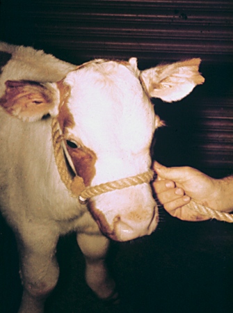
Congenitally Notched Ears.
Congenitally notched ears in an Ayrshire calf.
Roberts SJ (1973)
Syndactily.
Syndactily or mule foot in a Holstein calf. One or both front feet may be affected, or all four feet; more common in the front feet than in the hind feet.
Roberts SJ (1973)
Syndactily - Close Up.
Syndactily or mule foot in a Holstein calf. One or both front feet may be affected, or all four feet; more common in the front feet than in the hind feet.
Roberts SJ (1973)
Freemartin Vulva.
This is one of two heifers out of a set of quadruplets that shows an enlarged clitoris and a pronounced tuft of hair at the tip of the vulva. In utero, this heifer may have been positioned between her two male "womb mates" and have been subjected to a double dose of phenotypic adjustment.
Drost M (1980)
Freemartin Vulvar Tuft.
Typical long tuft of hair at the tip of the underdeveloped vulva in a freemartin heifer.
Roberts SJ (1973)
Freemartin Vulvar Tuft.
Frequently the hair at the ventral tip of the vulva forms a tuft in a freemartin heifer.
Drost M (1980)
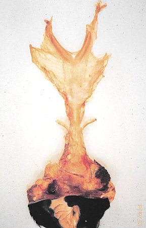
Freemartin Reproductive Tract.
Vestigial reproductive tract of a freemartin heifer. Note the absence of the cervix, the presence of bilateral vesicular glands (mesonephric duct remnants), rudimentary uterine horns, and vestiges of the ovaries. Note that, while the presence of the bilateral rudimentary seminal vesicular glands is a constant feature, the vestiges of the uterus vary considerable, from minuscule to fully identifiable by transrectal palpation. Ovaries are generally mere thickenings.
Roberts SJ (1986)
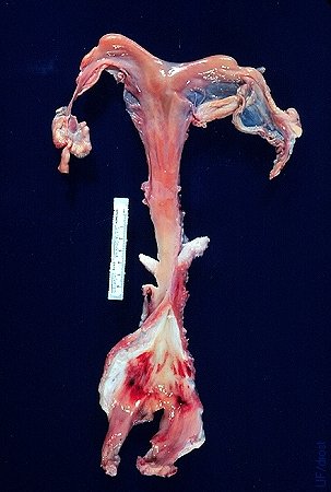
Freemartin - Infantile Reproductive Tract.
Underdeveloped reproductive tract of a heterosexual female twin. Note the vestigial ovaries, seminal vesicular glands and absence of the cervix. Trauma of the vestibular area is due to attempted vaginoscopy. (Courtesy of A de la Concha).
Drost M (1980)
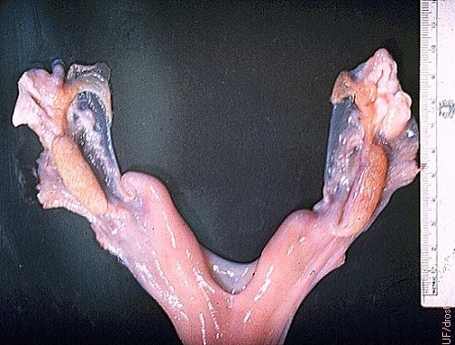
Hypoplasia of Female Reproductive Tract.
Hypoplasia of the uterus and the ovaries of an intact 2-year old Holstein heifer.
Roberts SJ (1973)
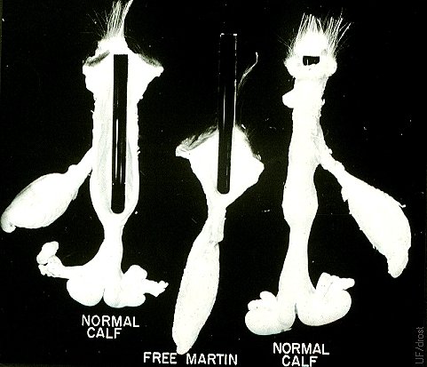
Freemartin Test.
Clinical diagnosis of a freemartin based on the length of the vagina in a freemartin as compared with a normal contemporary heifer. A test tube or a gloved finger may be used to gauge the length of the vaginal lumen.
Roberts SJ (1986)
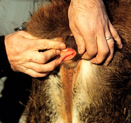
Vulvar Wattle in a Jersey Calf.
A vulvar wattle is an epithelial structure that resembles a small bull penis and is suspended from the dorsolateral wall of the vestibule in an occasional heifer. No other anomalies are associated with it. Surgical removal is straightforward but does require a ligature at its base because it usually has an active central artery. Owners sometimes mistakenly think that their animal is a hermaphrodite. The dorsal location readily distinguishes it from the clitoris which is present in its normal location in the ventral commissure of the vulva.
Roberts SJ (1973)
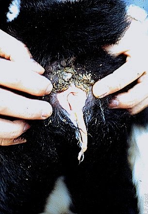
Vulvar Wattle in a Holstein Calf.
A vulvar wattle is an epithelial structure that resembles a small bull penis and is suspended from the dorsolateral wall of the vestibule in an occasional heifer. It may protrude from the vulva. Surgical removal is straightforward but does require a ligature at its base because it usually has an active central artery. Owners sometimes mistakenly think that their animal is a hermaphrodite. The dorsal location readily distinguishes it from the clitoris which is present in its normal location in the ventral commissure of the vulva.
Drost M (1979)
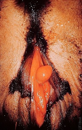
Male Pseudohermaphrodite.
Ambiguous genitalia of a male pseudohermaphrodite (phenotypically a female with the gonads of a male). A rudimentary penis is visible.
Roberts SJ (1973)
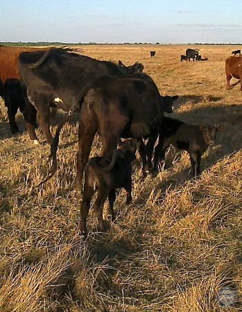
Male Pseudohermaphrodite.
Cow with twins. The one on the right is a normal bull calf. The one in front is a male pseudohermaphrodite. A drainage tube have been placed in the rudimentary vulva and vagina.
Fontaneto FJ (2013)
Male Pseudohermaphrodite.
Male pseudohermaphrodite calf. A drainage tube has been placed in the vestigial vulva and vagina.
Fontaneto FJ (2013)
Male Pseudohermaphrodite.
Male pseudohermaphrodite calf. Close-up of the urinary drainage tube placed in the vestigial vulva and vagina.
Fontaneto FJ (2013)
Male Pseudohermaphrodite.
Male pseudohermaphrodite calf. Close-up of the urinary drainage tube placed in the vestigial vulva and vagina. Healing has taken place.
Fontaneto FJ (2013)
Male Pseudohermaphrodite.
Male pseudohermaphrodite calf. Vestigial vulva and vagina have been repaired and sutures have been removed, and the urinary tube has been removed.
Fontaneto FJ (2013)
Twin Pseudohermaphrodite.
Male pseudohermaphrodite calf, in the lower half of the picture, twin to an intact normal bull calf. A urinary drainage tube has been inserted into its vestigial vulva.
Fontaneto FJ (2013)
Hypospadias.
Hypospadias. The urethral lumen is open along the ventral aspect of the penis in the perineal region. The area is wet and contaminated by free flowing urine.
Varsano S (2012)
Pulmonary Hamartoma.
Pulmonary hamartoma in a newborn calf. A hamartoma is a focal malformation that resembles a neoplasm, grossly and even microscopically, but resulted from faulty development.
Roberts SJ (1973)