The Visual Guide to
Bovine Reproduction
Reproductive Technology: In Vivo Embryos
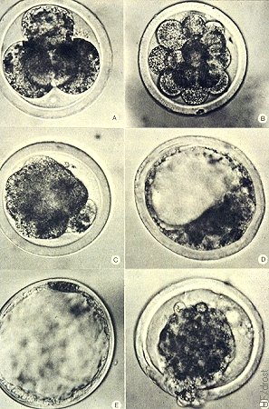
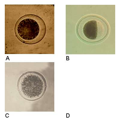
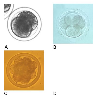
Embryo Chart.
Bovine embryos at different stages of development at 350 X. A) Day 3, 4-cell embryo; B) Day 5, 16-cell embryo; C) Day 6, Morula; D) Day 7, Early blastocyst; E) Day 10, Expanded blastocyst; F) Day 10, Hatching blastocyst.
Betteridge KJ (1977)
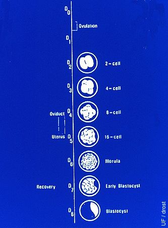
Timeline for Development.
Day 0 = estrus. Ovulation occurs 24 to 30 hours after the onset of estrus on Day 1. Cell division initially occurs at ~ 24-hour intervals, hence on Day 2 we observe 2-cell embryos, on Day 3, 4-cell embryos, on Day 4, 8-cell embryos, on Day 5, 16-cell embryos. The embryos descend from the oviduct into the uterus between Day 4 and 5 of the estrous cycle in the cow.
Drost M (1974)
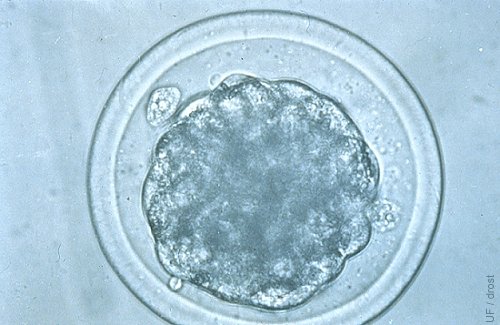
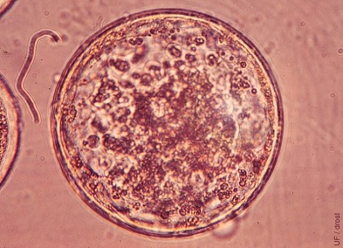
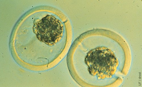
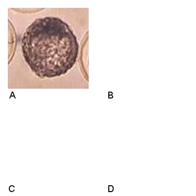
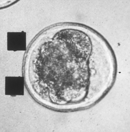
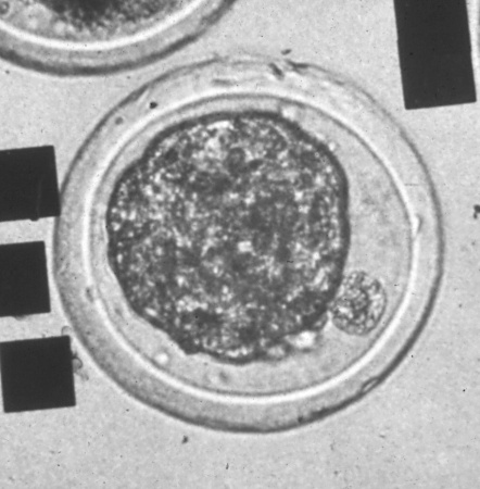
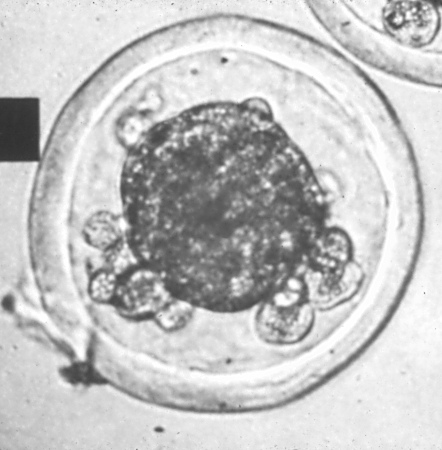
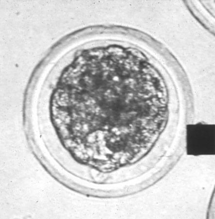
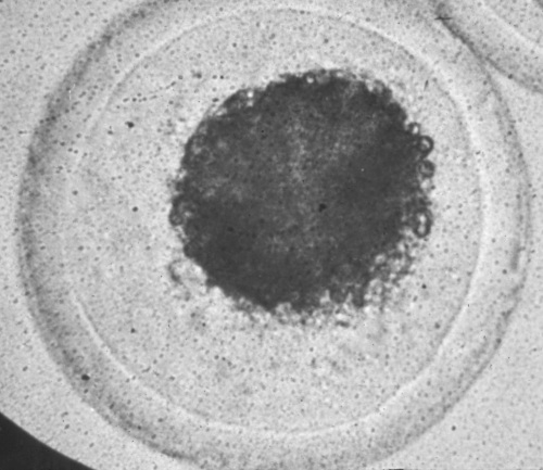
Compact Morula.
Compact Morula. IETS: Stage 4, Code 1, Cycle day 7.
Drost M (1980)
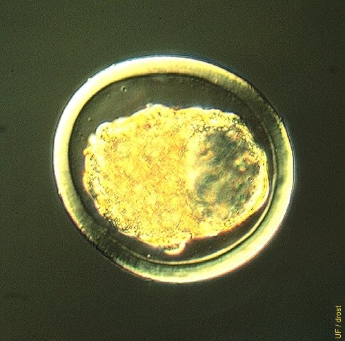
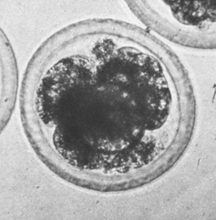
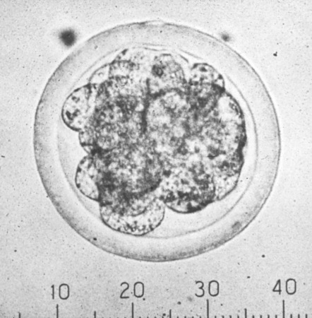
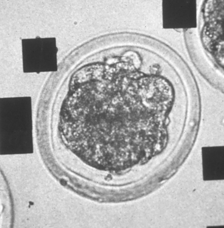
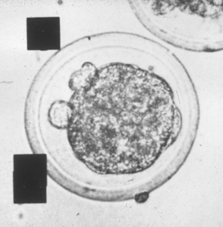
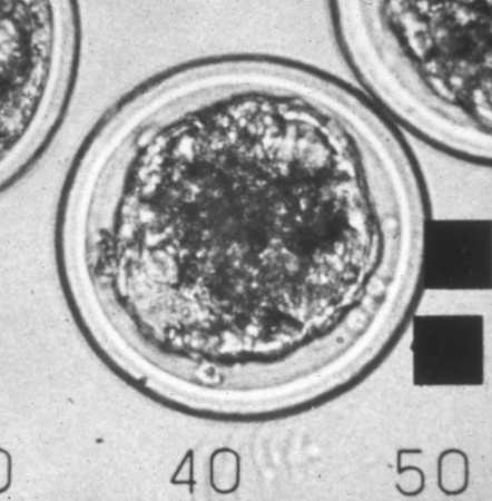
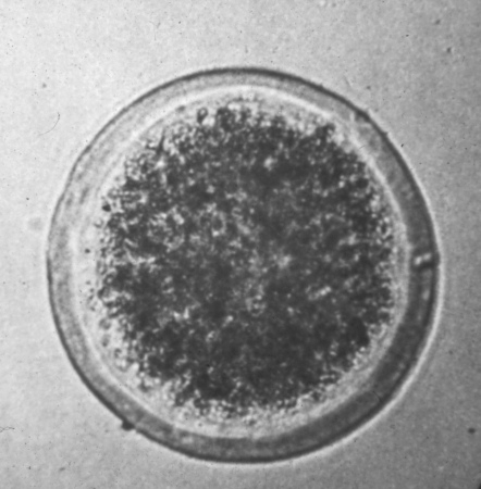
Early Blastocyst.
Early blastocyst. IETS: Stage 5, Quality 1, Cycle day 7.
Seidel GE (1991)
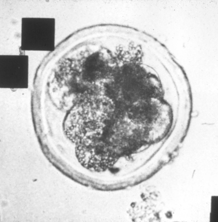
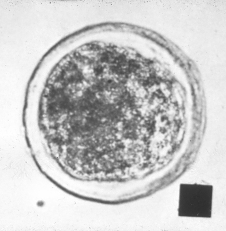
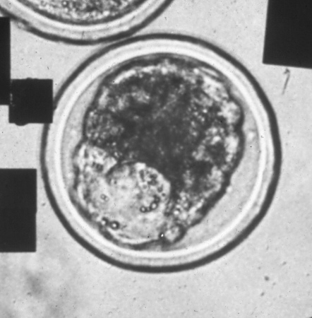
Blastocyst.
Blastocyst. IETS: stage 6, Quality 1, Cycle day 7.0 - 7.5.
Drost M (1980)
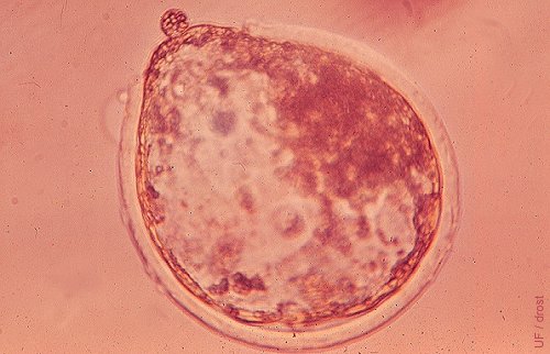
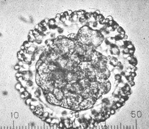
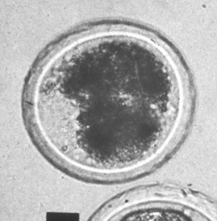
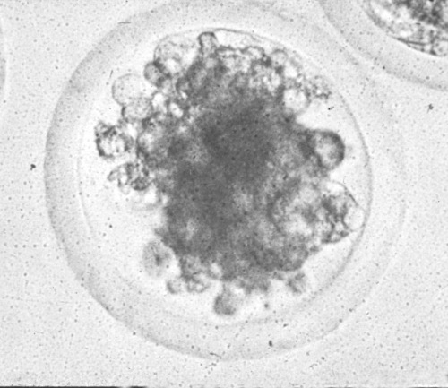
Expanded Blastocyst.
Expanded blastocyst. IETS: Stage 7, Quality 1, Cycle day 7.5.
Drost M (1980)
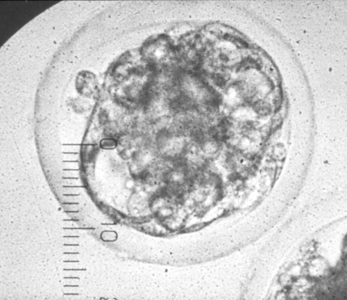
Hatching Blastocyst.
Hatching blastocyst. IETS: Stage 8, Quality 1, Cycle day 7.5 - 8.0.
Drost M (1980)
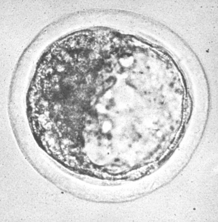
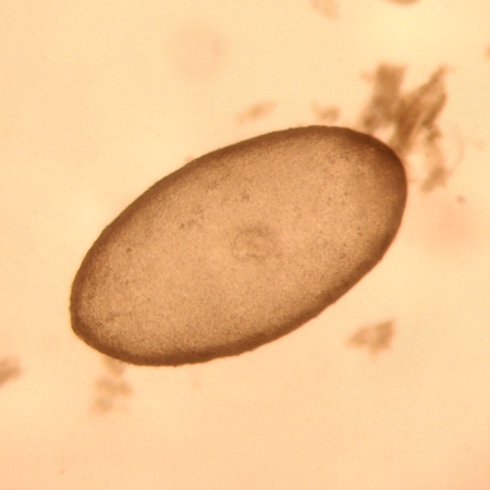
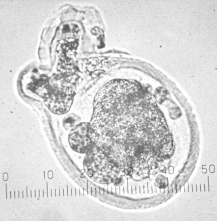
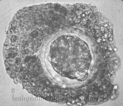
Day 13 Bovine Embryo.
Day 13 embryo. The conceptus is ovoid in shape.
Santos J (2009)
Day 13 Bovine Embryo.
Day 13 embryo. The conceptus is ovoid in shape.
Santos J (2009)
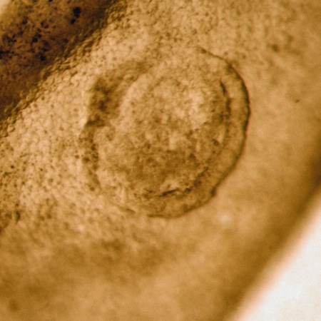
Day 14 Bovine Embryo.
Circular Day 14 embryo, seen within the elongated chorioallantoic membranes.
Santos J (2009)
Day 14 Bovine Conceptus.
Circular Day 14 embryo, seen within the elongated chorioallantoic membranes.
Santos J (2009)
Day 14 Bovine Embryo.
Bovine embryos rapidly elongate after hatching. After shedding of zona pellucida the conceptus becomes susceptible to trauma and contamination.
Seidel GE (1984)
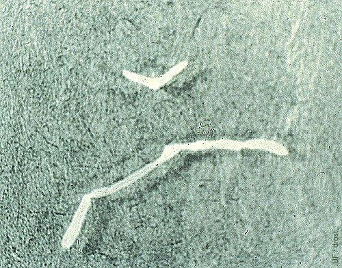
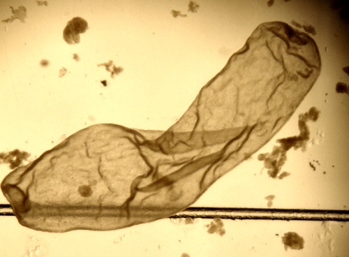
Day 14 Bovine Conceptuses.
The upper V-shaped conceptus is a normal Day 14 bovine conceptus (length 3.8 mm). The lower, longer (37.3 mm) conceptus is a Day 14 bovine conceptus from a cow treated with supplemental progesterone.
Geisert R (1983)
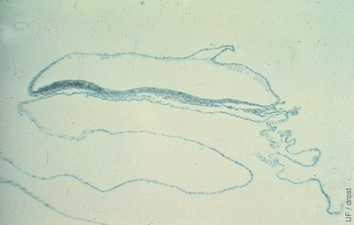
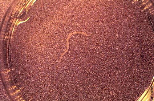
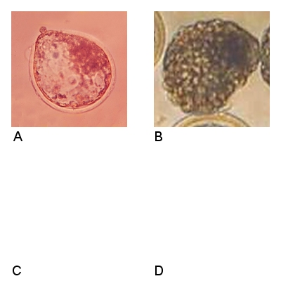
Day 19 Bovine Conceptus.
This long filamentous bovine conceptus was collected at slaughter from a cow known to be inseminated 19 days previously. The inner cell mass can be identified as a thicker, darkened area in the upper lefthand corner of the image.
Geisert R (1983)
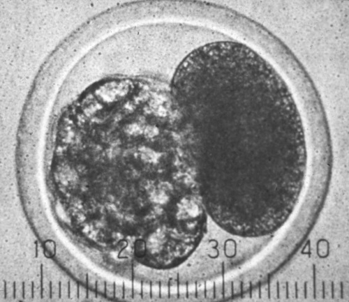
Embryo Splitting.
Two semi-embryos after splitting of a morula. One semi-embryo was placed in an empty surrogate zona pellucida. Notice the slits in the zonae pellucidae.
Seidel GE (1984)
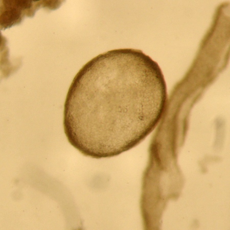
Stage 1, Unfertilized.
A. Single cell, unfertilized ovum (UFO) which may be collected on Day 7 from a superovulated donor along with embryos. The perivitelline membrane is intact and smooth. It is sometimes called a teddy bear eye. Comment: fertilization failure may be the result of asynchronous insemination, or insemination with dead or poor quality spermatozoa. B. Single cell, unfertilized oocyte in which the degenerated cellular material has pulled away from the vitelline membrane. It is sometimes referred to as a pseudoblastocyst which it may resemble when slightly out of focus or when the lenses are dirty. The contour of the vitelline membrane is smooth. C. Unfertilized ovum that has started to degenerate giving it a granular appearance. It is sometimes referred to as a salt and pepper egg.
Wright JM (2004)
Stage 2, 2- to 12-cell.
A. 2-cell embryo. B. 4-cell embryo. Note the spermatozoa present in the zona. C. 12-cell embryo.
Wright JM (2004)

Stage 3, Early Morula.
A. Quality 1, Excellent. Individual blastomeres of uniform size are still discernible. The cell mass occupies about 60% of the perivitelline space. This embryo was collected on Day 6.5. B. Quality 2, Fair. At least 50% of the cellular material is intact. One large extruded cell is present and three vacuolated cells may be discerned. C. Quality 3, Poor. About 30% of the cell mass appears intact and viable. The mass is accompanied by a number of degenerated cells. D. Quality 4, Degenerate.
Wright JM (2004)
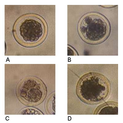
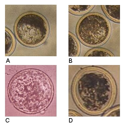
Stage 4, Morula.
A. Quality 1, Excellent, symmetrical embryo free from imperfections. Individual blastomeres may still be identified when focusing up and down. The outline of the embryo has scalloped edges. The cell mass of about 64 cells occupies approximately 80% of the perivitelline space. The zona pellucida is intact. Collected on Day 7. B. Quality 2, Fair. The cell mass still exceeds 50%. Several extruded cells are evident. C. Quality 3, Poor. Only about 25% of the intracellular cell mass appears to be intact and viable. There are a few extruded blastomeres and the remainder is fragmented material. D. Quality 4, degenerate embryo. There appear to be remnants of small blastomeres. The cellular material has degenerated and is non-viable. Collected on Day 6.
Wright JM (2004)
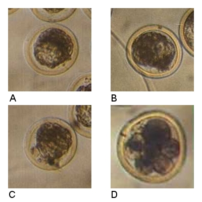
Stage 5, Early Blastocyst.
A. Quality 1, Excellent. The lightened area is a blastocele. The embryo is symmetrical and free from imperfections. The cell mass almost completely occupies the entire perivitelline space. B. Quality 2, Fair. About 75% percent of the cellular material is intact and viable. The outline of the embryo is irregular in the area of the blastocele. C. Quality 3, Poor. There is evidence of a blastocele and there appears to be an intact embryonic mass of 25%. A 3-dimensional image by virtue of focusing up and down under the microscope would aid in the evaluation of this embryo. D. Quality 4, degenerate. There are several early stage asynchronous blastomeres. All four embryos were collected on Day 7.
Wright JM (2004)
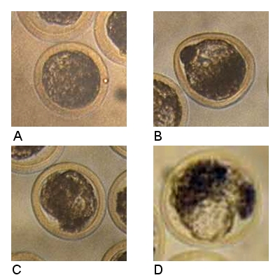
Stage 6, Blastocyst.
A. Quality 1, Good. The cellular material occupies the entire perivitelline space. There are no major imperfections. There is a slight tendency for the embryo to be pyknotic, which may also be due to the amount of transmitted light. B. Quality 2, Fair. A degenerative clump of cells is present near the blastocele. The zona pellucida is misshapen. There is evidence of pyknosis. C. Quality 3, Poor. There are several areas of irregular and extruded cells. The embryo is asymmetrical. There is at least a 25% intact, viable embryonic mass. D. Quality 4, Degenerate. Dark ragged debris is present, as well as a lighter blastocele-like space at 6 o'clock.
Wright JM (2004)
Stage 7, Expanded Blastocyst.
A. Quality 1, Good. The blastocele is clear for the most part and the intercell mass is well defined. The zona pellucida is thinner due to the expansion. B. Quality 2, Fair. The intercell mass is not well delineated which may be due extruded cells pressed against the zona pellucida. C. Poor. There is no clearly defined intercell mass. D. Degenerate. Cellular material has disintegrated and the embryo has collapsed within the perivitelline membrane.
Wright JM (2004)
Stage 8, Hatching Blastocyst.
A. Quality 1, Excellent. The expanding blastocyst is emerging from the fractured zona pellucida. The intercell mass is well delineated as is the layer of trophoblast cells lining the zona pellucida. B. Quality 2. Fair to Good. The collapsed embryo has shed the zona pellucida. Comment about C and D: Quality 3 and 4 hatched blastocysts, i.e. poor and degenerating hatched blastocysts are rarely identified as they resemble tissue fragments and debris. Suspicious material can always be incubated and potential further development monitored.
Wright JM (2004)
Stage 9, Expanded Hatched Blastocyst.
A. Quality 1, Excellent or Good. The hatched blastocyst has regained its spherical shape. A thicker layer of trophoblastic cells can be appreciated around the periphery.
Wright JM (2004)
4-Cell Embryo.
Degenerate 4-cell embryo recovered on Day 6.5 to 7.0. Not transferable. There are four large granular blastomeres.
Schneider HJ (1981)
Early Morula.
A severely retarded early morula with several large blastomeres. Recovered on Day 6.5 to 7.0.
Schneider HJ (1981)
Early Morula.
This early morula with nice even-sized blastomeres was recovered on Day 6.5 to 7.0, which makes it seem a bit retarded. It was transferred and a pregnancy resulted.
Schneider HJ (1981)
Early Morula.
A fair to poor quality early morula recovered on Day 6.5 to 7.0. The zona pellucida is surrounded by leucocytes.
Schneider HJ (1981)
Early Morula.
Recovered on Day 6.5 to 7.0, this poor quality early morula was transferred to a (well!) sychronized recipient and started a pregnancy.
Schneider HJ (1981)
Morula.
A compact morula. Individual blastomeres are no longer discernable. There is a polar body at the 4 o'clock position. Recovered on Day 6.5 to 7.0.
Schneider HJ (1981)
Morula.
A fair quality compact morula showing some extruded blastomeres. Recovered on Day 6.5 to 7.0.
Schneider HJ (1981)
Morula.
There is an intact cell mass in this morula but the zona pellucida appears to be disrupted and there are numerous extruded cells and there is some debris. Chances of establishing a pregnancy is less than 15%. One could culture the embryo to see if it continues development. Recovered on Day 6.5 to 7.0.
Schneider HJ (1981)
Morula.
The cell mass of this morula looks nice and symmetrical. One blastomere of the original 2-cell embryo is still present. The total number of cells is reduced. Recovered on Day 6.5 to 7.0.
Schneider HJ (1981)
Morula.
Well compacted morula with several small blastomeres not included. This reduced the total number of totipotent cells.
Schneider HJ (1981)
Morula.
A compact morula recovered on Day 6.5 to 7.0. The quality is fair (Code 2). The color is light and there are two extruded small blastomeres.
Schneider HJ (1981)
Morula / Early Blastocyst.
Good quality late morula / early blastocyst embryo. The zona pellucida is surrounded by fibrin like material which may prevent it from expanding. An uncommon occurrence. Recovered on Day 6.5 to 7.0.
Schneider HJ (1981)
Early Blastocyst.
A nice blastocyst that almost fills the entire perivitelline space.
Schneider HJ (1981)
Early Blastocyst.
Development of a small blastocele is evident. The quality is good (Code 1). Recovered on Day 6.5 to 7.0. 400 X
Schneider HJ (1981)
Early Blastocyst.
Not real uniform but a good quality embryo with a 60% chance of establishing a pregnancy in a synchronized recipient. Recovered on Day 6.5 to 7.0.
Schneider HJ (1981)
Early Blastocyst.
Excellent quality blastocyst. There is still some perivitelline space left. Recovered on Day 6.5 to 7.0.
Schneider HJ (1981)
Unfertilized Ovum.
A UFO recovered on Day 6.5 to 7.0. The cytoplasm is fragmented.
Schneider HJ (1981)
Unfertilized Ovum.
A degenerate, pyknotic oocyte recovered on Day 6.5 to 7.0.
Schneider HJ (1981)
Unfertilized Ovum.
An unfertilized egg recovered on Day 6.5 to 7.0. The perivitelline membranes is gone, giving the egg an granular appearance which leads to the vernacular "salt and pepper egg".
Schneider HJ (1981)
Early Morula, Frozen / Thawed.
A degenerate embryo recovered on Day 6.5 to 7.0. Not transferred.
Schneider HJ (1981)
Early Blastocyst, Frozen / Thawed.
A frozen-and-thawed early blastocyst with a fair quality rating. It was transferred and established a pregnancy. Recovered on Day 6.5 to 7.0.
Schneider HJ (1981)