The Visual Guide to
Bovine Reproduction
Abortion: Infectious Causes
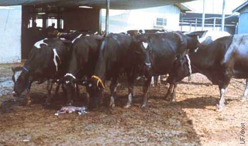
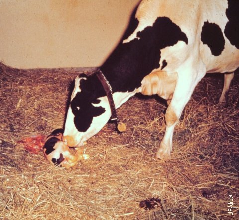
Brucella Abortion - Transmission.
Transmission of brucellosis is primarily by ingestion of Brucella organisms which typically occurs in this setting by snoopy cows "inspecting" an aborted fetus.
Nicoletti PL (1980)
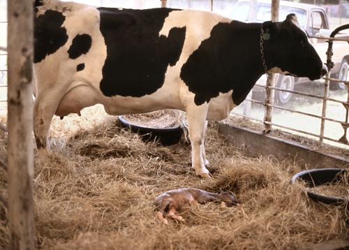
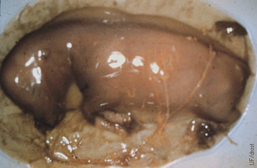
Brucella - Aborted Fetus.
Transmission of brucellosis is primarily by ingestion of Brucella organisms. Suspect cows should be isolated to prevent contact with the aborted fetus and the fetal membranes. Six month old fetus shown here.
Nicoletti PL (1980)
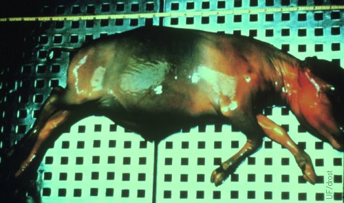
Brucella Abortus.
Autolytic aborted fetus as a result of brucellosis (5.5 months).
Drost M (1980)
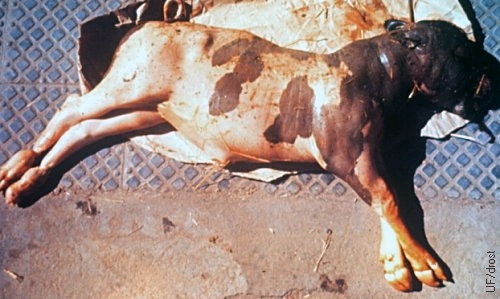
Brucella Abortion.
Autolysed aborted fetus as a result of brucellosis (6 months).
Nicoletti PL (1980)
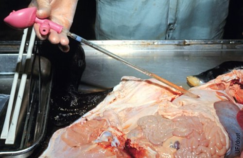
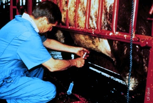
Diagnosis of Brucellosis.
Aspiration of abomasal contents for the purpose of recovery and identification of Brucella abortus from an aborted fetus.
Nicoletti PL (1980)
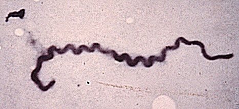
Leptospira Pomona.
This organism, a spirochete, is a common cause of abortion in cattle.
Drost M (1969)
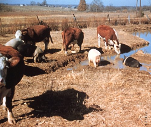
Transmission of Leptospirosis.
Optimal setting for the (interspecies) transmission of leptospirosis via urine, by running cattle and pigs together in an area with stagnant water.
Drost M (1969)
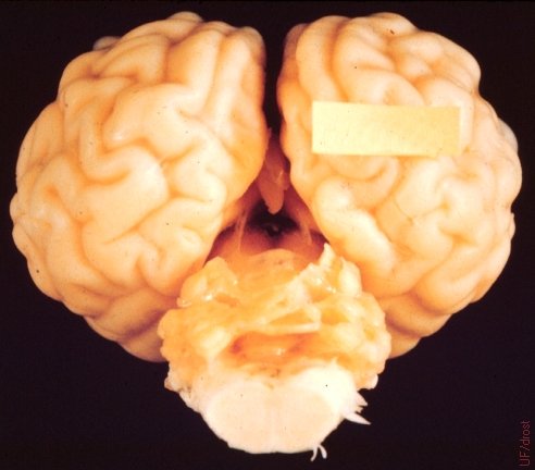
Cerebellar Hypoplasia.
This is the brain of a newborn calf showing the lack of development of the cerebellum, caused by BVD virus.
Roberts SJ (1973)
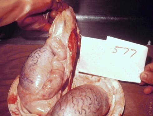
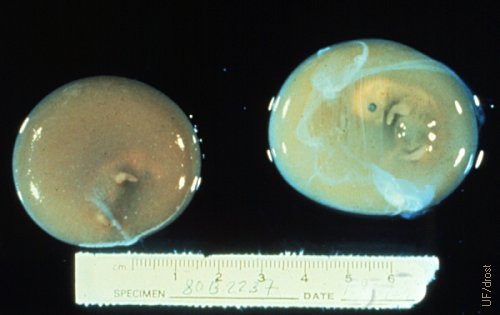
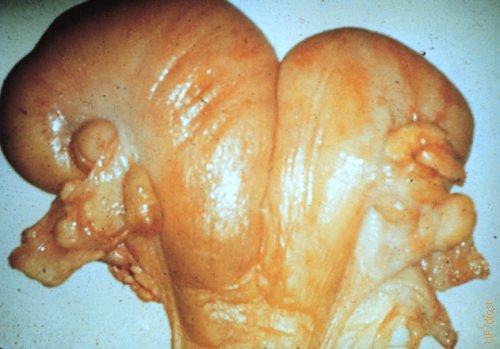
Bovine Epididymitis.
Bovine epididymis showing enlargement due to infection with Brucella abortus.
Nicoletti PL (1980)
Campylobacter Abortion.
Aborted fetus as a result of campylobacteriosis (45 days).
Drost M (1969)
Campylobacter Fetus Subspecies Fetus.
Campylobacter fetus subspecies fetus, a bacterium, causes infertility as a result of early embryonic death and occasional (5%) abortion.
Roberts SJ (1973)
Aborted Fetus due to Leptospirosis.
Abortion of a 5.5 month old fetus due Leptospira pomona.
Drost M (1969)
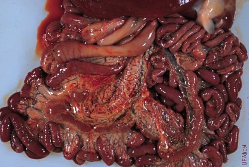
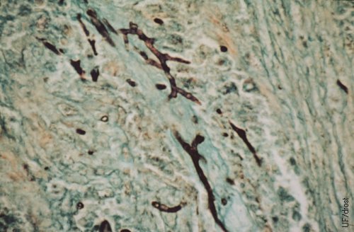
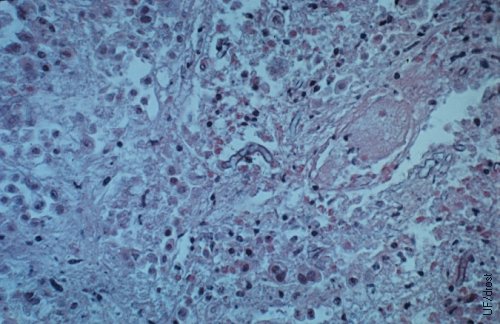
Epizootic Bovine Abortion.
Epizootic bovine abortion, or Foothill Abortion, appears to be geographically limited to the western United States. Live fetuses may be aborted, and most dead fetuses are fresh. Fetal lesions include granulomatous hepatitis and lymphoid hyperplasia in the lymph nodes. Once a heifer or a cow aborts and remains in the area she will be immune.
Drost M (1969)
Epizootic Bovine Abortion.
Epizootic bovine abortion, or Foothill Abortion, appears to be geographically limited to the western United States. Live fetuses may be aborted and most dead fetuses are fresh. Fetal lesions include granulomatous hepatitis and lymphoid hyperplasia in the lymph nodes. Once a heifer or a cow aborts and remains in the area she will be immune.
Drost M (1969)
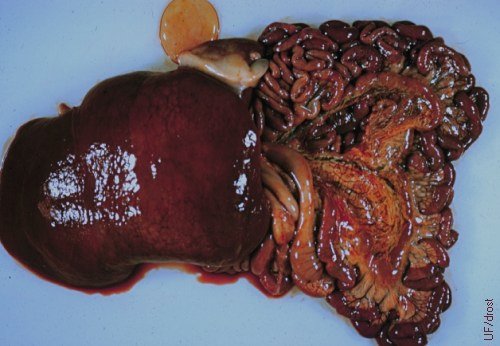
Epizootic Bovine Abortion.
Epizootic bovine abortion, or Foothill Abortion, appears to be geographically limited to the western United States. Live fetuses may be aborted, and most dead fetuses are fresh. Fetal lesions include granulomatous hepatitis, which is generally periportal, and central veins are dilated.
Drost M (1969)
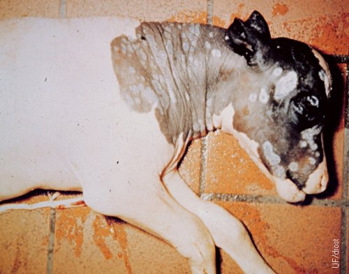
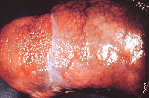
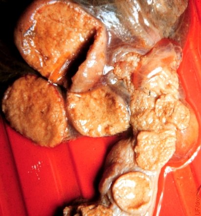
Mycotic Abortion.
Mycotic lesions on a 6.5 month old fetus. The incidence of fetal lesions is 30 %; 75-80 % are caused by Aspergillus sp. (septate) and 10-15% by Mucorales sp. (nonseptate).
Roberts SJ (1973)
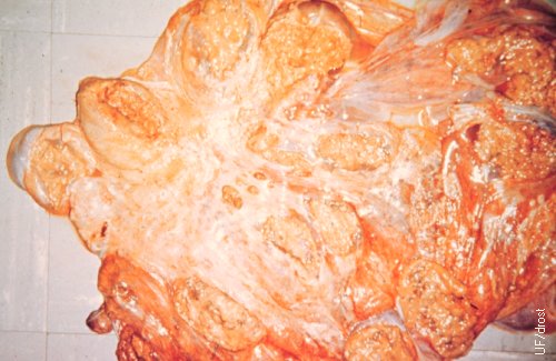
Necrotic Placentitis Caused by Mycotic Infection.
Mycotic infection of the placenta grossly resembles that of brucellosis and campylobacteriosis. Abortion occurred around 6 months of gestation.
Roberts SJ (1973)
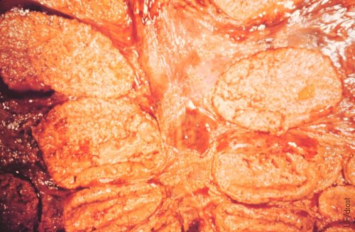
Necrotic Placentitis.
Placental dysfunction due to necrosis of placentomes leads to asphyxia and demise of the fetus.
Roberts SJ (1973)
Necrotic Placentitis.
Placental dysfunction due to necrosis of placentomes leads to asphyxia and demise of the fetus.
Roberts SJ (1973)
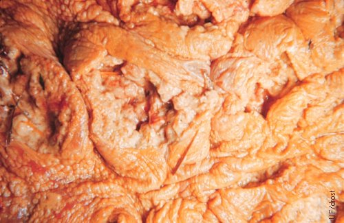
Severe Necrotic Placentitis.
Placental dysfunction due to necrosis of caruncles leads to asphyxia and demise of the fetus and abortion.
Roberts SJ (1973)
Mycotic Abortion.
Aspergillus fumigatus, the mold responsible for 75-80 per cent of fungal abortions in cattle.
Roberts SJ (1973)
Fungal Abortion.
Histologic section of the placenta with mold infection.
Roberts SJ (1973)
Diagnosis of Trichomoniasis in the Bull.
Collection of smegma sample for the detection of Tritrichomonas foetus in the bull.
Chenoweth PJ (1985)
Embryonic Death.
Embryonic death due to Tritrichomonas foetus infection (~50 day conceptuses).
Drost M (1980)
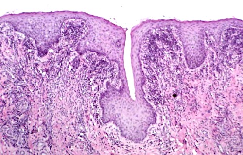
Preputial Epithelial Crypt in the Bull.
Protozoa like Tritrichomonas foetus are protected by, and proliferate in the epithelial crypts of the preputial lining of the bull. These crypts are deeper and more extensive in older (>4 years) bulls.
BonDurant RH (2008)
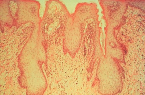
Epithelial Crypts in the Preputial Lining of the Bull.
Protozoa like Tritrichomonas foetus are protected by, and proliferate in the epithelial crypts of the preputial lining of the bull. These crypts are deeper and more extensive in older (>4 years) bulls.
McEntee K (1973)
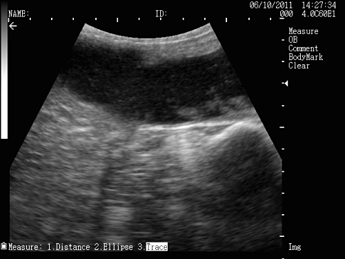
Tritrichomoniasis Abortion.
Dispersed flocculent material in the fluid filled lumen of the uterus in a cow that tested positive for Tritrichomoniasis.
Bolinger JD (2011)
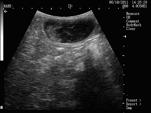
Fetal Death.
Remnants of a 60-day fetus in the uterus of a cow that tested positive for Tritrichomoniasis.
Bolinger JD (2011)
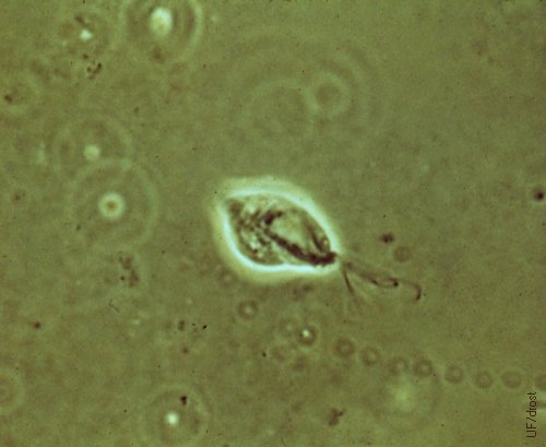
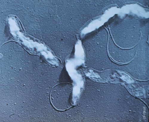
Tritrichomonas Foetus.
Causative organism of trichomoniasis, Tritrichomonas foetus, a protozoon. Three flagella are distinguishable. The live organism has a tumbling motion.
Drost M (1980)
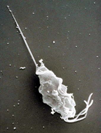
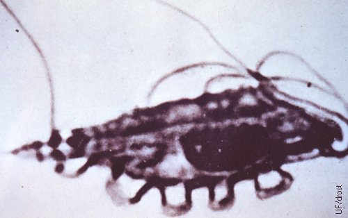
Scanning Electron Micrograph of T Foetus.
Scanning Electron Micrograph of Tritrichomonas foetus, a protozoon. Three flagella are distinguishable.
BonDurant RH (2008)
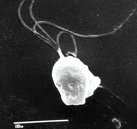
Scanning Electron Micrograph of T Foetus.
Scanning Electron Micrograph of Tritrichomonas foetus, a protozoon.
BonDurant RH (2008)
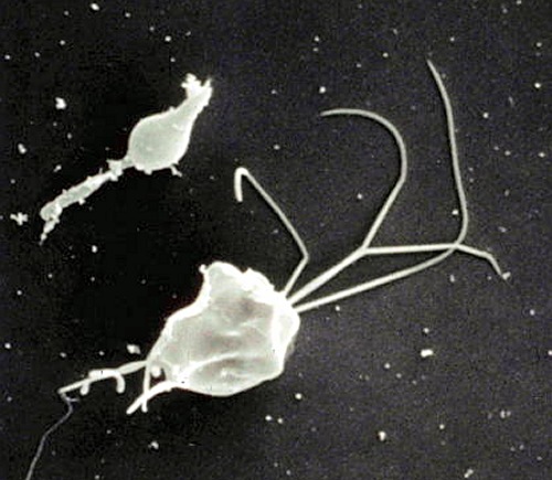
Miscellaneous Trichomonads.
Scanning Electron Micrograph of miscellaneous non-Tritrichomonas foetus organisms.
BonDurant RH (2008)
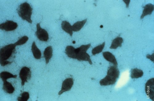
Tritrichomonas Foetus Organisms.
Causative organisms of trichomoniasis (Tritrichomonas foetus), protozoa. Live organisms display a constant tumbling motion.
Drost M (1980)
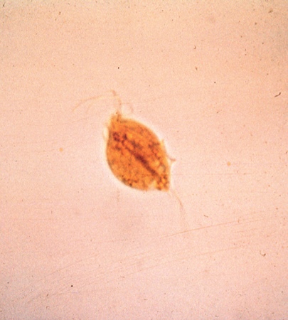
Tritrichomonas Foetus Organism.
Causative organism of trichomoniasis (Tritrichomonas foetus), a protozoon. Three flagella and an undulating membrane are faintly distinguishable. The live organisms display a constant tumbling motion.
Drost M (1980)
Tritrichomonas Foetus Organism.
Causative organism of trichomoniasis (Tritrichomonas foetus), a protozoon. Note three flagella and an undulating membrane.
Drost M (1980)
Postcoital Pyometra.
Postcoital pyometra is pathognomonic for trichomoniasis in cattle.
Drost M (1980)
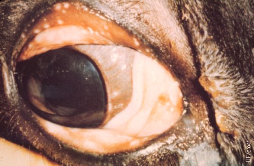
Bovine Herpes Eye Lesion.
Ocular lesions associated with bovine herpes virus infection (BHV1) or infectious bovine rhinotracheitis (IBR).
Roberts SJ (1973)
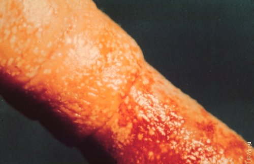
Bovine Herpes Penile Lesions.
Infectious pustular balanoposthitis (IBPP). Lesions on the penis associated with bovine herpes virus infection.
Roberts SJ (1973)