The Visual Guide to
Bovine Reproduction
Ultrasonography: Pregnancy Diagnosis
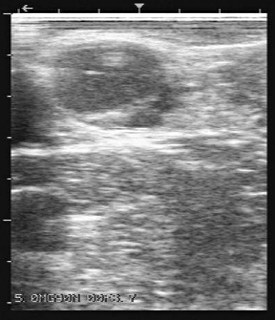
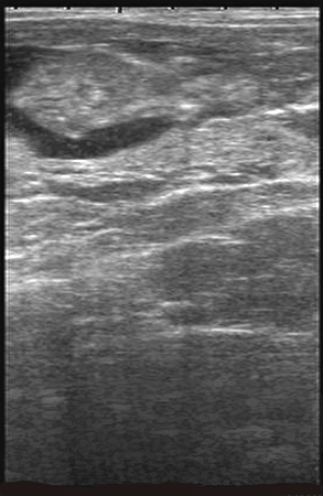
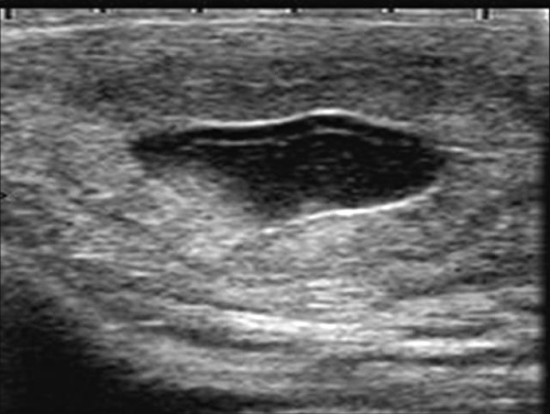
21-Day Pregnancy.
The mature CL (left) and small pocket of uterine fluid (right) at 21 days after documented breeding indicate likely pregnancy. However, the embryo cannot yet be seen so it is unwise to make diagnosis of pregnancy this early.
Colloton J (2006)
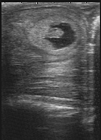
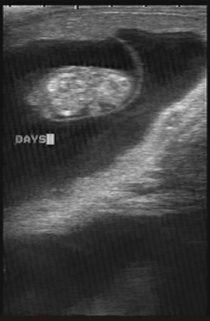
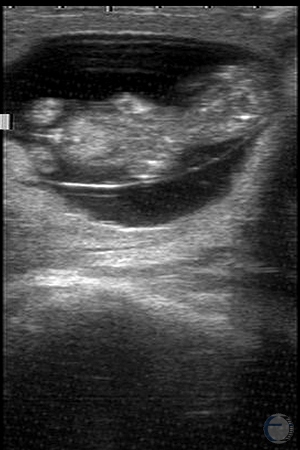
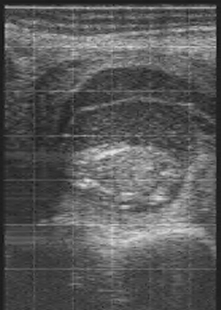
26-Day Twins.
At 26 days these embryos are readily visible and heartbeats can be observed. A definitive diagnosis of twin pregnancy versus open can be made at this stage.
Colloton J (2006)
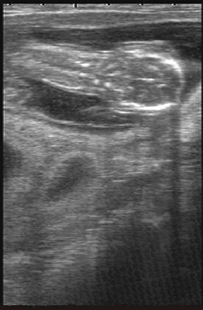
28-Day Pregnancy.
The embryo can be seen on the left. Crown / rump length (CRL) = 9 mm. A mature CL is visible on the ipsilateral ovary.
Colloton J (2006)
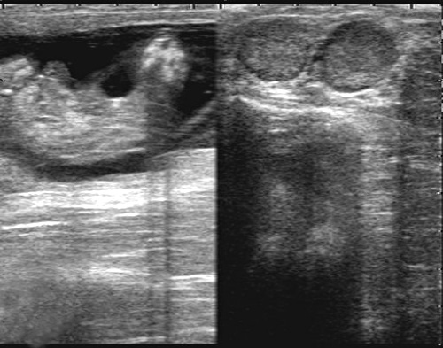
28-Day Pregnancy.
In the right panel the 28-day old embryo can readily be recognized. In the left panel, the 28-day old embryo is located close to the endometrium and more difficult to identify.
Bartolome J (2006)
29-Day Pregnancy.
The developing embryo is clearly delineated and partially surrounded by fetal fluids in this cross section of the gravid uterine horn.
Colloton J (2006)
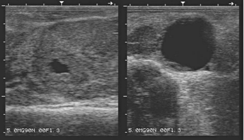
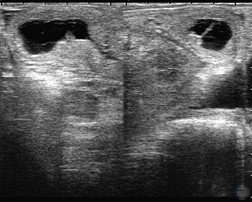
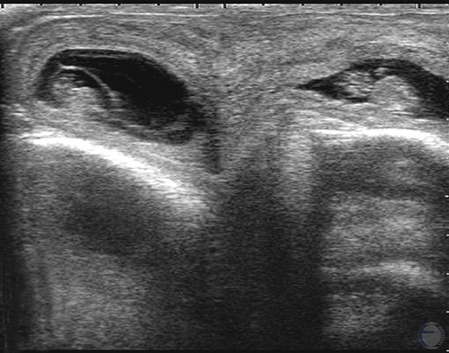
29-Day Pregnancy.
Left panel: Fluid in the uterine horn. Right panel: corpus luteum next to a large follicle, at 29 days of gestation.
Bartolome J (2006)
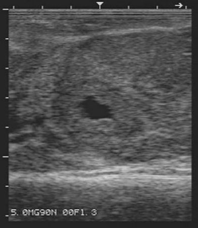
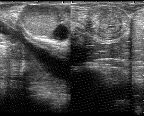
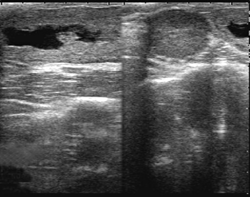
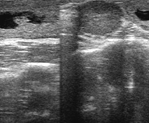
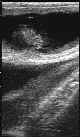
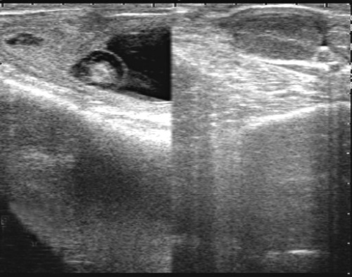
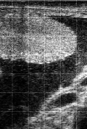
29-Day Pregnancy / Fluid.
Cross section of a gravid uterus at 29 days. Accumulation of fluid can readily be seen in the cross section of the uterine horn. In the absence of a view of the embryo, a presumptive diagnosis of pregnancy can be made, but the diagnosis must be confirmed at a later date.
Bartolome J (2006)
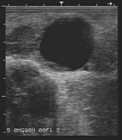
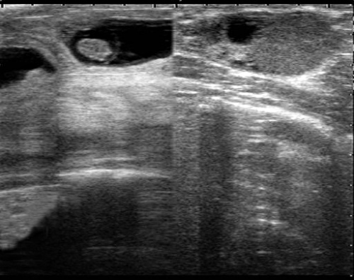
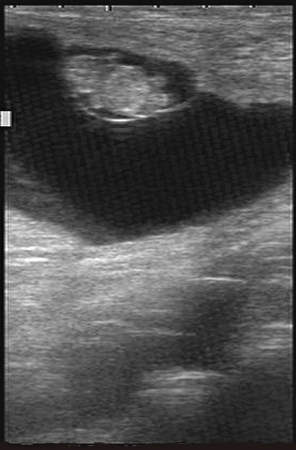
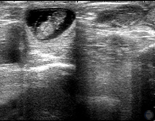
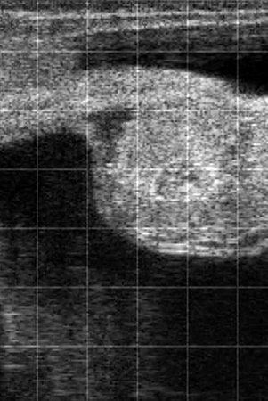
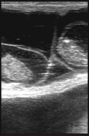
29-Day Pregnancy / CL.
Corpus luteum (upper left) adjacent to a large 27 mm follicle, 29 days after insemination.
Bartolome J (2006)
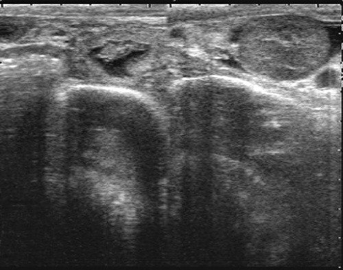
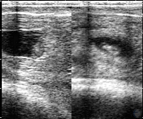
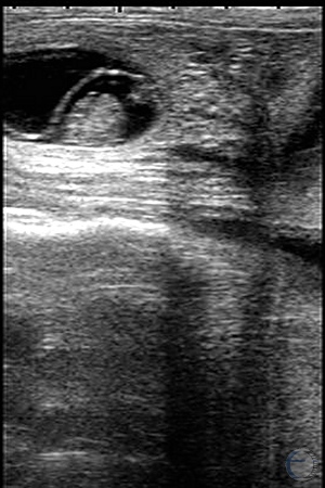
30-Day Pregnancy.
The left panel shows a cross section of the gravid horn. The ipsilateral CL of pregnancy is shown in the right panel.
Colloton J (2006)
31-Day Pregnancy.
A 31-day pregnancy. The embryo has a crown / rump length (CRL) of 11 mm.
Colloton J (2006)
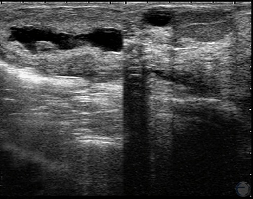
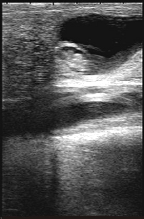
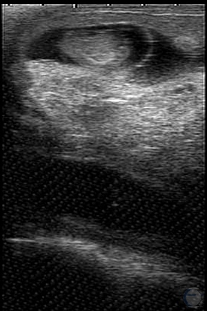
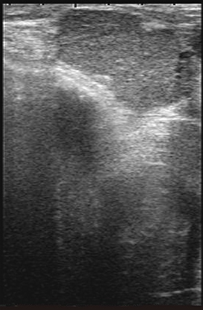
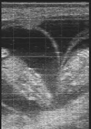
31-Day Pregnancy / Uterus.
Nice cross section of the uterine horn. The 31-day embryo is outlined by the surrounding fluid.
Bartolome J (2006)
31-Day Pregnancy / CL.
Gravid corpus luteum at Day 31 of gestation.
Bartolome J (2006)
33-Day Pregnancy.
A 33-day pregnancy. Embryo has a crown / rump length (CRL) of 13 mm. There is a CL on the ipsilateral ovary.
Colloton J (2006)
33-Day Pregnancy.
A 33-day pregnancy. Embryo has a crown / rump length (CRL) of 13 mm. There is a CL on the ipsilateral ovary.
Colloton J (2006)
35-Day Pregnancy.
A 35-day pregnancy. Embryo has a crown / rump length (CRL) of 16 mm.
Colloton J (2006)
38-Day Pregnancy.
There are two cross sections of the curled gravid uterine horn. Right panel: Follicular waves continue during pregnancy, particularly during the first trimester.
Colloton J (2006)
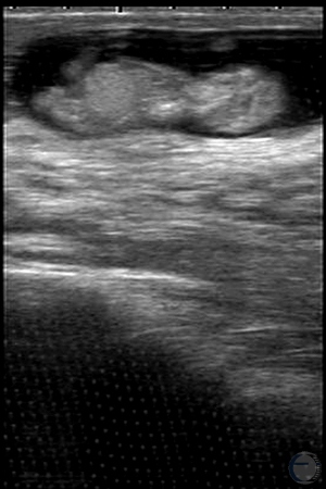
39-Day Twin Pregnancy.
These 39-day old twin embryos are in opposite uterine horns and have a crown / rump length (CRL) of 21 mm.
Colloton J (2006)
40-Day Pregnancy.
This is a 40-day pregnancy. Note the thin amniotic membrane surrounding the embryo. Allantoic fluid surrounds the amniotic vesicle.
Colloton J (2006)
41-Day Pregnancy.
The embryo is surrounded by the amniotic vesicle. The fluid to the right is allantoic fluid. The CL of pregnancy is shown in the right panel.
Colloton J (2006)
41-Day Pregnancy.
Note the delineation of the amniotic vesicle, which is approximately 15 mm long on palpation.
Colloton J (2006)
42-Day Pregnancy.
On the right is a 42-day fetus. In this view the spine is clearly visible. The fetus has a crown / rump length (CRL) of 26 mm.
Colloton J (2006)
43-Day Pregnancy.
The head, body and four appendages are clearly visible in this early fetus.
Colloton J (2006)
45-Day Pregnancy.
A 45-day fetus on the right. Note the thin amniotic membrane. The fetus has a crown / rump (CRL) of 29 mm.
Colloton J (2006)
48-Day Pregnancy.
The early fetus is curled up and neatly surrounded by its amniotic membrane. The longitudinal diameter of the vesicle is 35 mm on palpation.
Colloton J (2006)
49-Day Pregnancy.
This is a 49-day pregnancy. The fetus has a crown / rump length (CRL) = 36 mm.
Colloton J (2006)
54-Day Pregnancy.
This is a 54-day pregnancy. The fetus has a crown / rump (CRL) = 48 mm.
Colloton J (2006)
58-Day Pregnancy.
This is a 58-day pregnancy. At this stage the fetus may be in a curled position, which makes measuring fetal length difficult. Head diameter, head length, or thoracic diameter should be used instead.
Colloton J (2006)
60-Day Pregnancy.
This is a 60-day pregnancy. By this stage fetal sex can be determined. The male genital tubercle is visible on the left. The fetal head diameter = 17 mm.
Colloton J (2006)
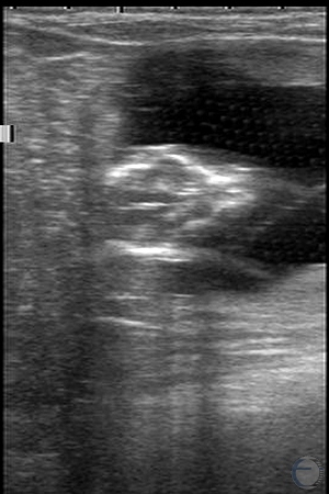
64-Day "Twin" Pregnancy.
This is a 64-day pregnancy. Note the double CL on the right. A thorough examination for twins revealed only a single fetus.
Colloton J (2006)
68-Day Pregnancy.
This is a 68-day pregnancy. This is a view of a cross section of the head which has a diameter of 20 mm.
Colloton J (2006)
68-Day Pregnancy.
This is a 68-day pregnancy with a longitudinal section of the fetal head. The head length = 29 mm.
Colloton J (2006)
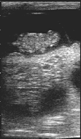
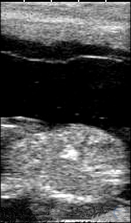
71-Day Fetal Head.
Bone density is gradually increasing and easier to visualize.
Colloton J (2006)
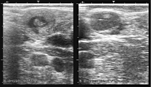
80-Day Fetus.
Normal fetus in the left horn of a Holstein cow. In the right horn there was an empty amniotic vesicle. There was a CL on the left ovary, and no CL on the right ovary.
Bartolome J (2009)
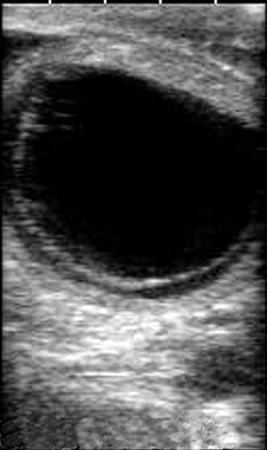
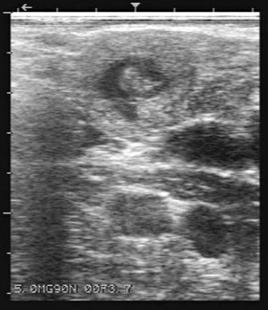
Empty Amniotic Vesicle.
Empty amniotic vesicle in the right horn of a Holstein cow. Normal fetus in the left horn. There was a CL on the left ovary, and no CL on the right ovary.
Bartolome J (2009)
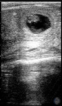
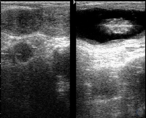
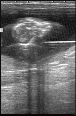
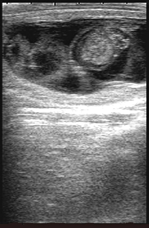
Two CL of Pregnancy.
Fifty percent of pregnant cows with 2 corpora lutea will have twins. Monozygotic twins in cattle are rare so examining the ovaries helps to determine if it is necessary to look for twins.
Colloton J (2006)
Early Embryonic Death.
There are two cardinal signs of embryonic death in this image: The chorioallantois is separating from the endometrium, and the fluid of pregnancy is flocculent. In a live scan it would also be apparent that the embryo has no heartbeat. Note the amorphous appearance of the embryo.
Colloton J (2006)
Fetal Death.
The extreme flocculence in the pregnancy fluid indicates that this fetus died some time ago. A dead fetus can remain in the uterus for days to weeks before being expelled. Note that the fetus and amniotic membrane are still easy to identify. This pregnancy would feel normal on palpation.
Colloton J (2006)
Fetal Demise.
Note the flocculence in the amniotic fluid and the lack of form in the 54-day fetuses. Limbs, heads, abdominal organs, hearts and ribs could readily be seen. This pregnancy felt normal on palpation.
Colloton J (2006)
Dead Twins.
Note the flocculence in the amniotic fluid and the lack of form in these 54-day fetuses. By 54 days, limbs, head, abdominal organs, heart and ribs should be readily seen. This pregnancy felt normal on palpation. Although fetal size and shape appear normal, thorough examination revealed lack of fetal heart beats or movement. The amniotic fluid is also abnormally cloudy.
Colloton J (2006)
One Dead Twin.
The fetus on the right had a heartbeat. To the left are the remains of a dead twin. This pregnancy is very high-risk and should be re-checked during the next herd visit.
Colloton J (2006)
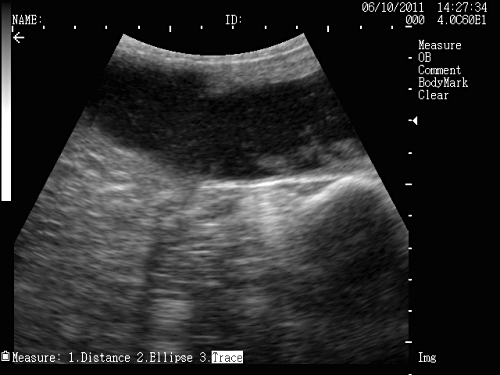
Tritrichomoniasis Abortion.
Dispersed flocculent material in the fluid filled lumen of the uterus in a cow that tested positive for Tritrichomoniasis.
Bolinger JD (2011)
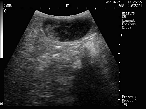
T. Foetus Abortion.
Remnants of a 2-month old fetus in the uterus of a cow that tested positive for Tritrichomoniasis.
Bolinger JD (2011)