The Visual Guide to
Bovine Reproduction
Ultrasonography: Cyclic Activity
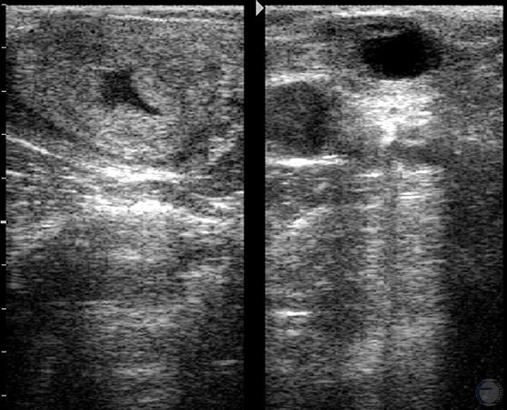
Proestrus.
Regressing CL on the left ovary. Ovulatory follicle on the contralateral ovary.
Bartolome J (2006)
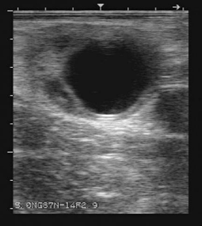
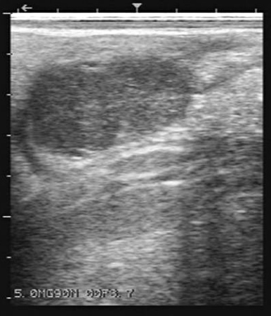
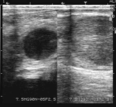
Preovulatory Follicle.
A 2 cm preovulatory follicle during proestrus.
Bartolome J (2006)
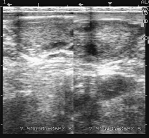
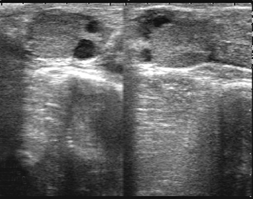
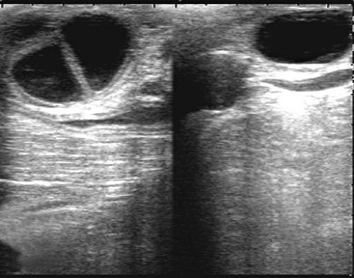
Uterine Edema.
Edema of the uterus is characteristic of cows and heifers during estrus and metestrus. Shown are the transverse and longitudinal views of a uterine horn.
Bartolome J (2006)
Estrous Uterus.
Clear fluids such as mucus and follicular fluid appear black when scanned. Do not mistake the mucus of estrus for pregnancy. There is no CL during estrus.
Colloton J (2006)
Left Ovulation / Right Follicle.
Small ovaries. Ovulation in the left ovary. Growing follicle in the right ovary.
Bartolome J (2006)
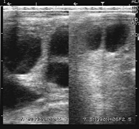
Multiple Follicles.
Multiple follicles in both ovaries as in a cow or heifer during the process of superovulation.
Bartolome J (2006)
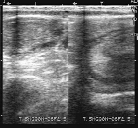
Metestrus.
Ovulation in the ovary in the left panel. Edema of the uterus at that time (during metestrus) in the right panel.
Bartolome J (2006)
Diestrous CL.
A typical mid-cycle CL and a small follicle. Follicles occur in waves throughout the estrous cycle and pregnancy, hence are almost always seen on ovarian scans in cycling cows.
Colloton J (2006)
Diestrous Uterus.
To the left is a longitudinal section of one uterine horn during diestrus. Note the lack of fluid in the lumen. To the right is a normal mid-cycle 3 cm corpus luteum with a small lacuna.
Colloton J (2006)

Mid-cycle CL.
To the left is a normal mid-cycle corpus luteum. To the right is a cross section through both uterine horns near the body of the uterus.
Colloton J (2006)
Corpus Luteum and Follicle.
Regressing corpus luteum and small follicle. The regressing CL would be firm on transrectal palpation.
Bartolome J (2006)
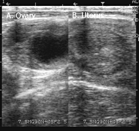
Dominant Follicle.
A. Ovary with a dominant follicle. B. Cross section of the uterine horn.
Bartolome J (2006)
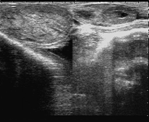
Left Dominant Follicle / Right CL.
Dominant follicle in the left ovary and a mature corpus luteum in the right ovary. Diestrus.
Bartolome J (2006)
Contralateral CLs.
A CL on each ovary suggests the possibility of twins in contralateral uterine horns. The presence of several follicles does not preclude pregnancy. Follicular waves continue during pregnancy, especially during the first trimester.
Colloton J (2006)
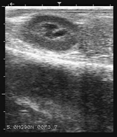
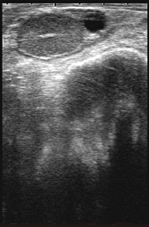
Corpus Luteum with Cavity.
Corpus luteum with a cavity (lacuna) sometimes referred to as a hollow corpus luteum, or misleadingly as a "cystic CL". These corpora lutea are functionally normal. They produce a comparable amount of progesterone to that of a normal diestrous CL or a gravid CL.
Bartolome J (2006)
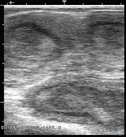
Flaccid Uterus.
Homogeneous appearance of the wall of a flaccid uterus and an inactive ovary in the left panel. Anovulatory follicle in the contralateral ovary in the right panel.
Bartolome J (2006)
Anovulation.
Inactive ovary with some 2 mm follicles in the left panel. Large anovulatory follicle on the ovary in the right panel.
Bartolome J (2006)
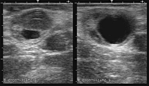
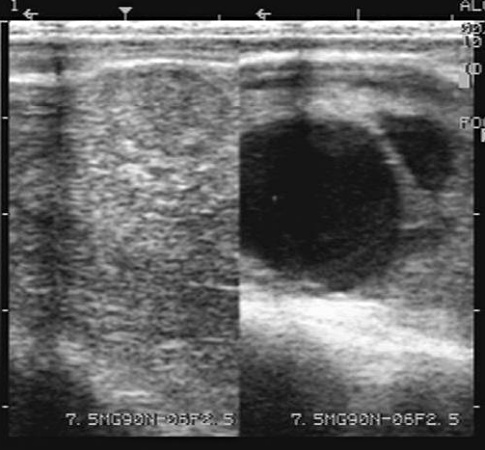
Large Follicular Cyst.
There is a large follicular cyst on the left ovary and a normal follicle on the right ovary. It is entirely possible the normal follicle will ovulate and lead to development of a corpus luteum.
Colloton J (2006)
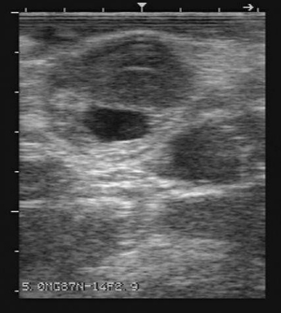
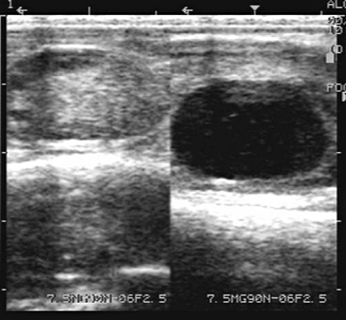
Follicular Cyst.
Two small CLs on the left ovary and a follicular cyst on the right ovary. Many follicular cysts are benign. They are merely large anovulatory follicles. Note the fluid centers in the CLs. This is normal and occurs in about 80% of CLs at some point in their development.
Colloton J (2006)
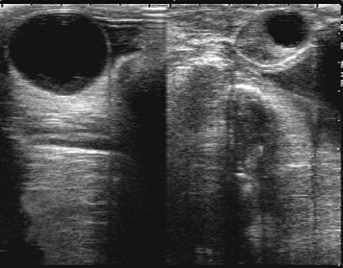
Follicular Cysts.
Follicular cysts are thin-walled, greater than 2.5 cm in diameter, and tend to be irregular in shape. Based on a single examination it is impossible to determine if these cysts are pathologic or benign.
Colloton J (2006)
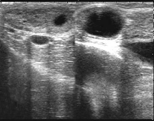
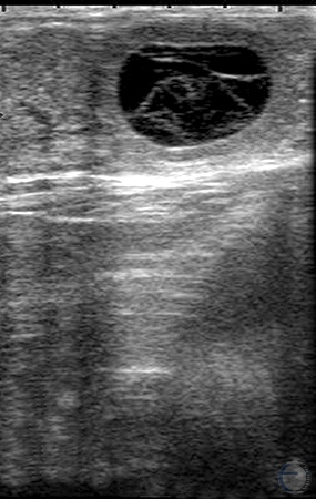
Luteal Cyst.
Anovulatory follicle with a luteinized wall and internal trabeculae. There is no ovulatory papilla.
Bartolome J (2006)

Luteinized Cyst.
The wall of the ovarian cyst is luteinized and there is some floccular material inside the cyst. The luteal tissue has the appearance of a shell.
Bartolome J (2006)
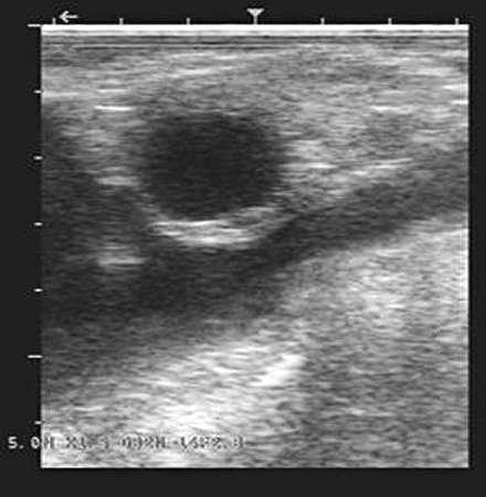
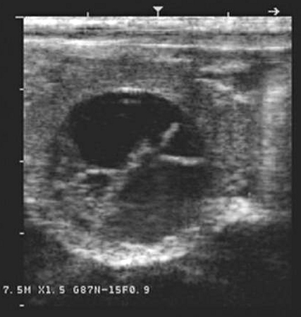
Luteinizing Cyst.
This fluid-filled structure has a thick luteal wall, but may be misdiagnosed as a follicular cyst on palpation. This may be a variation of a normal fluid-filled corpus luteum or it may be a luteinizing follicular cyst. The fine lines are trabeculae representing reflections.
Colloton J (2006)