The Visual Guide to
Bovine Reproduction
- Uterine Torsion
- Strangulated Umbilical Cord
- Pelvic Subluxation
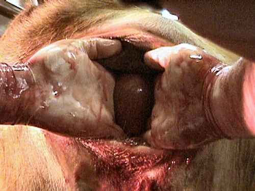
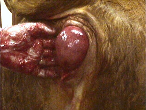
- Prolapsed Vagina
- Prolapse of the Bladder
- Ruptured Prepubic Tendon
- Prolonged Gestation
- Hydrops Allantois
- Hydrops Amnii
- Mummification
- Maceration
Accidents of Gestation: Mummification
Mummy with Membranes.
Completely dry, papyraceous mummy with membranes. Age of the fetus 3 to 4 months.
Drost M (1979)
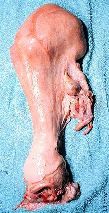
Mummified Fetus in Utero.
Firm mass in the absence of fluid in the right horn, suggestive of a small mummified fetus. Palpation of an eye socket would confirm the diagnosis. Note persistent ipsilateral corpus luteum.
Drost M (1979)
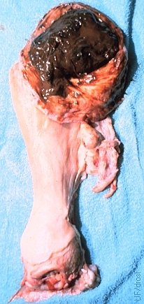
Opened Uterus with Mummy.
Mummification of a 3-month old fetus. Mummification most commonly occurs at this fetal age.
Drost M (1979)
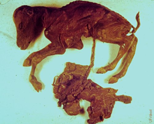
4 Month Old Mummy with Membranes.
Complete 4-month old mummy with membranes. Note characteristic bird-like head with deep sockets. Death and mummification with the result of the displacement of the umbilical cord around the neck of the fetus.
Drost M (1979)
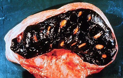
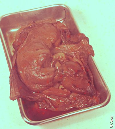
Mummification of 7.5 Month Old Fetus.
Hematic mummification of a 7.5 month old fetus. Mummification more commonly occurs in 3 to 4 month old fetuses. Note the black hemoglobin staining. These large fetuses require surgical removal.
Drost M (1979)
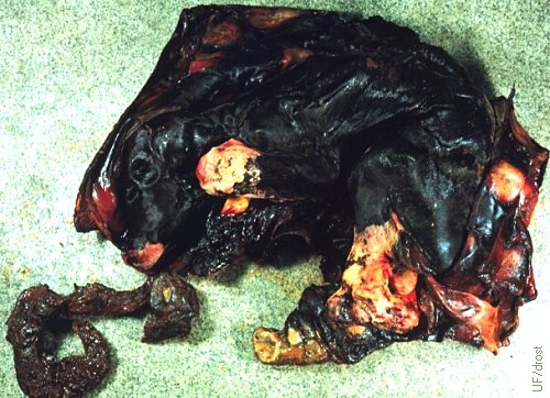
Mummification of 7.5 Month Old Fetus.
Mummification of a 7 month fetus is relatively rare. Mummification usually starts around 3 months of gestation. Note inspissated fetal membranes in the lower left hand corner of the image.
Drost M (1979)
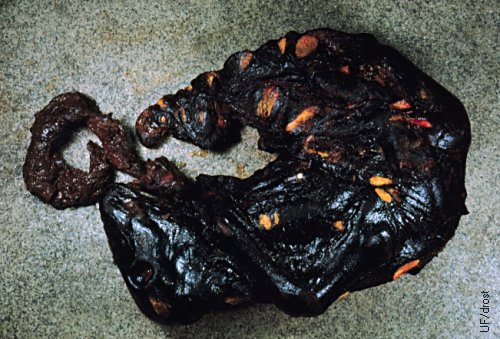
Large Mummy with Membranes.
Seven-month old mummified fetus that was delivered by cesarean section. The mummy is still tightly surrounded by its membranes. Note the (yellow) cotyledons.
Drost M (1979)
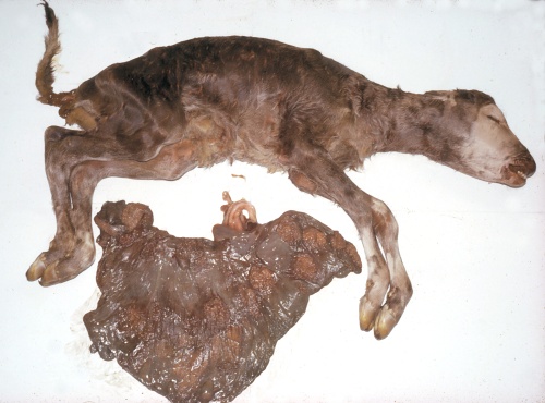
8.5 Month Old Mummifying Fetus.
This 8.5 month old fetus is in the process of mummification. The fetal membranes have not yet become totally dessicated and parchment-like.
Roberts SJ (1973)
Mummified Twin.
This cat-size (gestational age approximately 4 months) mummified was delivered at term when parturition and labor were initiated by the normal twin. This is a rare occurrence in cattle in contrast with the delivery of occasional mummified fetuses in litter bearing species such as the pig. If the surviving fetus is a female she may be a freemartin if the mummy is a male.
Roberts SJ (1973)
Twin Mummy.
This cat-size (gestational age approximately 4 months) mummified was delivered at term when parturition and labor were initiated by the normal twin. This is a rare occurrence in cattle in contrast with the delivery of occasional mummified fetuses in litter bearing species such as the pig. If the surviving fetus is a female she may be a freemartin if the mummy is a male.
Roberts SJ (1973)
Mummectomy.
When expulsion with the aid of prostaglandin fails the colpotomy approach for removal of a mummy is feasible. It is performed under epidural anesthesia. Exposure varies with the flexibility of the broad ligaments hence is better in older cows.
Hopper RM (2007)
Delivery of a Mummy per Vaginam.
Colpotomy approach for removal of a mummy per vaginam. It is performed under epidural anesthesia. Exposure varies with the flexibility of the broad ligaments hence is better in older cows. Uterine contents are sterile.
Hopper RM (2007)
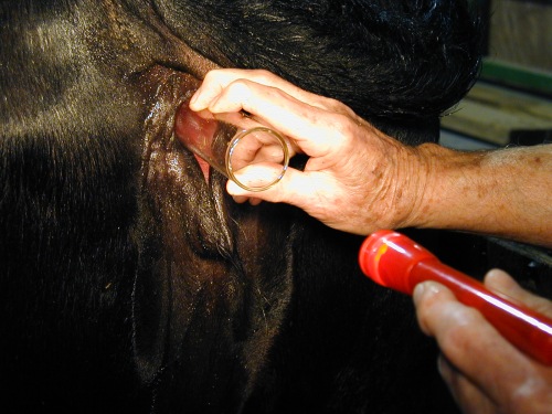
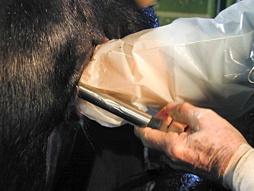
Colpotomy 1.
Prior to the transvaginal procedure the cervix and vagina are examined with a speculum for evidence of pus or contamination. Here, a cylindrical glass speculum [length 24 cm, diameter 4 cm] is used.
Drost M (2009)
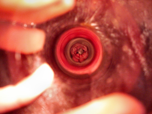
Colpotomy 2.
View of the cervix through a cylindrical glass speculum. A small piece of bone can be identified in the external os (it turned out to be the tip of a claw).
King AC (2009)
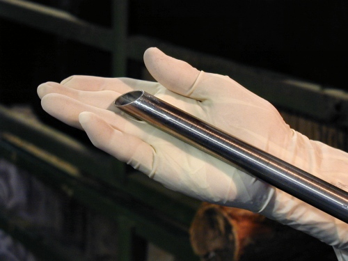
Colpotomy 3.
The anterior cutting end of a sterile stainless steel shaft [length 50 cm, diameter 2 cm] is shown prior to insertion into the vagina.
Drost M (2009)
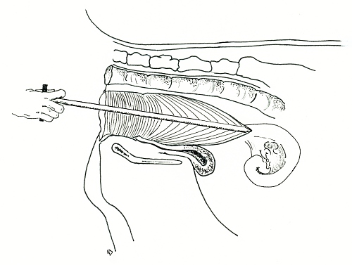
Colpotomy 4.
Placement of the tip of the colpotomy spear is at the 2 0'clock position in relationship to the cervix and aimed slightly ventrally. This minimizes the risk of puncturing a distended rumen.
Lunsford ND (2008)
Colpotomy 5.
The cutting end of the shaft is advanced into the fornix, at a 2 o'clock position in relationship to the external os of the cervix. The wall of the fornix is pushed forward and with a quick thrust the shaft enters the peritoneal cavity. After removal of the shaft the incision is enlarged by inserting first one finger, then two, and subsequently the entire hand.
Drost M (2009)
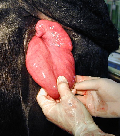
Colpotomy 6.
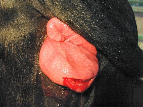
The uterus is exteriorized by retracting it into the vagina and exposing it at the vulva.
Drost M (2009)
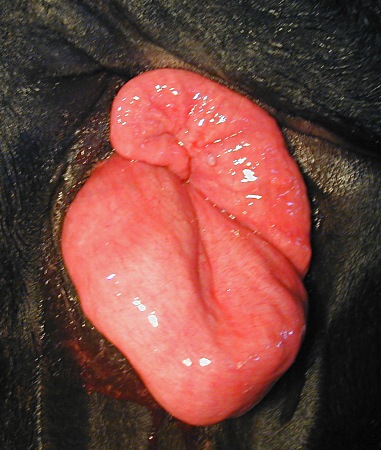
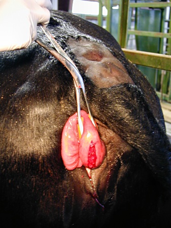
Colpotomy 7.
Exposure of the uterus via colpotomy under epidural anesthesia.
Drost M (2009)
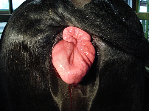
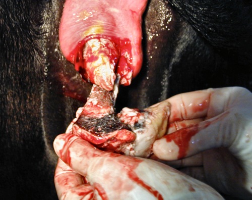
Colpotomy 8.
The enlarged uterine horn (containing a small mummy, or in this case several macerated bones) is positioned for the removal of its contents. The mass was curved and felt dry without crepitus.
King AC (2009)
Colpotomy 9.
An 8 cm incision has been made along the greater curvature.
Drost M (2009)
Colpotomy 10.
Several bones and small hooves are removed. There was no odor, no pus, nor fluid.
Drost M (2009)
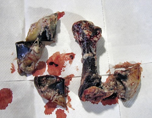
Colpotomy 11.
Several bones, small hooves, and teeth, covered with some inspissated tissue, were removed. There was no fetid odor.
Drost M (2009)
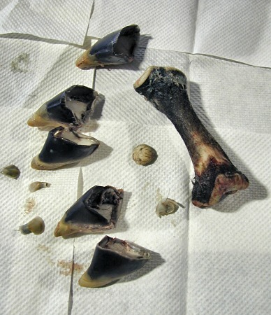
Colpotomy 12.
After cleansing, a femur, five claws, a carpal bone, and three small teeth, compatible with 7-month old fetus were identified.
Drost M (2009)
Colpotomy 13.
Positioning of the uterine horn with a uterine forceps for closure of the incision.
Drost M (2009)
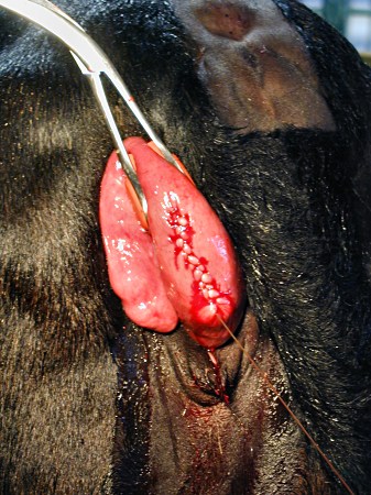
Colpotomy 14.
Closure of the uterine incision with the Utrecht pattern. After rinsing with sterile saline the uterus was returned to the abdominal cavity. The incision in the fornix was not sutured.
Drost M (2009)
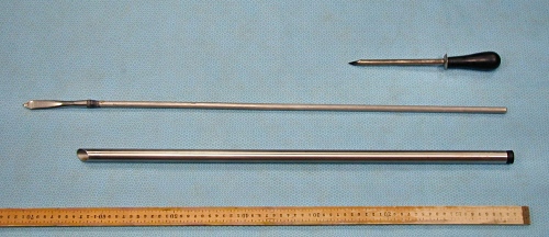
Colpotomy 15.
Homemade stainless steel shaft with a sharp beveled cutting end [length 50 cm, diameter 2 cm], patterned after the Kimberling-Rupp instrument below.
Drost M (2009)
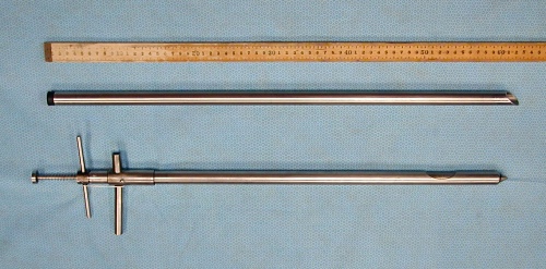
Colpotomy 16.
A bloat trocar (top), after sterilization, can be used to make a stab incision in the fornix. In the middle, a home made spear with a pointed V-shaped tip. At the bottom, a homemade stainless steel shaft with a sharp beveled cutting end [length 50 cm, diameter 2 cm]; made from the shaft of a defunct IV stand. The legs of a retired small animal examination table can also be used for this purpose.
Drost M (2009)