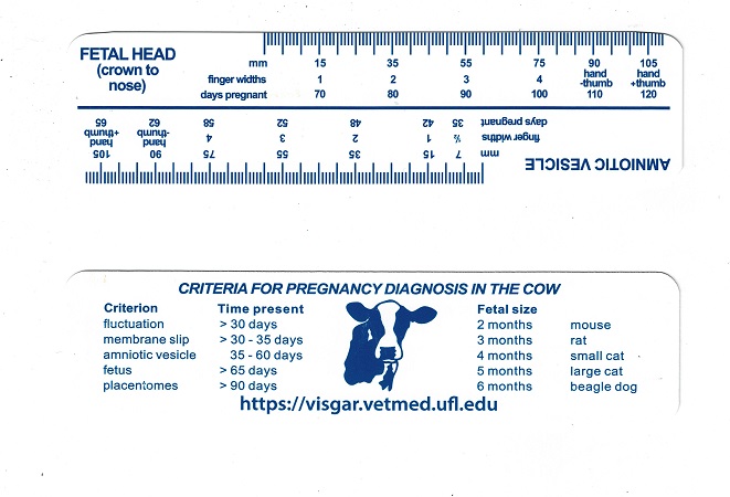
Pregnancy Diagnostic.
Guidelines listed on a portable, practical, plastic ruler for determination of the stage of pregnancy in the bovine.
Drost M (2021)
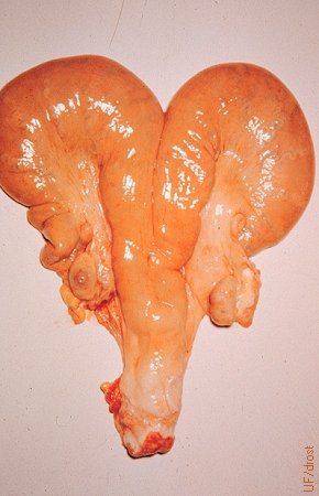
Asymmetry of the Uterine Horns.
The earliest presumptive diagnosis of pregnancy by transrectal palpation can be made on the basis of asymmetry of the horns and the presence of fluid in the larger horn on the side of a fully developed corpus luteum. This is particularly valid in heifers.
Drost M (1982)
Early Asymmetry of the Horns.
The earliest presumptive diagnosis of pregnancy by transrectal palpation can be made on the basis of asymmetry of the horns and the presence of fluid in the larger horn on the side of a fully developed corpus luteum. This is particularly valid in heifers. This is an image of a 32-day gravid uterus.
Drost M (1982)
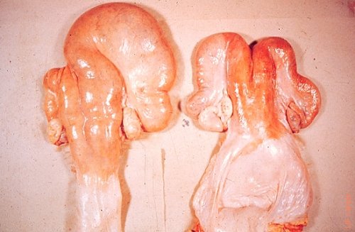
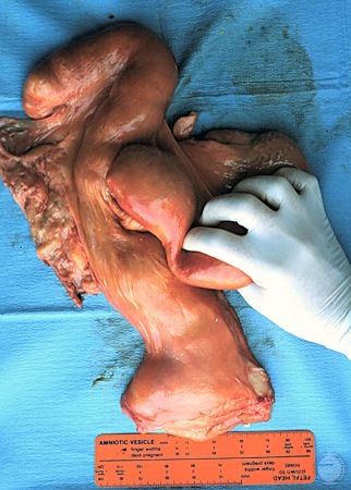
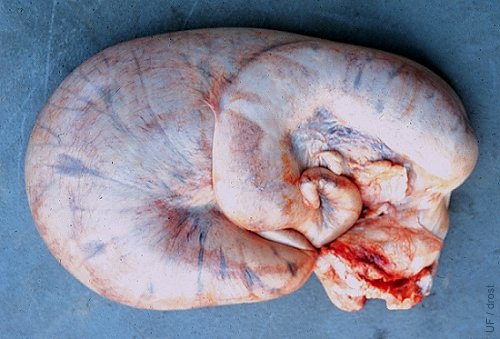
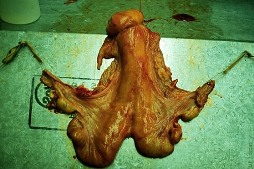
Triplets.
Due to multiple conceptuses the asymmetry is lost which serves as an early indication. Multiple corpora lutea are also suggestive. Twin or triplets can be confirmed by the palpation, or ultrasonography, of two or three amniotic vesicles.
Drost M (1982)
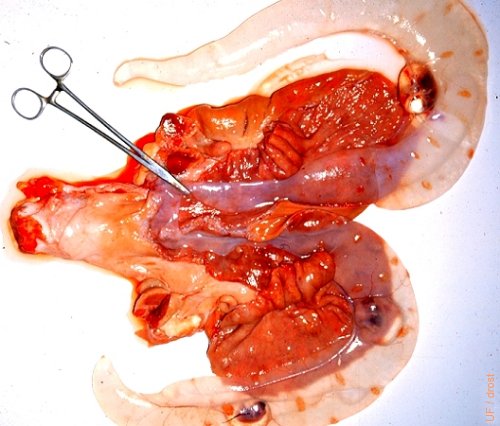
Triplet Conceptuses.
Triplets. One amniotic vesicle is located in the left horn, the other two are located in the right horn of the uterus.
Drost M (1982)
Slipping the Chorio-Allantoic Membranes.
The earliest positive sign of pregnancy in the cow, by palpation per rectum, is slipping the chorio-allantoic membranes, generally along the greater curvature.
Drost M (1982)
Slipping Membranes.
The earliest positive sign of pregnancy in the cow, by palpation per rectum, is slipping the chorio-allantoic membranes, generally along the greater curvature. After 50 days of gestation, membranes can be slipped in the contralateral horn as well. In case of bicornual twins, membranes may be slipped as early as 35 days in the cow, and 30 days in the heifer.
Drost M (1982)
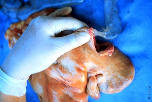
Amniotic Vesicle.
The amniotic vesicle can first be palpated at 35 days when it is 7.5 mm in diameter; at 42 days the diameter is 15 mm, at 48 days 35 mm, at 52 days 55 mm, at 58 days 75 mm, at 62 days 90 mm, and at 65 days 105 mm. Initially the vesicle is turgid. After 60 days the vesicle becomes more flaccid which allows direct palpation of the small fetus.
Drost M (1982)
Localizing the Amniotic Vesicle.
The amniotic vesicle is gently surrounded by placing all four fingers dorsally and the thumb ventrally along the greater curvature of the horn.
Drost M (1982)
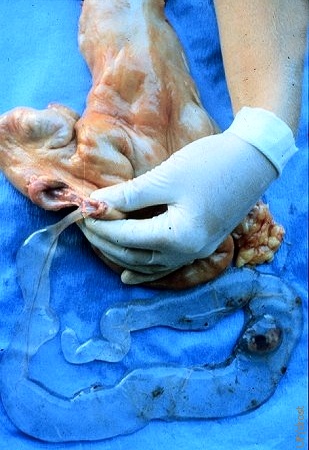
Trauma to the Blood Supply.
The blood supply to the amniotic vesicle along the lesser curvature is relatively fragile, especially for the early vesicle. Once disrupted the embryo / fetus loses its blood supply and dies. Rough handling can also lead to complete rupture of the amniotic vesicle. This is sometimes used as a method for terminating pregnancy in feedlot heifers.
Drost M (1982)
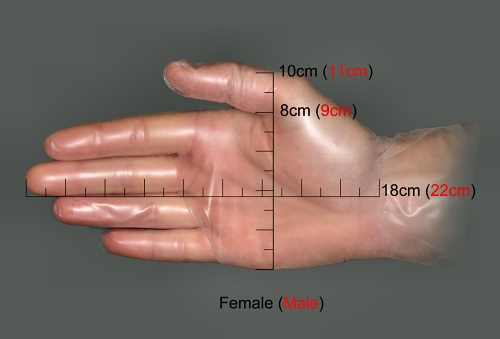
Hand Measurements.
Average measurements of the male and female hand to size up the reproductive tract or fetus per rectum. CRL (crown rump length) approximately 9 cm at 2 months, 22 cm at 4 months, 44 cm at 6 months, 80 cm at 8 months, and 100 cm at 9 months.
Löfstedt RM (2010)

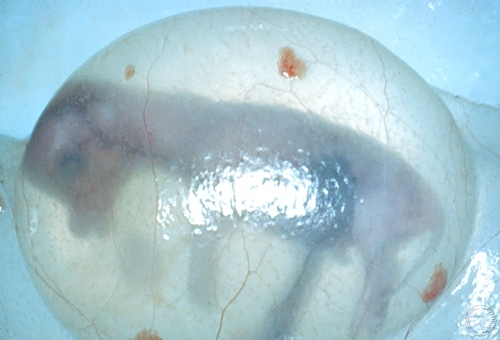
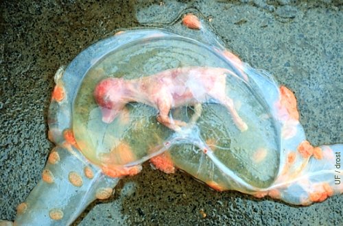
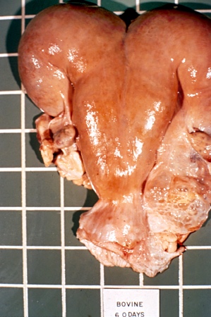
60-Day Fetus.
Around 60 days of gestation the amniotic sac is flaccid enough to permit direct palpation of a small, mouse-size fetus.
Drost M (1982)
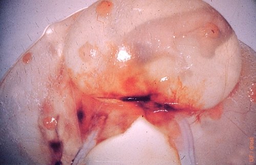
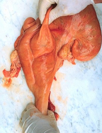
2.5-Month Conceptus.
The chorio-allantoic membranes have been removed over the amniotic vesicle. Even visually it can be appreciated that the vesicle has become more flaccid. The cotyledons are also readily visible.
Drost M (1982)
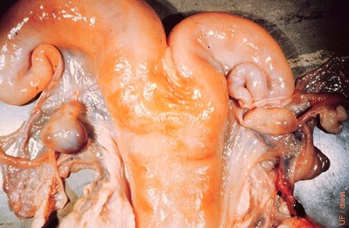
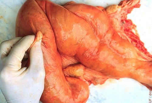
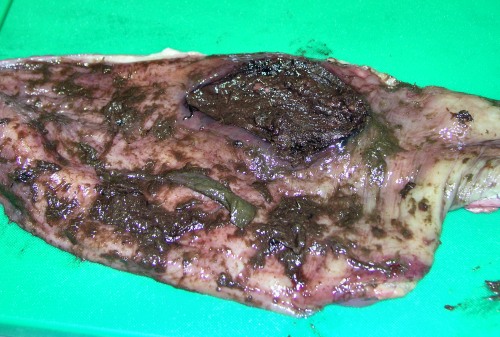
CL of Pregnancy on the Contralateral Ovary.
Only on rare occasions will the corpus luteum of pregnancy be found on the contralateral ovary in a singleton pregnancy. Here a singleton fetus, the size of a large cat, was found in the right horn while the CL was on the left ovary. Slaughterhouse specimen.
Drost M (1982)
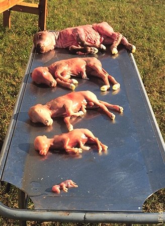
Fetal Sizes.
At 2 months of gestation the fetus is the size of a mouse, at 3 months the size of a rat, at 4 months a small cat, at 5 months a large cat, and at 6 months the size of a Beagle dog.
Drost M (1982)
Palpation of the Fetal Head.
The fetal head can be palpated directly through the uterine wall and the flaccid membranes. Measurement of the tip of the nose to the forehead can assist in determining the approximate age of the fetus. At 70 days of gestation the distance from the tip of the nose to the top of the forehead is 15 mm, at 80 that distance is 35 mm, at 90 days 55 mm, at 100 days 90 mm, and at 120 days the distance is 105 mm.
Drost M (1982)
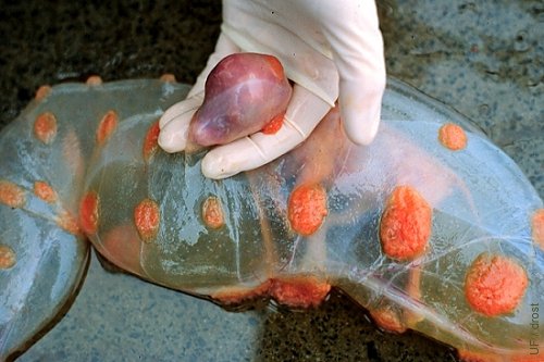
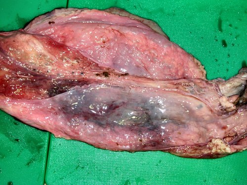
Palpation of a Placentome.
Placentomes can first be palpated per rectum around 3 months of gestation. Composed of the maternal caruncle and the fetal cotyledon, placentomes vary considerably in size. They are largest in the gravid horn near the fetus.
Drost M (1982)
Exposed Placentome.
The uterine wall is cut to expose a caruncle in the gravid horn. The size of the placentomes varies with their location within the uterus.
Drost M (1982)
Rectal Trauma.
External view of the rectum showing trauma and bruising incurred during a transrectal examination.
Cavalieri J (2009)
Rectal Edema.
External view of the rectum showing edema and bruising incurred during a transrectal examination.
Cavalieri J (2009)
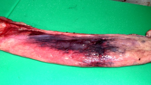
Non-perforating Mucosal Tear.
Serious, 11 cm, mucosal tear in the rectum, sustained during a transrectal examination. Fortunately it was a non-perforating tear.
Cavalieri J (2009)
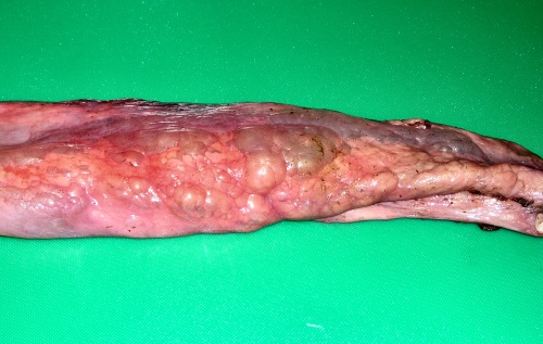
Severe Mucosal Trauma.
Mucosal view of a severe, non-perforating, mucosal tear and surrounding mucosal trauma. Near the center is a necrotic strip of mucosa. The injuries were sustained during a rough, transrectal, examination.
Cavalieri J (2009)
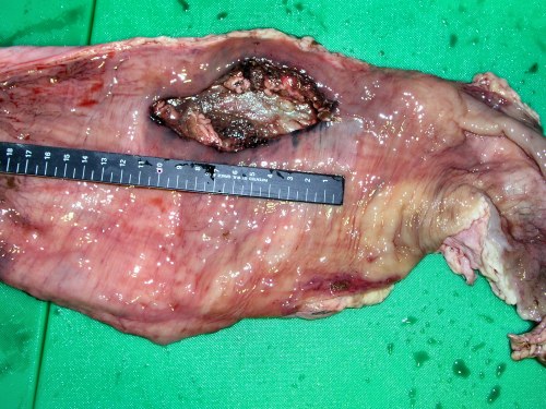
Damaged Rectal Wall.
Injuries sustained during a rough, transrectal, ultrasound examination, showing bruising and edema of the rectal wall. The rectal wall was not perforated. External view.
Cavalieri J (2009)
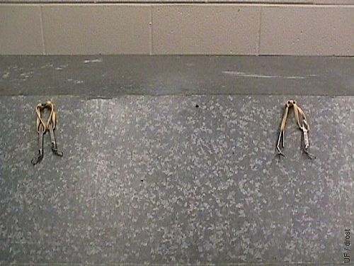
Normal Uterus on Palpation Table.
This normal pluriparous uterus is suspended on the sloping surface of a palpation table. The mesovaria are connected by rubber bands to small hooks, allowing retraction onto the horizontal surface of the table, which mimics the pelvic floor.
Drost M (1982)

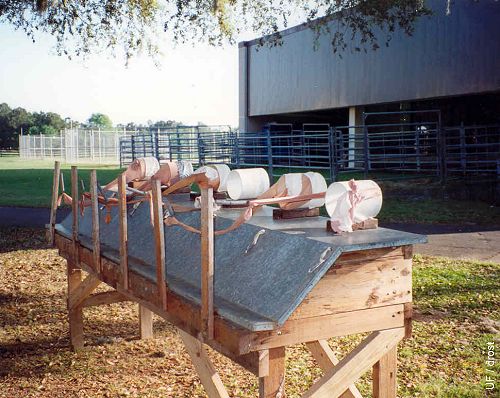
Pregnant Uteri on Palpation Table.
Gravid uteri from the slaughterhouse are suspended on the sloping surface of the palpation table for demonstration and practice of palpation.
Drost M (1982)
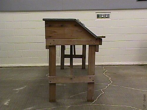
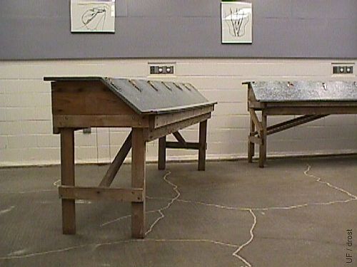
Palpation Table - Side View.
The height of the table is 110 cm. The top level is 42.5 cm deep. The slanting section measures 32.5 cm. The "gutter" measures 10 cm.
Drost M (1982)
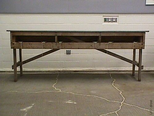
Palpation Table - Rear View.
This is a rear view of the table. The palpater stands on this side of the table and reaches across for the specimens suspended on the other side. The table is 400 cm long, and 110 cm high. The frame of the table is constructed with exterior 2x4s and marine plywood. The top surface is covered with galvanized sheet metal.
Drost M (1982)
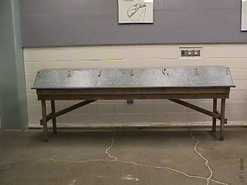
Palpation Tables - Overview.
Overview of two palpation tables as seen from the front where the specimens can be suspended.
Drost M (1982)
Palpation Table - Front View.
This is the front view of a 4-meter long palpation table. The slaughterhouse specimens are suspended on this side during use.
Drost M (1982)
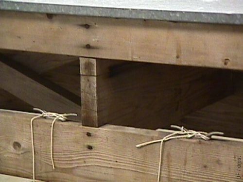
Palpation Table - Cleats.
The 4-meter long table will accommodate six reproductive tracts. One set of cleats (shown here) is placed at the rear of the table for each of the tracts. The string attached to the cervix is secured here.
Drost M (1982)
Palpation Table - Towel Clamps.
O-ring screws are placed 66 cm apart to accommodate six reproductive tracts along the front of the table. Towel clamps are connected to the O-rings with rubber bands. The (Jones) towel clamps are attached to the mesovarium near the ovarian bursa to suspend the tract and to give it flexibility during the practice of retraction.
Drost M (1982)
Palpation Table - Rectal Wall.
A leg of a panty hose is used to simulate the feel of the hand in the rectum while palpating a slaughterhouse tract on the palpations table. The toe-end of the panty hose is attached to a spring on a vertical board. The upper end of the leg of the panty hose is taped to a PVC ring (diameter 15 cm) through which the palpater inserts the hand and arm.
Rathwell AC (1986)
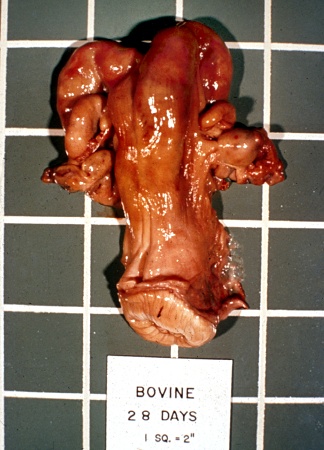
Pregnant Uterus - 28 Days.
Early pregnancy asymmetry of the horns. The larger horn is ipsilateral to the corpus luteum. [5 centimeter squares]
Chenoweth PJ (2012)
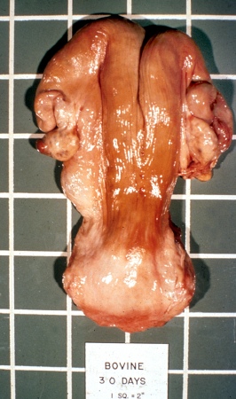
Pregnant Uterus - 30 Days.
Early pregnancy asymmetry of the horns. The larger horn is ipsilateral to the corpus luteum. In heifers the fetal membranes can be slipped. [5 centimeter squares]
Chenoweth PJ (2012)
Pregnant Uterus - 40 Days.
Early pregnancy asymmetry of the horns. The larger horn is ipsilateral to the corpus luteum. Fetal membranes can be slipped and a small amniotic vesicle can be recognized in the gravid horn. [5 centimeter squares]
Chenoweth PJ (2012)
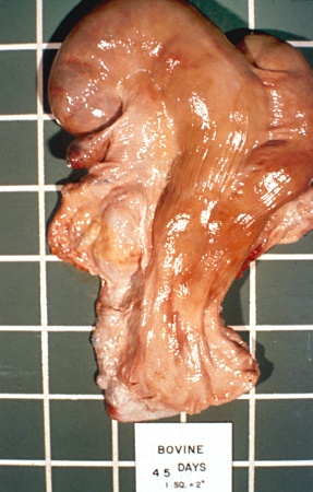
Pregnant Uterus - 45 Days.
Pregnancy asymmetry of the horns. The larger horn is ipsilateral to the corpus luteum. Fetal membranes can be slipped in the gravid horn and a 2 cm amniotic vesicle recognized. [5 centimeter squares]
Chenoweth PJ (2012)
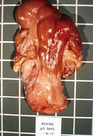
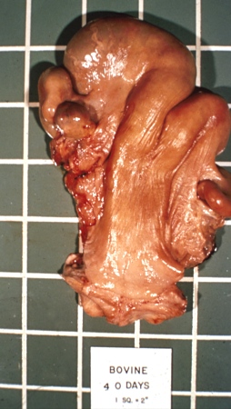
Pregnant Uterus - 50 Days.
Fetal membranes can be slipped in both horns. A 45 mm amniotic vesicle felt in the gravid horn is ipsilateral to the CL. [5 centimeter squares]
Chenoweth PJ (2012)
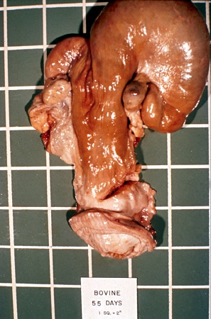
Pregnant Uterus - 55 Days.
Fetal membranes can be slipped in both horns and a 6.5 cm amniotic vesicle can be palpated in the gravid horn that is ipsilateral to the CL. [5 centimeter squares]
Chenoweth PJ (2012)
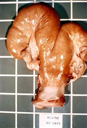
Pregnant Uterus - 60 Days.
Fetal membranes can be slipped in both horns and a small fetus, the size of a mouse, can be palpated in the gravid horn that ipsilateral to the CL. [5 centimeter squares]
Chenoweth PJ (2012)
Pregnant Uterus - 60 Days Twins.
Fraternal twins. Membranes can be slipped in both horns and two small fetuses, each the size of a mouse, can be palpated (one in each horn). There are 2 corpora lutea with one on each ovary. [5 centimeter squares]
Chenoweth PJ (2012)
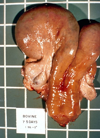
Pregnant Uterus - 75 Days.
A small fetus, the size of a large mouse or of a small rat, can be palpated in the larger horn. Fetal membranes can be slipped in both horns. [5 centimeter squares]
Chenoweth PJ (2012)
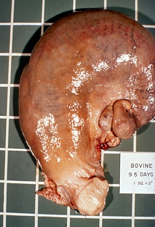
Pregnant Uterus - 95 Days.
A small fetus, the size of a rat, can be palpated in the larger horn. The ipsilateral uterine artery is identifiable and is 3 mm in diameter. [5 centimeter squares]
Chenoweth PJ (2012)
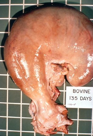
Pregnant Uterus - 135 Days.
A fetus the size of a cat can be balloted in the larger horn. There is fremitus in the ipsilateral uterine artery which is 7.5 mm in diameter. Placentomes are palpable. [5 centimeter squares]
Chenoweth PJ (2012)
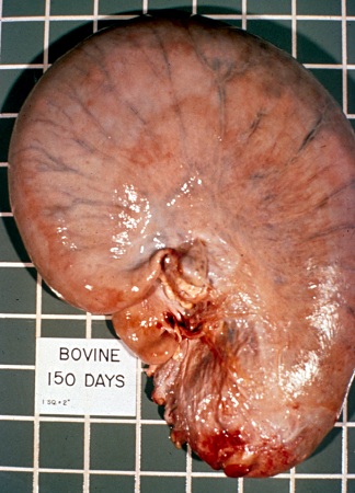
Pregnant Uterus - 150 Days.
A fetus the size of a large cat can be balloted in the larger horn. There is fremitus in the ipsilateral uterine artery which is 9 mm in diameter. Placentomes can be palpated. [5 centimeter squares]
Chenoweth PJ (2012)
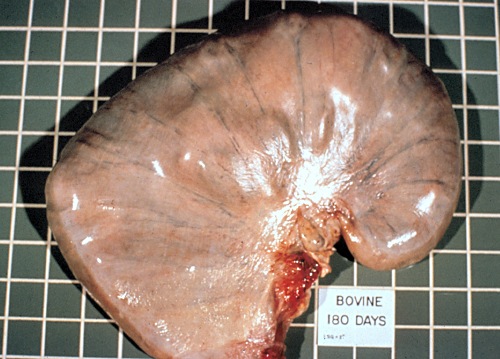
Pregnant Uterus - 180 Days.
A fetus the size of a Beagle dog can be balloted in the larger horn. There is fremitus in the ipsilateral uterine artery which is 12 mm in diameter. Multiple placentomes can be palpated. [5 centimeter squares]
Chenoweth PJ (2012)
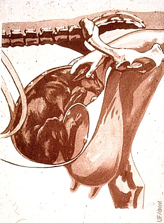
Palpation of the Near-Term Fetus.
Generally, during the last two months of gestation the fetus can be routinely palpated directly (again) per rectum. Frequently the interdigital space of the claws can be pinched to determine whether the fetus is alive or not (pedal reflex).
Drost M (1976)
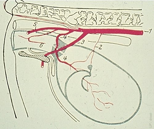
Palpation of the Uterine Artery.
The character of the blood flow in the uterine artery changes from a pulse to fremitus (Latin: murmur - roar), which is palpable per rectum from the third month of gestation on. The diameter of the uterine artery on the gravid side increases from 3 mm at 3 months, 6 mm at 4 months, 9 mm at 5 months, to 12 mm at 6 months. At 7 months of gestation the contralateral uterine artery also begins to enlarge and become readily palpable. Legend: 1 = dorsal aorta; 2 = ovarian A; 3 = internal iliac A; 4 = uterine A; 5 = pudendal A; 6 = vaginal A.
Source unknown (German textbook)