Double Cervical Os.
Double external cervical os. Frequently there is asymmetry between the two ora. The cervical canal assumes the configuration of the letter Y.
Roberts SJ (1973)
Double Pseudo Cervices.
During embryonic development the anterior portions of the paramesonephric ducts failed to fuse, creating two pseudo external cervical ora. They connected via an antichamber with the normal external os of the cervix. A rare specimen.
Tanabe TY and Almquist GO (1966)
Double Cervix.
Double cervix during the first stage of parturition, during the process of cervical relaxation. Degree of development and degree of relaxation of the double cervical openings may be different.
Richter J and Goetze R (1978)
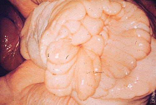
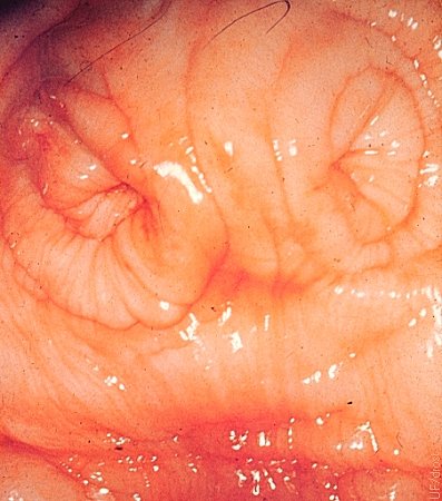
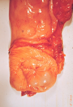
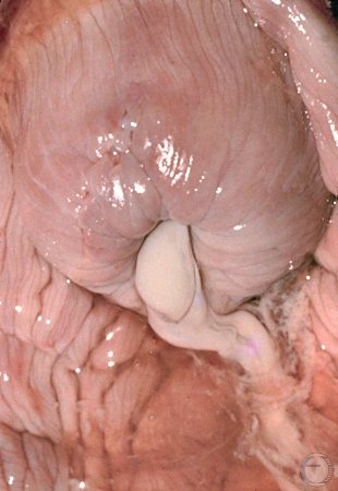
Cervical Ectropion.
Cervical ectropion, characterized by hypertrophy of the cervical folds and prolapse through the external os.
Drost M (1970)
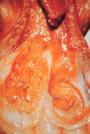
Cervical Anomaly.
There are only two cervical rings and there is a persistent median fold (remnant of the mesonephric duct system) between the markers.
Roberts SJ (1973)
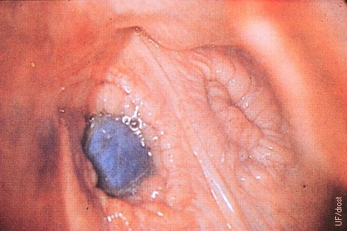
Cervical Cysts.
Cervical cysts are usually retention cysts of (occluded) cervical glands acquired secondary to trauma. An eponym is nabothian cyst in honor of Martin Naboth.
Roberts SJ (1973)
Cervical Adhesion.
Adhesion of the cervix causing obstruction and secondary mucometra. The adhesion is a result of trauma (usually during delivery).
Roberts SJ (1973)
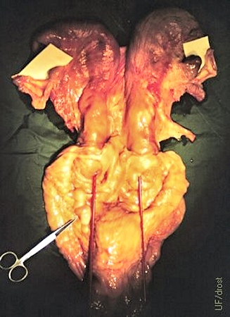
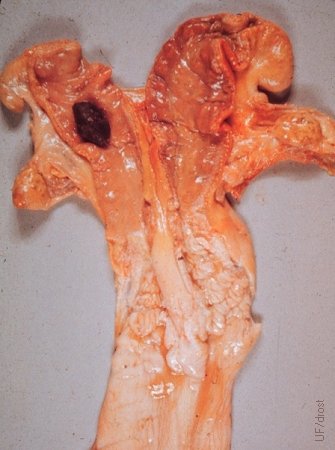
True Double Cervix.
In a rare true double cervix or uterus didelphys there are two cervices each connected to its respective uterine horn. A red pipet is inserted in each of the two cervical canals. Gartner's ducts are indicated by the scissors.
Goemann GG (1975)
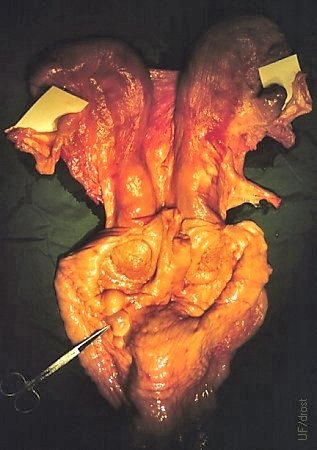
True Double Cervix.
In a rare true double cervix or uterus didelphys there are two cervices each connected to its respective uterine horn. The scissors are pointing to two mesonephric duct cysts (Gartner's ducts), remnants of the male duct system.
Goemann GG (1975)
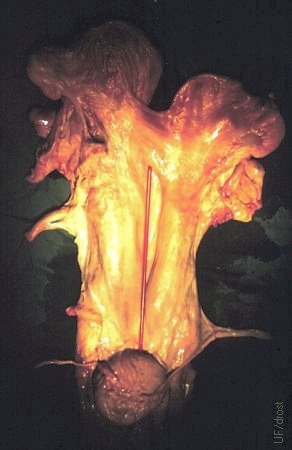
True Double Cervix.
In a rare true double cervix or uterus didelphis there are two cervices each connected to its respective uterine horn. In this ventral view a septum is visible between the two cervices as delineated by a red pipet.
Goemann GG (1975)
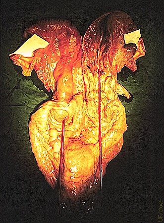
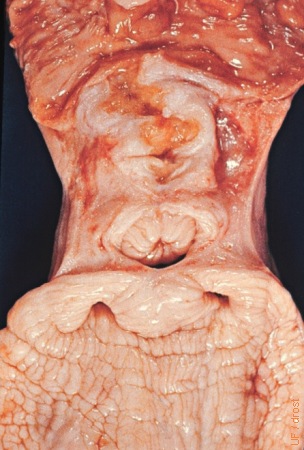
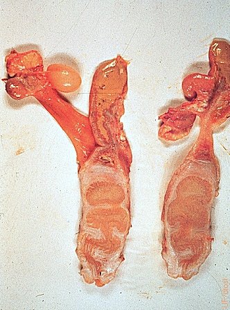
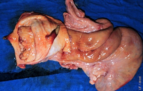
Uterus Didelphys.
True double cervix (uterus didelphys), two individual cervices each connected to its respective horn. One red pipet is inserted into the left cervix, the other in the opened right cervix and horn.
Goemann GG (1975)
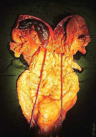
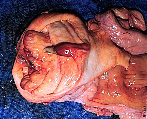
Uterus Didelphys.
True double cervix (uterus didelphys), two individual cervices each connected to its respective horn. Red pipets are inserted into each of the cervices and corresponding horns. Note inspissated, brown (retained?) mucus in the left horn.
Goemann GG (1975)
Lack of Transverse Folds.
Cervix of a heifer born without transverse folds (rings) of the cervix.
Roberts SJ (1973)
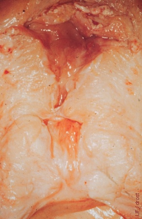
Cervical Obstruction.
The white scar represents a cervical obstruction which can lead to secondary mucometra.
Roberts SJ (1973)
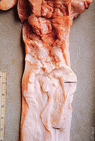
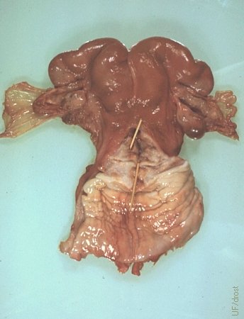
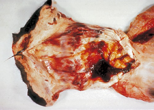
Segmental Aplasia.
Segmental aplasia of the left uterine horn and complete duplication of the cervix (uterus didelphys). There is an occluding obstruction at the base of the left horn which traps uterine secretions. Here the secretions are inspissated and have formed a hysterolith (uterine stone; uterine "pebble" in this case?). The corpus luteum is retained for lack of a signal from the ipsilateral horn (prostaglandin F2alpha) to regress. The cow appears to have been pregnant in the right horn.
Roberts SJ (1973)
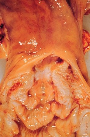
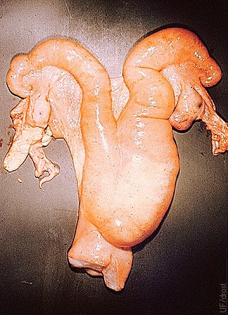
Segmental Aplasia.
Segmental aplasia of the uterus and cervix. Inspissated mucus fills the occluded areas of the cervix and inspissated uterine secretions are present in the right uterine horn. The left uterine horn is missing making this also an example of uterus unicornis.
Roberts SJ (1973)
Cervical Discharge.
Cervical discharge of mucopus indicating either a uterine or a cervical infection.
Roberts SJ (1973)
Cervical Stenosis.
Cervical stenosis without occlusion. Owner had difficulty passing the insemination syringe through the internal os of the cervix of this Shorthorn heifer. In addition the right oviduct was blocked leading to hydrosalpinx. Stenosis was probably due to pipet trauma.
Drost M (1973)
Hydrometra.
Fluctuant thin walled uterus with hydrometra as a result of occlusion by an anomalous cervix.
Roberts SJ (1973)
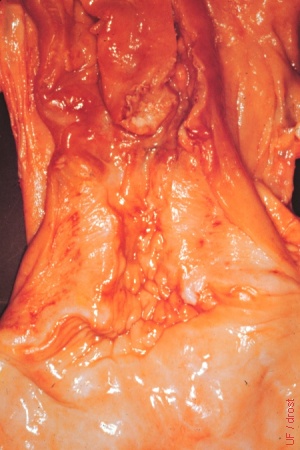
Persistent Median Band.
The small, 15 mm wide vertical median band was snapped by the fetus during parturition. The cow died from unrelated causes 2.5 weeks post partum.
Drost M (1971)
Persistent Median Band.
The small, 15 mm wide vertical median band was snapped by the fetus during parturition. The cow died from unrelated causes 2.5 weeks post partum.
Drost M (1971)
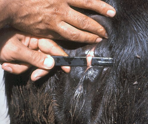
Persistent Median Band.
These cord-like structures are considered to be vestiges of arrested development of the paramesonephric ducts [Mullerian ducts]. The small vertical median band may delay delivery of a calf or may be snapped by the calf during parturition.
Nicoletti PL (1980)
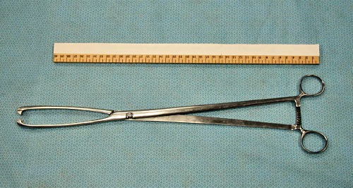
Cervical Forceps.
Cervical forceps are used to retract the cervix to aid in retraction of a pendulous uterus. One jaw is inserted into the external os, the other external to the cervical os. The instrument can also aid in passing a catheter through the cervix when the latter is large and far forward.
Drost M (2009)
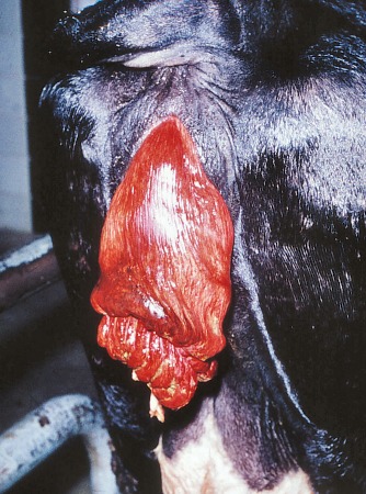
Postpartum Cervix.
Enlarged hyperemic cervical folds are prolapsed.
Chenoweth PJ (2012)
Cervical Prolapse.
Enlarged hyperemic cervical folds are prolapsed.
El Naggar MA (2005)
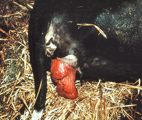
Prolapse of the Cervix.
The enlarged hyperemic cervix can prolapse due to poor vestibular tone and abdominal pressure, when the cow lies down.
El Naggar MA (2005)