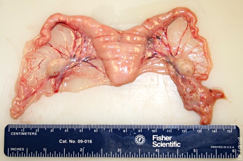
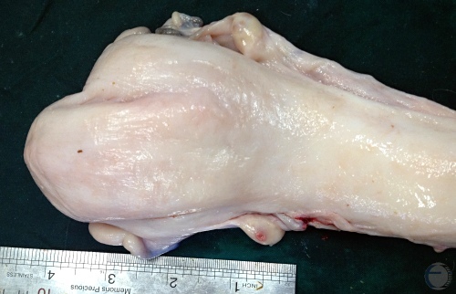
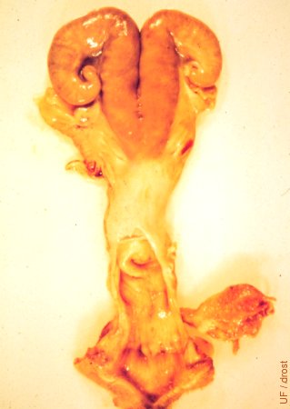
Normal Reproductive Tract.
Normal uterus, inactive ovaries, and normal oviducts. Also included are the cervix and the anterior vagina.
Smith MC (2006)
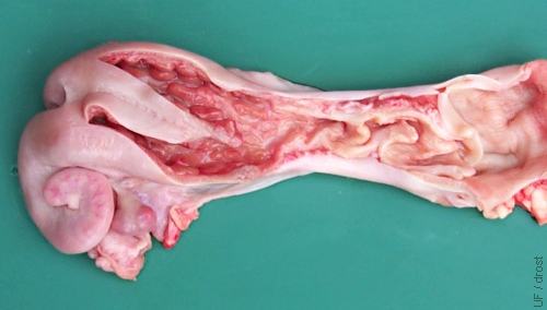
Uterus Opened Up.
Normal uterus of a cycling (2 CL) ewe opened up to show the caruncles, the short uterine body, and the cervix.
Smith MC (2006)
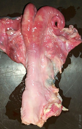
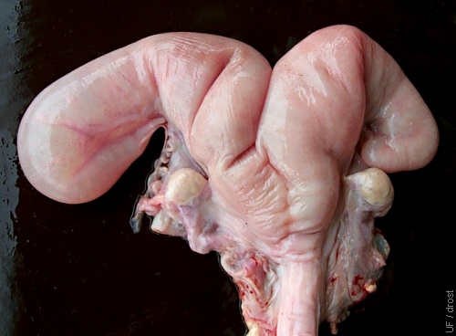
Normal Uterus.
Pluriparous uterus. Slight asymmetry of the horns indicates that the ewe has been pregnant before in the larger right horn.
Smith MC (2006)
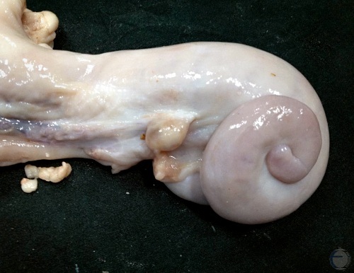
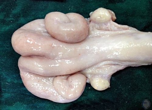
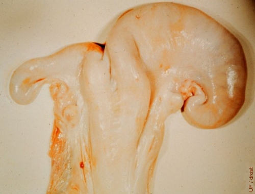
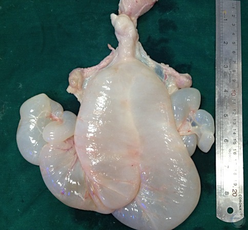
Normal Nongravid Uterus.
Normal non-pregnant uterus. Coiled right horn.
Mogheiseh A (2013)
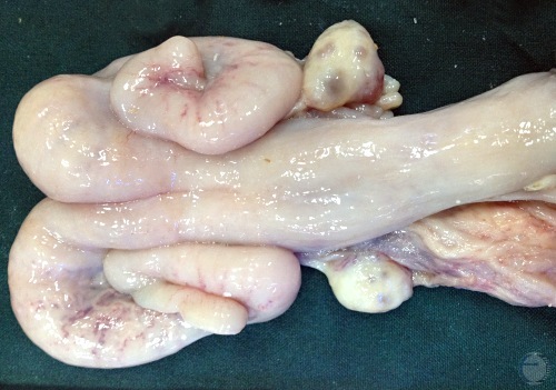
Normal Anestrous Uterus.
Normal non-pregnant uterus during anestrus. Inactive ovaries.
Mogheiseh A (2013)
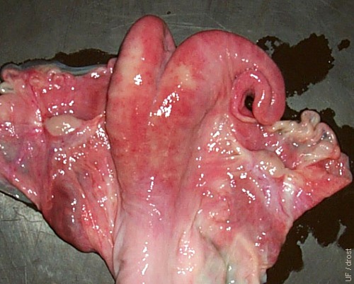
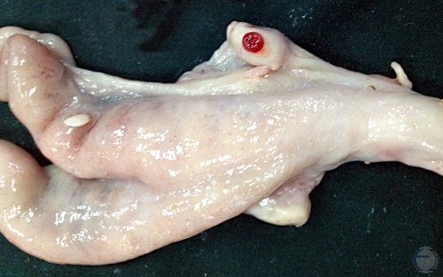
Normal Estrous Uterus.
Normal uterus during estrus. Follicular activity is most noticeable on the right ovary.
Mogheiseh A (2013)
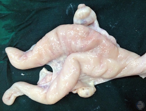
Normal Metestrous Uterus.
Normal uterus during metestrus. Prominent corpus hemorrhagicum on the right ovary.
Mogheiseh A (2013)
Normal Diestrous Uterus.
Normal uterus during diestrus. Corpus luteum on the right ovary with a prominent ovulation papilla (crown).
Mogheiseh A (2013)
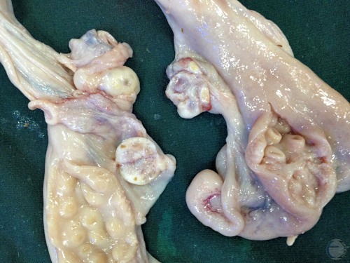
Ovine versus Caprine Uterus.
Uteri at approximately 1 month post partum. Caprine endometrium on the left shows shallow dish-shaped caruncles. Ovine endometrium on the right shows cup-shaped caruncles.
Mogheiseh A (2013)
Non-pregnant Uteri and Cervices.
Normal uteri with opened cervices. Note annular folds slanted towards the fornix.
Mogheiseh A (2013)
Prepuberal Uterus.
Normal uterus and inactive ovaries from a 7-month old, 55 kg, prepuberal lamb during the non-breeding season.
Smith MC (2006)
Postpartum Uterus.
The reproductive tract has been opened to show the folds of the cervix that are directed towards the vagina. Note the caruncles in the uterus of the ewe that lambed 2 to 3 weeks prior to the necropsy.
Smith MC (2006)
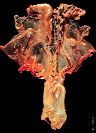
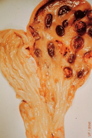
Pigmented Caruncles.
Normal uterus of a pluriparous, nonpregnant Suffolk ewe. The body and the right horn have been opened up to show the endometrium and the pigmentation of the caruncles.
Drost M (1974)
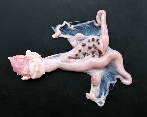
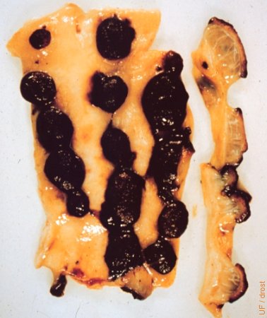
Caruncular Pigment.
Normal uterus of a blackfaced virgin lamb. The left horn has been opened up to show the endometrium and the pigmentation of the caruncles.
Smith MC (2010)
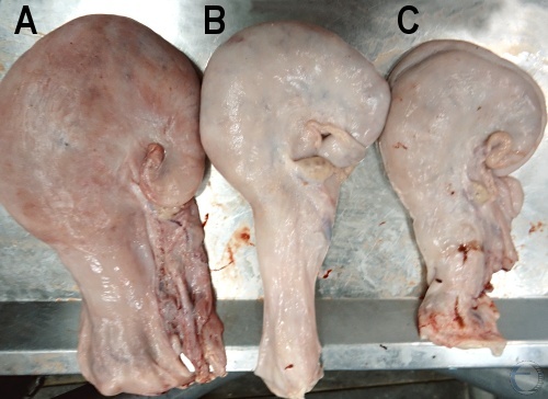
Uterine Involution.
A = 1 week post partum. B = 2 weeks post partum. C = 3 weeks post partum. There is a gradual involution.
Mogheiseh A (2013)
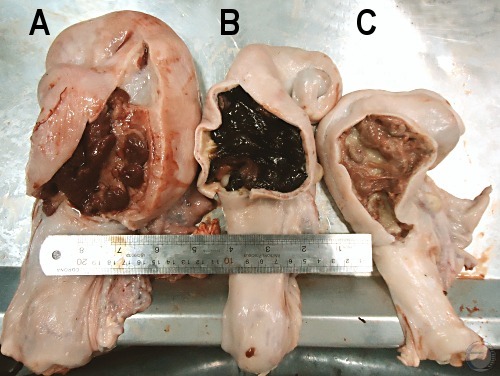
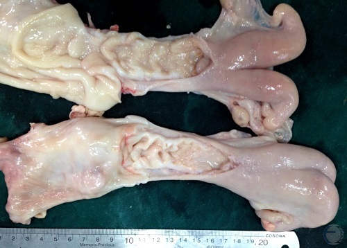
Uterine Involution.
A = 1 week post partum. B = 2 weeks post partum. C = 3 weeks post partum. There is a gradual and progressive involution.
Mogheiseh A (2013)
Uterine Involution.
Left horn is enlarged and firm and there is no ovarian activity. Anestrus.
Mogheiseh A (2013)
Uterine Involution at 2 Weeks.
Normal uterus at 2 weeks post partum with inspissated lochia.
Mogheiseh A (2013)
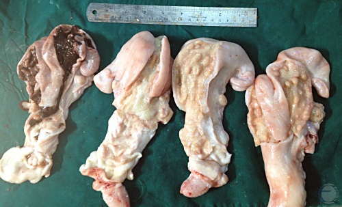
Persistent Lochia at about 1 Month.
Uteri at 3 to 4 weeks post partum. Uterus at the far left still has noticeable lochia due to infection.
Mogheiseh A (2013)
Uterus 17 Hours Post Partum.
Cup-shaped caruncles 17 hours post partum. Some of the chorio-allantois is still present on the right.
Roberts SJ (1973)
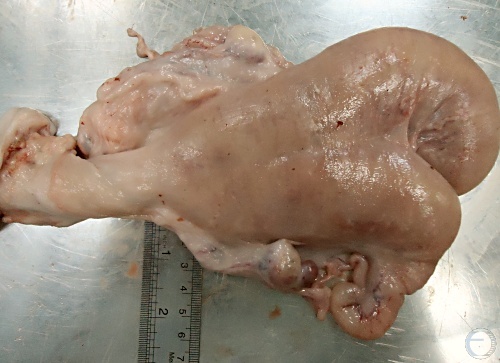
Uterus 8 Days Post Partum 1.
Asymmetrical uterus of a ewe 8 days after she gave birth to a singleton lamb.
Roberts SJ (1973)
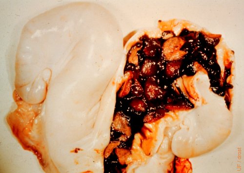
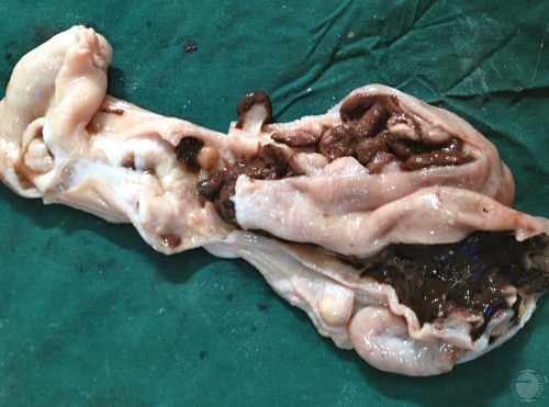
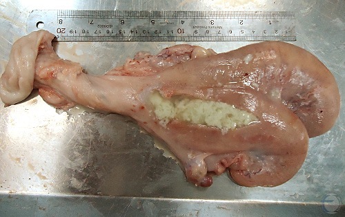
Uterus 8 Days Post Partum 2.
Thick-walled uterus opened to expose inspissated lochia (primarily necrotic caruncles).
Roberts SJ (1973)
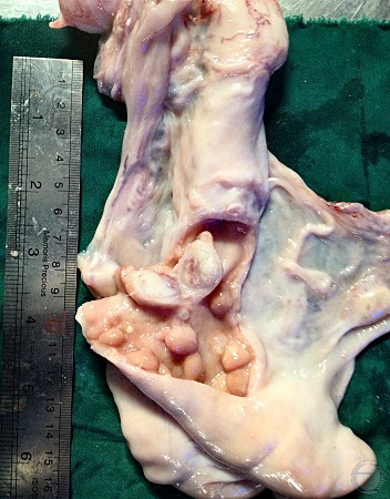
Uterus 8 Days Post Partum 3.
The previously gravid horn has been opened up and washed out to highlight the involuting caruncles. Normal.
Roberts SJ (1973)
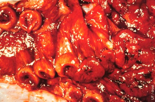
Uterus 14 Days Post Partum.
The surfaces of the caruncles have become necrotic in the normal process of involution.
Roberts SJ (1973)
Uterus 4 Weeks Post Partum.
Uterus of a ewe that delivered twins, which accounts for the symmetry of the horns. Involution is 90 per cent complete. Structure on the lower right is the bladder. Anestrous (inactive ovaries).
Roberts SJ (1973)
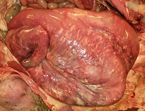
Postmortem Autolysis.
This postpartum uterus shows discoloration due to postmortem autolysis. Lighter colored, still enlarged, uterine blood vessels are also visible.
Smith MC (2006)
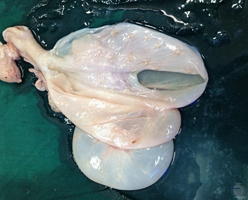
Hydrometra.
Accumulation of serous fluid in a nongravid uterus as a result of blockage. In this case an imperforate cervix. The wall of the uterus is thin.
Smith MC (2006)
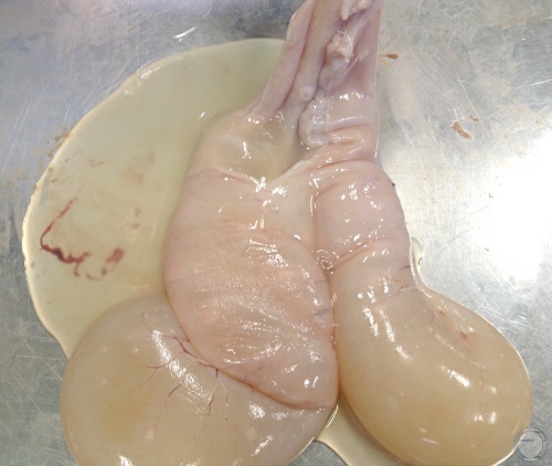
Mucometra or Hydrometra.
Accumulation of sterile fluid in the uterine lumen can be associated with persistence of the corpus luteum. This may lead to anestrus and pseudopregnancy. The uterus may be extremely thin walled.
Mogheiseh A (2013)
Hydrometra Fluid.
Incision of the uterus reveals clear, sterile, watery fluid and a thin uterine wall. No placentation.
Mogheiseh A (2013)
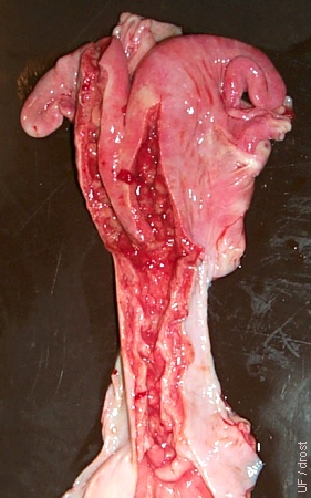
Pyometra.
Thick-walled pus-filled uterus and a retained corpus luteum on the right ovary.
Mogheiseh A (2013)
Pyometra - Pus.
Thick-walled pus-filled uterus and a retained corpus luteum on the right ovary. Incision in the uterus confirms the presence of pus.
Mogheiseh A (2013)
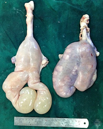
Pyometra versus Pregnancy.
Compared with a gravid uterus on the right the pus filled uterus on the left does not contain a fetus and does not show placental development.
Mogheiseh A (2013)
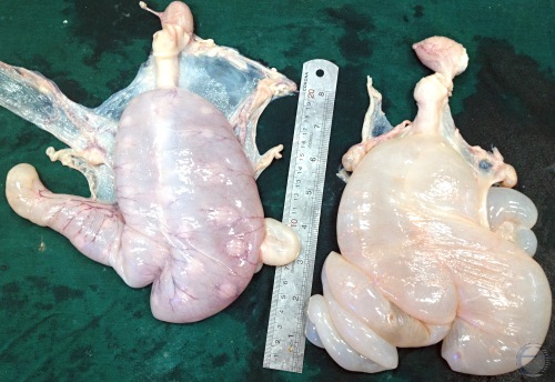
Hydrometra versus Pregnancy.
Compared with a gravid uterus on the left the fluid filled uterus (hydrometra) on the right does not contain a fetus and does not show placental development.
Mogheiseh A (2013)
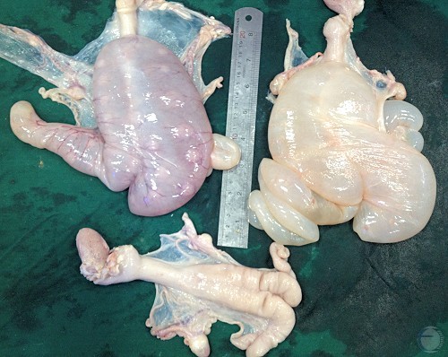
Hydrometra vs Pregnancy vs Nonpregnancy.
Compared with a gravid uterus on the left the fluid filled uterus (hydrometra) on the right does not contain a fetus and does not show placental development. No fluctuance is evident in the nonpregnant uterus.
Mogheiseh A (2013)
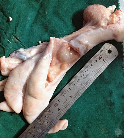
Endometrial Mass.
Small firm endometrial mass between caruncles in a previously gravid horn. It may contain some fluid.
Mogheiseh A (2013)
Endometrial Mass - Cross-section.
Cross section of a small, firm endometrial mass in a previously gravid horn. No evidence of infection. Adjacent to involuted caruncles.
Mogheiseh A (2013)