The Visual Guide to
Ovine Reproduction
Abortion: Infectious Causes
Brucella abortus in Sheep.
Abortion due to Brucella abortus is rare in sheep in the United States. The bacterium was recovered from the fetal fluids. Sheep are quite resistant to B abortus. Not to be confused with brucellosis due Brucella ovis (Ram Epididymitis Organism).
Drost M (1975)
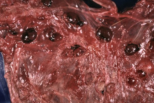
Placental Lesions - Brucella ovis.
Placental lesions as a result of Brucella ovis infection. Placental edema and caruncular necrosis.
Nicoletti PL (1980)
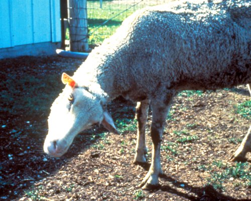
Torticollis.
Spasmodic contraction of the neck muscles, chiefly those supplied by the spinal accessory nerve. The head is drawn to one side and usually rotated so that the chin points to the other side. This was caused by listeriosis.
Shipley C (2006)
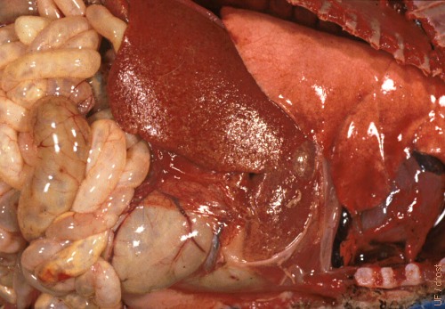
Listeria Liver Lesions.
Liver lesions in a lamb dying at a few days of age due to Listeria monocytogenes. There was milk in the digestive tract and the lungs were inflated.
Smith MC (2006)
Chlamydial Lesions.
Placenta (chorio-allantois) from a ewe that aborted due to Chlamydophila abortus, previously Chlamydia psittaci.
Drost M (1974)
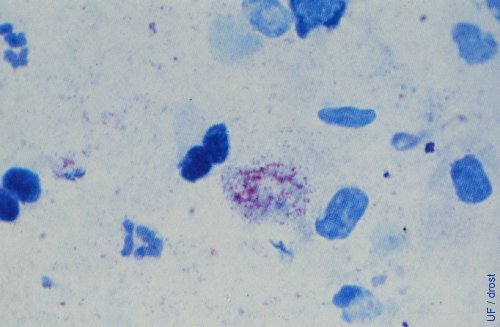
Chlamydial Abortion.
Stained placental smear shows coccoid elementary bodies. Chlamydiosis is characterized by abortion during the last month of gestation. The disease is also called Enzootic Abortion of Ewes (EAE) and is caused by a bacterium of the Chlamidiaceae family, Chlamydophila abortus (previously Chlamydia psittaci). The agent is not a virus, but it does have a virus-like life cycle.
Drost M (1974)
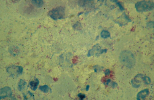
Chlamydia.
Stained placental smear showing intracellular acid-fast bacteria as coccoid elementary bodies. Chlamydiosis is characterized by abortion during the last month of gestation. The disease is also called Enzootic Abortion of Ewes (EAE) and is caused by Chlamydophila abortus, previously known as Chlamydia psittaci.
Smith MC (2006)
EAE Placentitis.
Both cotyledonary and intercotyledonary areas are severely thickened and covered with exudate. Chlamydiosis is characterized by abortion during the last month of gestation. The disease is also called Enzootic Abortion of Ewes (EAE) and is caused by an intracellular bacterium, Chlamydophila abortus.
Smith MC (2006)
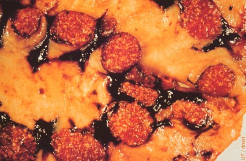
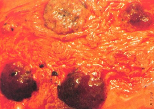
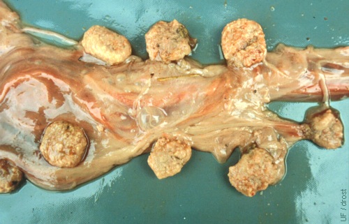
Toxoplasma Lesions.
Typical "pepperoni-like" lesions of the caruncles of a ewe infected with Toxoplasma gondii.
Drost M (1974)
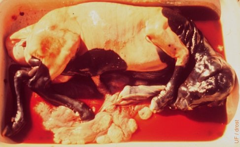
Toxoplasmosis.
The dead fetus is still contained within the fetal membranes. Toxoplasma gondii was recovered from the necrotic cotyledons.
Drost M (1974)
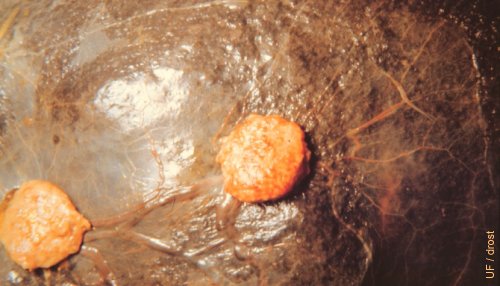
Toxoplasmosis / Cotyledon.
Lesions on a cotyledon typical of toxoplasmosis. The mineralized foci can be demonstrated by compression with a glass slide.
Smith MC (2006)
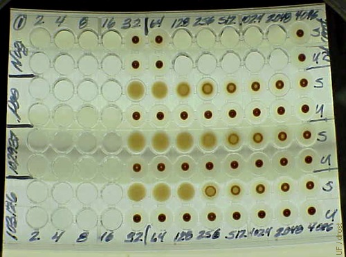
Test Plate for Toxoplasmosis.
IHA (Indirect hemagglutination) test plate for IgG antibodies directed against Toxoplasma gondii. The upper two lines of wells are the negative control, with the positive control and two positive samples below, at various dilutions. In wells labeled S, the red blood cells have been sensitized with T gondii antigen. The U rows of cells (unsensitized erythrocytes) are a further control that antibodies are directed at T gondii, not the sheep erythrocytes themselves - all the U wells on this plate are negative.
Smith MC (2006)
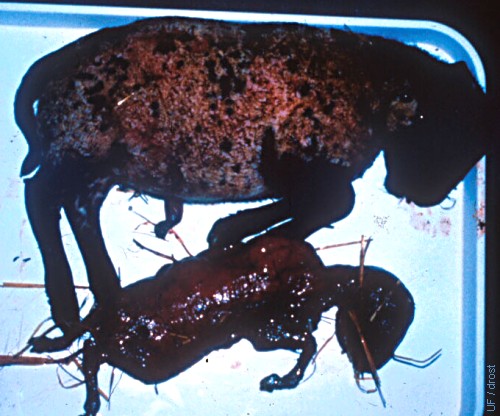
Toxoplasmosis / Fetus.
Decomposing fetus and mummy aborted due to infection with Toxoplasma gondii. The cat is a common intermediary host.
Smith MC (2006)
Toxoplasmosis / Placenta.
Necrotic cotyledons on the membranes from a ewe that aborted due to toxoplasmosis. Intercotyledonary areas are normal.
Smith MC (2006)
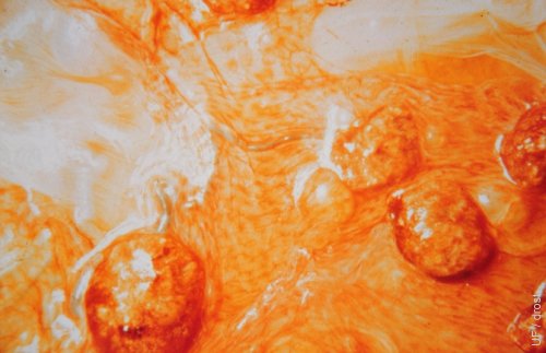
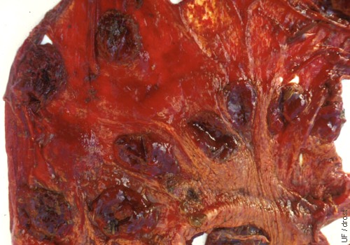
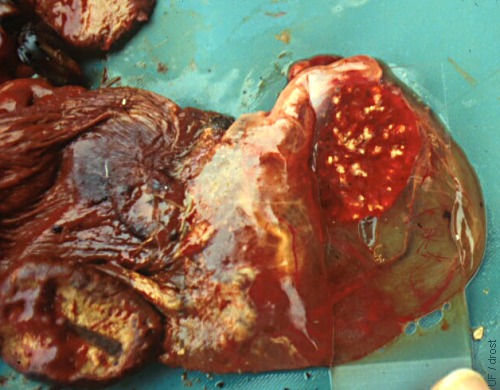
T gondii Infected Placentomes.
Placentomes infected with Toxoplasma gondii.
Drost M (1974)
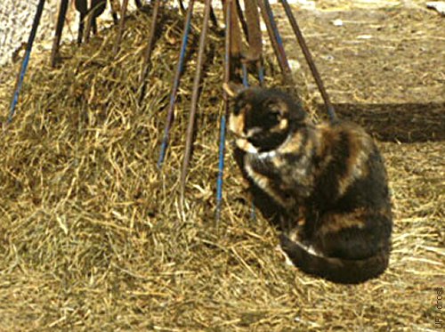
Toxoplasmosis / Cat.
Toxoplasmosis is a zoonotic disease. Cats (especially young kittens) are an intermediate host for Toxoplasma gondii infections in sheep as well as in pregnant women.
Smith MC (2006)
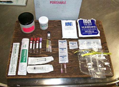
Abortion Kit.
Kit supplied by the Animal Health Diagnostic Center at Cornell for acquiring appropriate test samples for abortion diagnosis, including fresh and formalinized samples of various fetal organs and placenta and fetal fluids.
Smith MC (2006)