The Visual Guide to
Ovine Reproduction
Reproductive Technology: Laparoscopy
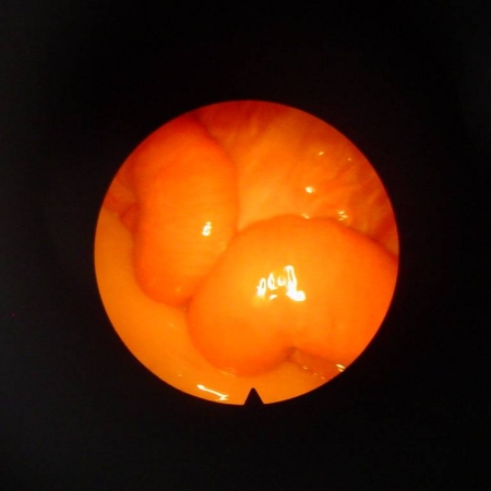
Laparoscopic View of Uterus.
Laparoscopic view of the uterine bifurcation in a nonpregnant ewe.
Shipley C (2006)

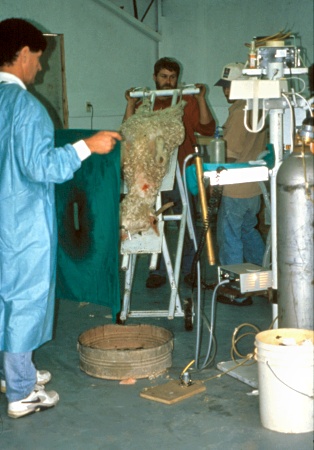
Laparoscopy.
The anesthetized ewe is suspended at a near vertical angle to allow the abdominal viscera to drop down, and the uterus and ovaries to be suspended for easier viewing with the laparoscope.
Drost M (2006)
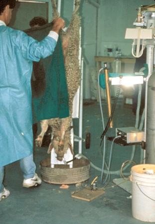
Laparoscopy.
After surgical preparation of the posterior abdominal site for laparoscopy the area is draped to maintain asepsis.
Drost M (2006)
Laparoscopy.
After surgical preparation of the posterior abdominal site for laparoscopy the area is draped to maintain asepsis.
Drost M (2006)
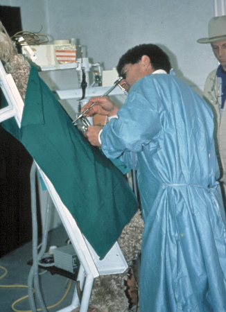
Laparoscopy.
The sterile laparoscope has been inserted to view the uterus and the ovaries.
Drost M (2006)
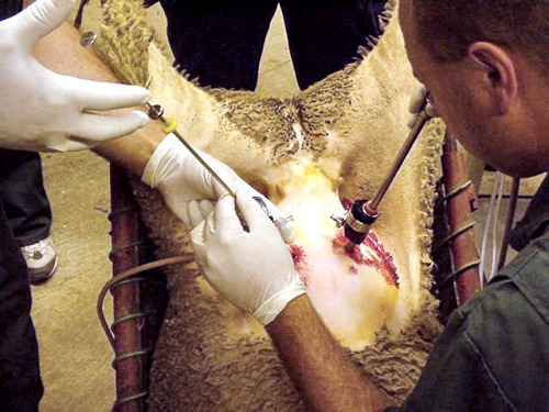
Laparoscopic Insemination.
The sterile laparoscope has been inserted to view the uterus and the ovaries. Via a second portal, the insemination pipet is inserted to inject the semen directly into the uterine horn.
Shipley C (2006)
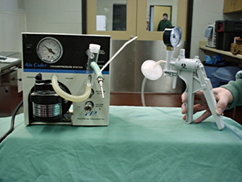
Carbon Dioxide Dispenser.
A sterile needle may be inserted into the abdominal cavity to deliver carbon dioxide to distend the abdomen and facilitate the viewing of the internal organs.
Shipley C (2006)