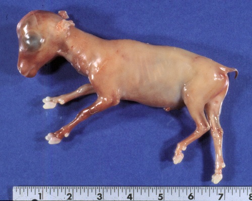
Triplets at about 2 Months.
Three amniotic vesicles. Gestational age approximately 2 months. An ultrasonogram with a sector scanner would permit counting of fetuses and thus adjustment of the nutrition for ewes carrying multiples.
Smith MC (2006)
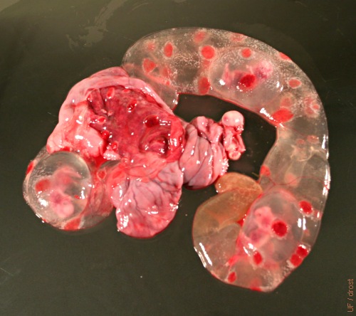
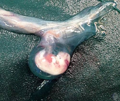
Fetus - 81 Days.
Crown Rump length of 8 cm at 81 days of gestation.
Shipley C (2006)
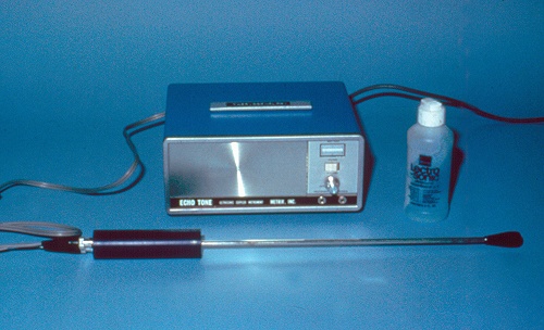
Ultrasound Unit.
Ultrasound unit with a transrectal probe primarily for pregnancy diagnosis.
Shipley C (2006)
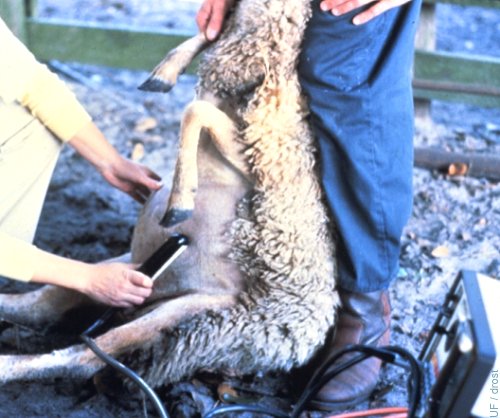
Transabdominal Ultrasound.
The ewe may be placed on her rump while the transducer scans the lower abdomen for the presence of the fetus or fetuses and fetal fluids.
Drost M (1980)
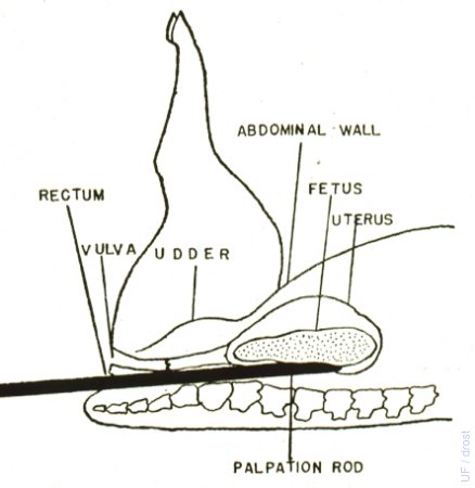
Rectal Abdominal Palpation 1.
With the ewe in dorsal recumbency, a 15 cm diameter rod is gently inserted into the rectum. Proper restraint is essential. The potential for rectal trauma is considerable. The procedure is no longer recommended.
Hulet CV (1972)
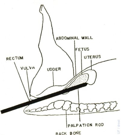
Rectal Abdominal Palpation 2.
The rectal probe is angled upwards towards the udder. When the ewe is pregnant, the rod presses the gravid uterus against the ventral abdominal wall where it can be identified by the hand of the operator which has been placed on the abdomen in front of the udder.
Hulet CV (1972)
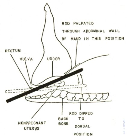
Rectal Abdominal Palpation 3.
When the ewe is not pregnant (or very early pregnant) the firm rod can be distinctly felt below the abdominal wall in front of the udder.
Hulet CV (1972)
Pregnant 25 Days.
Transparent chorioalloantoic membranes and a 1 cm amniotic vesicle. Caruncles are beginning to enlarge.
Mogheiseh A (2013)
Pregnant 26 Days.
Cloudy chorioalloantoic membranes and a small amniotic vesicle. Embryonic death.[bladder off to the right].
Mogheiseh A (2013)
Pregnant 27 Days.
Asymmetrical horns. Right horn enlarged. CL on right ovary.
Mogheiseh A (2013)
Pregnant 28 Days.
Fluid filled membranes. Amniotic vesicle in the center. Small caruncles.
Mogheiseh A (2013)
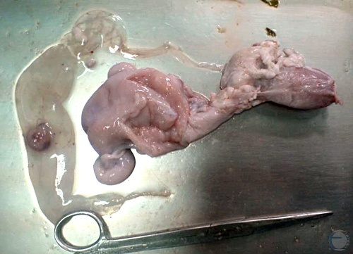
Pregnant 30 Days.
Amniotic vesicle inside the chorioallantois. There is some postmortem cloudiness.
Mogheiseh A (2013)
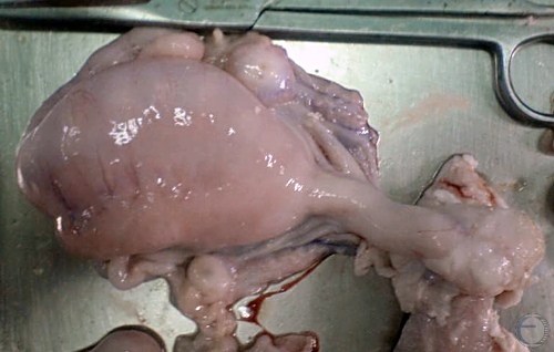
Pregnant 30 Days.
Amniotic vesicle. Caruncles are becoming cup shaped.
Mogheiseh A (2013)
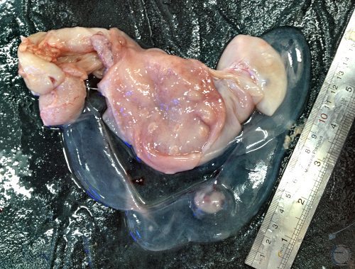
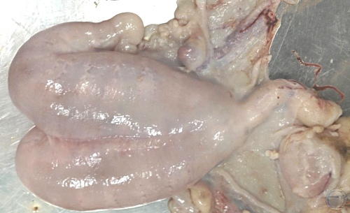
Pregnant 30 Days.
Asymmetry. Left horn pregnancy with CL on the left.
Mogheiseh A (2013)
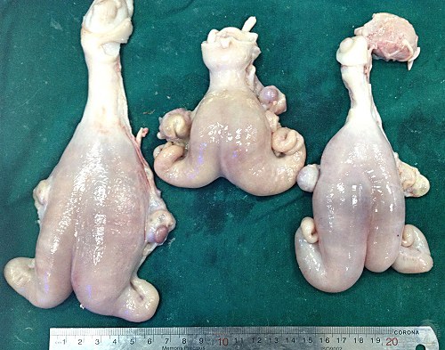
Pregnant 30 to 35 Days.
Gravid uteri. Uterus on the far right may contain twins as its horns are symmetrical.
Mogheiseh A (2013)
Pregnant 30 and 40 Days.
Uterus at the top: right horn singleton pregnancy. Uterus at the bottom: bilateral twin pregnancy, based on symmetry of the horns, and bilateral corpora lutea.
Mogheiseh A (2013)
Twins vs Singleton at 30 and 40 Days.
Uterus at the left: bilateral pregnancy with twins at 30 days of gestation. Uterus on the right: right horn singleton pregnancy at 40 days of gestation.
Mogheiseh A (2013)
Twins at 30 and 40 Days.
Uterus at the left: bilateral pregnancy with twins at 30 days of gestation. Uterus on the right: bilateral pregnancy at 40 days of gestation.
Mogheiseh A (2013)
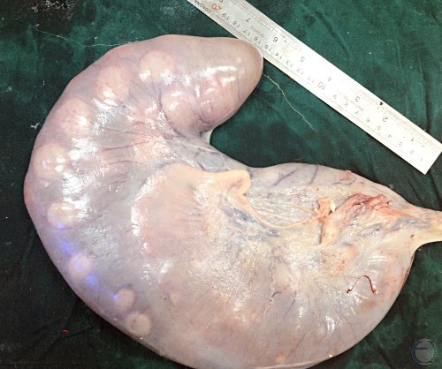
Pregnant 45 Days.
Right horn pregnancy at 45 days. There is a CL on the right ovary.
Mogheiseh A (2013)
Twins at 45 Days.
Unilateral twin pregnancy at 45 days. Two amniotic vesicles are seen in the same horn. Two corpora lutea are on the ipsilateral ovary.
Mogheiseh A (2013)
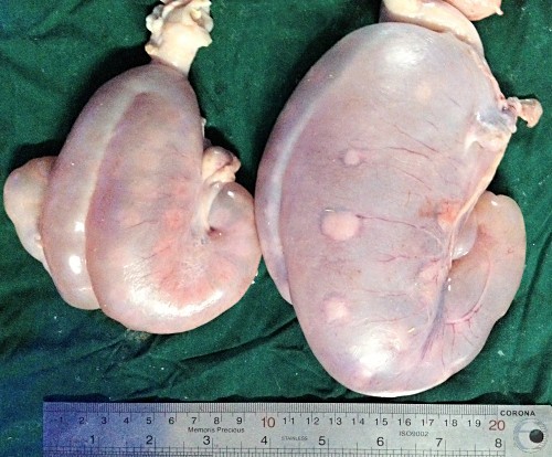
Pregnant at 45 and 50 Days.
Left: singleton at 45 days of gestation. Right: twins at 50 days of gestation.
Mogheiseh A (2013)
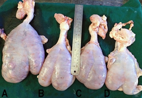
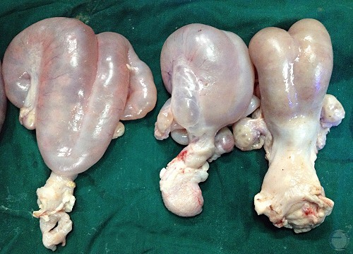
Pregnant Uteri: 65, 55, 45, 35 Days.
A. 65 days, B. 55 days, C. 45 days, D. 35 days.
Mogheiseh A (2013)
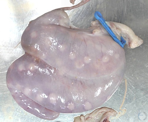
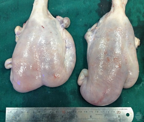
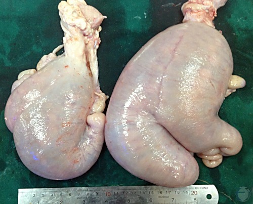
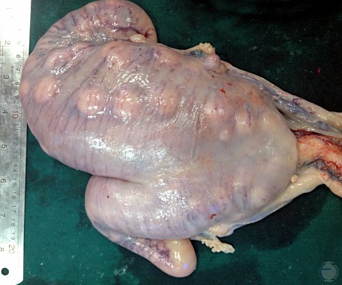
Twins 60 Days.
Symmetrical bilateral distension of the uterine horns. Placentomes are prominent.
Mogheiseh A (2013)
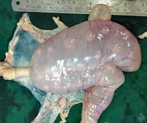
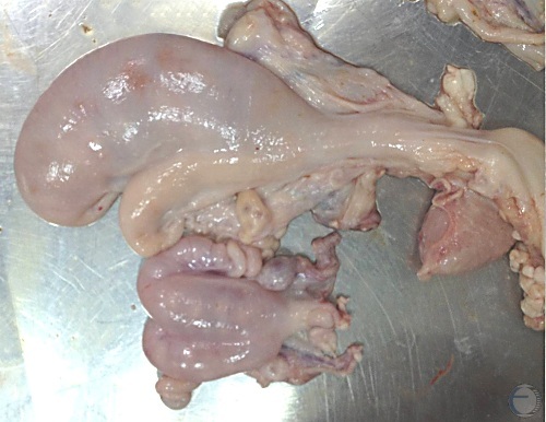
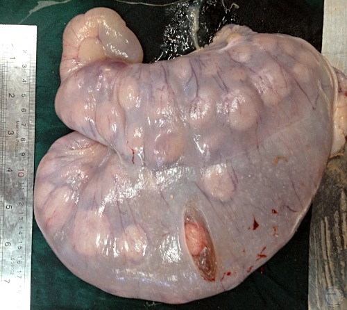
Pregnant 65 Days.
Right horn pregnancy at 65 days of gestation.
Mogheiseh A (2013)
Allantoioc Fluid Color.
Normal allantoic fluid tends to have a pale urine-like color.
Mogheiseh A (2013)
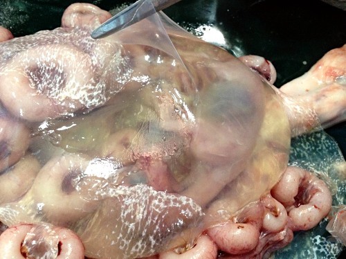
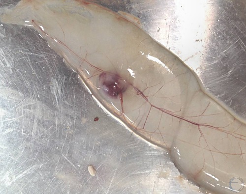
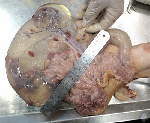
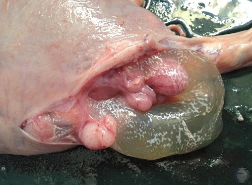
Clear Amniotic Membrane.
The allantoic membranes has been opened up to show the clear intact amniotic vesicle with its transparent membrane.
Mogheiseh A (2013)
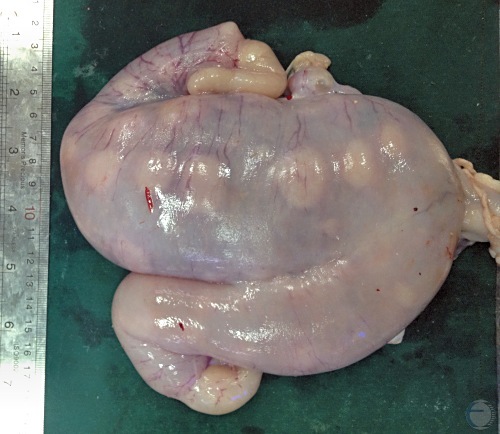
Pregnant 90-100 Days.
Left horn pregnancy at 90 to 100 days gestation.
Mogheiseh A (2013)
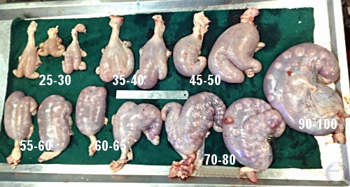
Pregnant 25 to 100 Days.
Pregnant uteri ranging from 25 to 100 days of gestation.
Mogheiseh A (2013)