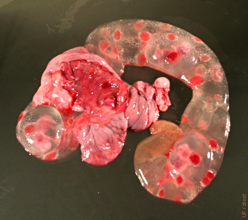
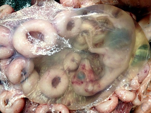
Triplets.
Three amniotic vesicles can be seen inside the chorioallantois. The red cotyledons are arranged in four rows. The normal total number of cotyledons is 90 to 100.
Smith MC (2006)
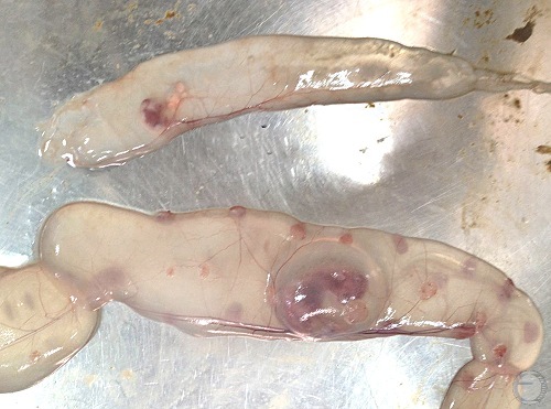
30- and 40-Day Old Amniotic Vesicles.
Amniotic vesicles can be seen inside the chorioallantois. The small cotyledons are arranged in four rows. The normal total number of cotyledons is 90 to 100. The gestational age of the top conceptus is 30 days and that of the bottom conceptus 40 days.
Mogheiseh A (2013)
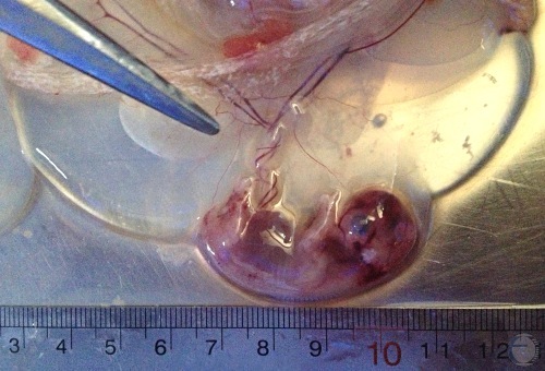
30- and 35-Day Old Amniotic Vesicles.
Amniotic vesicles exposed. Gestational ages: left is 30 and right is 35 days.
Mogheiseh A (2013)
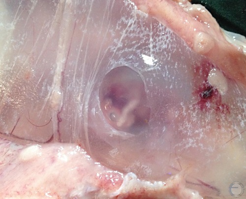
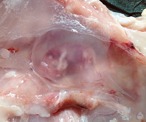
35-Day Old Conceptus.
Window (tear) in the chorionic membranes exposes the amnion.
Mogheiseh A (2013)
Exposed 35-Day Amnion.
The chorionic mebranes have been opened up to expose the 35 day old amniotic vesicle and its fetus.
Mogheiseh A (2013)
Exposed 35-Day Amnion.
The chorionic mebranes have been opened up to expose the 35 day old amniotic vesicle and its fetus. The white "scum" on the chorioallantoic membranes is epithelial plaque formation.
Mogheiseh A (2013)
35-Day Conceptus.
Complete conceptus with amniotic vesicle and the extended chorionic membranes of both horns plus the endometrium showing small caruncles.
Mogheiseh A (2013)
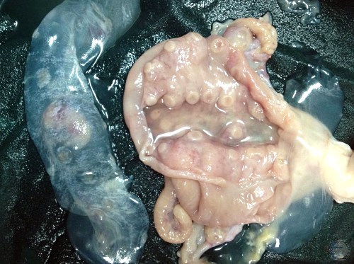
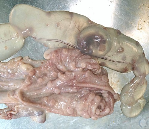
35-Day Conceptus and Placenta.
The gravid horn has been laid open to show the 35 day old conceptus plus the endometrium with rows of small caruncles.
Mogheiseh A (2013)
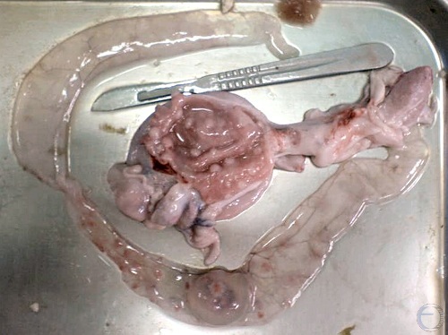
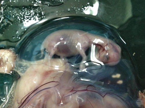
40-Day Fetus.
Note the umbilical cord of this 40-day old fetus in the amniotic sac.
Mogheiseh A (2013)
40-Day Conceptus.
Complete conceptus: fetus, amnion, and chorion at 45 days of gestation.
Mogheiseh A (2013)
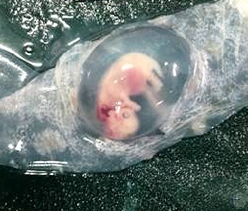
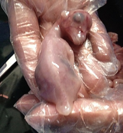
45-Day Fetus.
45-day female fetus in its amnion. Note the genital tubercle underneath the tail.
Mogheiseh A (2013)
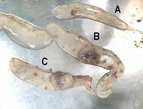
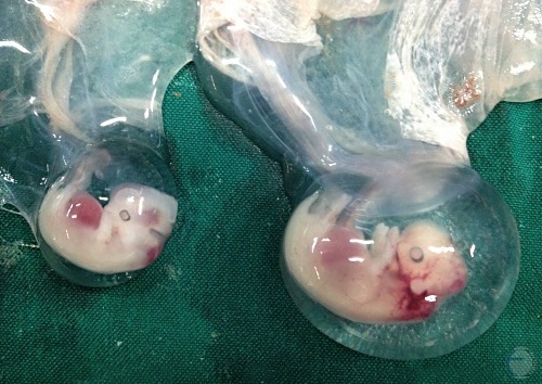
30- to 50-Old Conceptuses.
A. 30-day old conceptus. B. 50-day old conceptus. C. 40-day old conceptus.
Mogheiseh A (2013)
65-Day Old Conceptus.
The chorionic membranes have been opened to show the amnion containing a 65-day old fetus. Note the cup shaped caruncles (doughnuts).
Mogheiseh A (2013)
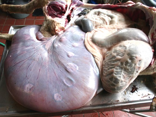
Uterus with Sextuplets.
Uterus with sextuplets at necropsy. This Finn x Dorset ewe also had a fatty liver, suggestive of pregnancy ketosis (also erroneously referred to as pregnancy toxemia though no toxins are involved).
Smith MC (2006)
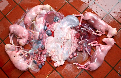
Sextuplets.
Sextuplets at 4 months of gestation. The Finn x Dorset ewe also had a fatty liver, suggestive of pregnancy ketosis [size of the tiles is 15 cm square]
Smith MC (2006)
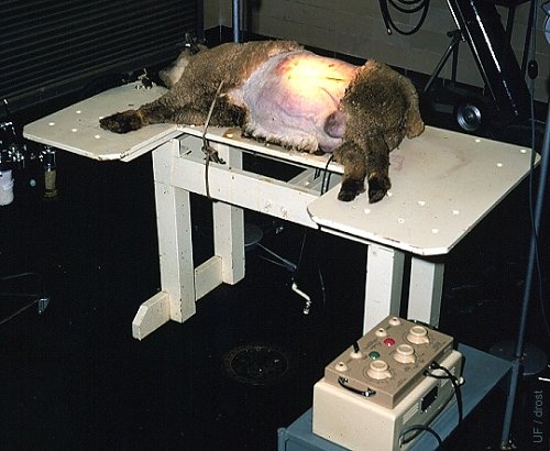
Fetal Surgery.
This pregnant ewe is positioned for intrauterine fetal surgery. The ewe is under general inhalation anesthesia. An electrocautery unit is shown in the right foreground.
Drost M (1968)
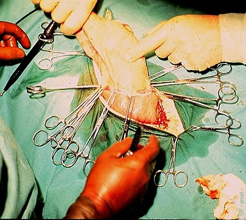
Fetal Surgery.
Fetal adrenalectomy. A 120-day old fetal lamb is shown partially exteriorized in preparation for surgery. The uterine wall and the fetal membranes are marsupialized and clamped to the skin of the fetus to minimize loss of fetal fluids.
Drost M (1968)
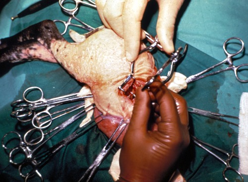
Fetal Adrenalectomy.
The right fetal adrenal gland is exposed with the aid of a small retractor.
Drost M (1968)
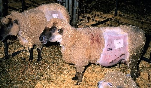
Fetal Canulation.
The 4-month old fetuses of these ewes have been cannulated via their carotid artery. The cannulas have been exteriorized via the flank of the ewe and placed in a protective pouch. Fetal blood can be sampled at predetermined times and frequency. Notice the orientation of the diagonal incision in the lower left flank in ewe 134.
Drost M (1968)