The Visual Guide to
Ovine Reproduction
- Lambing Facilities
- Normal Lambing
- Early Signs of Labor
- Presentation
- Lambing Injuries
- Cesarean Section
- Fetotomy
- Neonate
Obstetrics: Cesarean Section
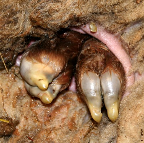
Hind Feet at the Vulva.
Dystocia due to feto-maternal disproportion. Frequently the legs are crossed over when the lamb is large. The hooves show prominent eponychia (soft protective tips).
Smith MC (2006)
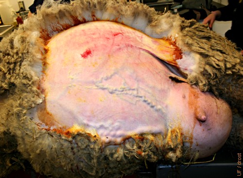
Prepped for Surgery.
With the ewe in dorsal recumbency the abdomen has been clipped, prepared for cesarean section, and the line of incision has been topically anesthetized. Note the large mammary veins and the swollen udder.
Smith MC (2006)
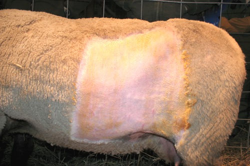
Left Flank Approach.
The left flank has been clipped and prepared for surgery. An inverted L block is generally used for anesthesia.
Smith MC (2010)
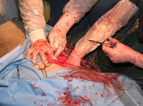
Abdominal Incision.
A 15 to 20 cm midline skin incision is made in front of the udder.
Smith MC (2006)
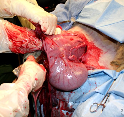
Extracting the Lamb.
The uterine horn has been incised along the greater curvature and the lamb is ready for delivery (lower two hands), while an assistant holds on to the uterus. Ewe in dorsal recumbency.
Smith MC (2006)
Surgical Delivery of the Lamb.
The lamb is grasped by the forelimbs and slowly extracted, while an assistant hangs on to the uterus. The head is resting on the forelimbs.
Smith MC (2006)
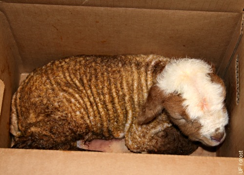
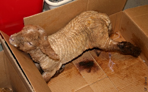
Surgically Delivered Lamb.
Newly delivered lamb with swollen head from dystocia is placed in a draft free clean container, under a heat lamp if necessary.
Smith MC (2006)
Twin Lamb.
Newborn twin lamb placed in a clean draft free environment.
Smith MC (2006)
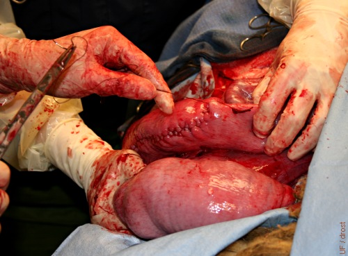
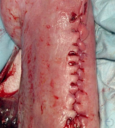
Uterine Closure.
The uterine incision was closed with a Utrecht pattern, a single suture pattern that simultaneously aligns the endometrial edges and inverts the myometrium.
Smith MC (2006)
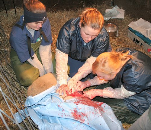
Suturing the Uterus.
These students are closing the uterine incision with a Utrecht pattern, a single suture pattern that simultaneously aligns the endometrial edges and inverts the myometrium.
Smith MC (2010)
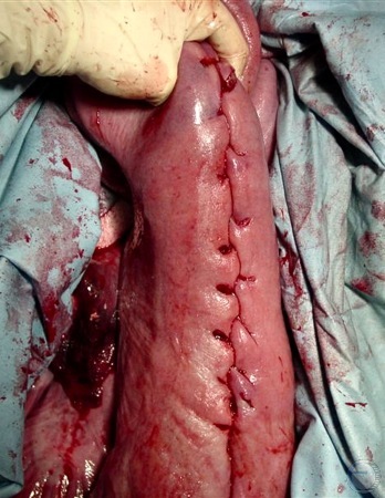
Uterine Incision Line.
This uterine incision was closed with an inverting Utrecht suture pattern, creating a nice seal. The ewe was euthanized 2 days after delivery of quadruplets, because she remained recumbent.
Smith MC (2006)
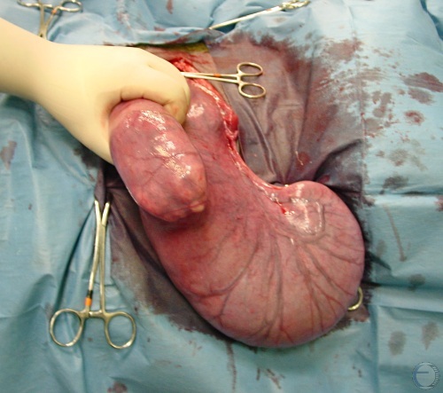
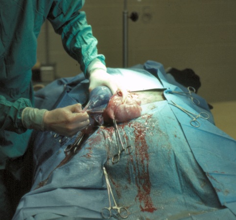
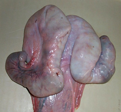
Midline Approach.
Gravid uterus exteriorized via a midline incision by grasping two legs of the lamb.
Villarroel A (2013)
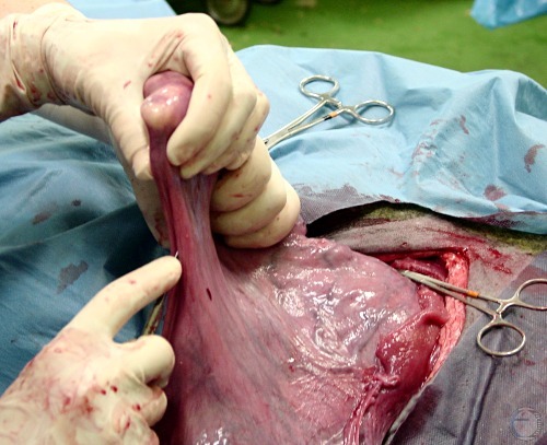
Uterine Incision.
Incision is made along the greater curvature of the uterus which is an area devoid of placentomes and away from major blood vessels.
Villarroel A (2013)
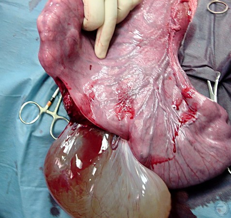
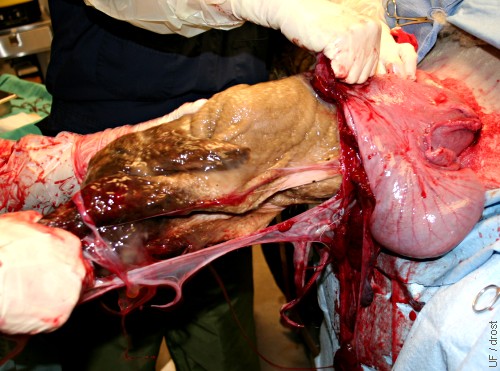
Amnion Appears.
Fluid filled amnion appears (no evidence of cotyledons). Fetus still held by the hindlegs.
Villarroel A (2013)
Utrecht Pattern.
Closure of the uterine incision with the Utrecht pattern which achieves a simultaneous alignment of the cut edges of the incision and infolding of the serosal surfaces.
Villarroel A (2013)
Utrecht Pattern.
Closure of the uterine incision with the Utrecht pattern which achieves a simultaneous alignment of the cut edges of the incision and infolding of the serosal surfaces.
Villarroel A (2013)
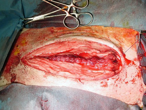
Midline Closure.
Midline incision after c-section in a ewe is ready for closure.
Villarroel A (2013)
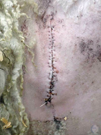
Seroma of the Incision.
Seroma and swelling of the skin incision.
Villarroel A (2013)
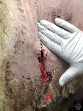
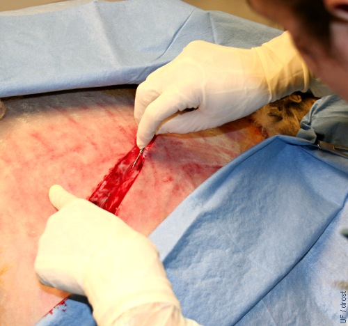
Draining the Seroma.
Draining of the seroma after removal of the bottom suture.
Villarroel A (2013)