The Visual Guide to
Ovine Reproduction
Postpartum Problems: Prolapsed Uterus
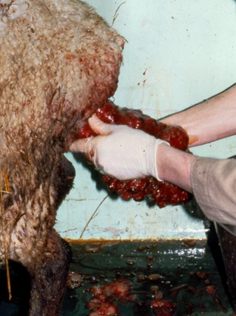
Uterine Prolapse.
Uterine prolapse immediately post partum. The fetal membranes are gone. Shown are the dark caruncles. Hypocalcemia may be a contributing cause. Calcium treatment should be given after the uterus has been replaced. A suture across the vulvar lips is commonly used to prevent recurrence.
Smith MC (2006)
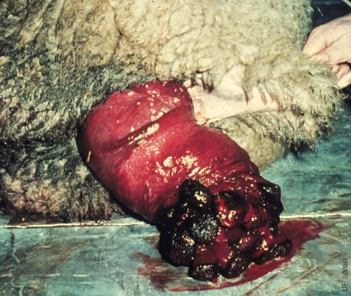
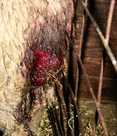
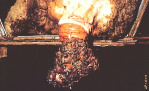
Partial Prolapse of the Uterus.
Partial prolapse of the uterus of relatively recent origin.
Utrecht (1976)
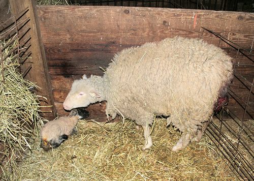
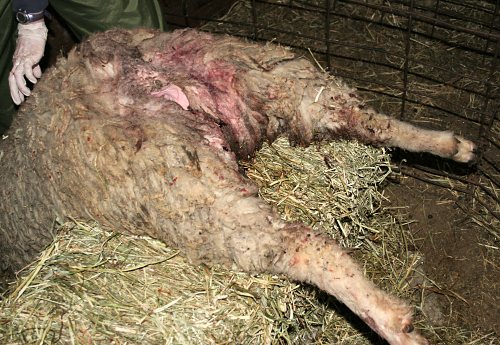
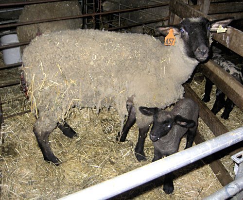
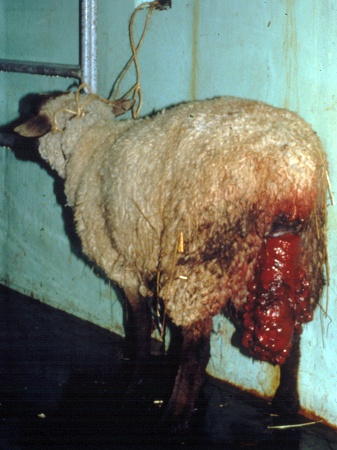
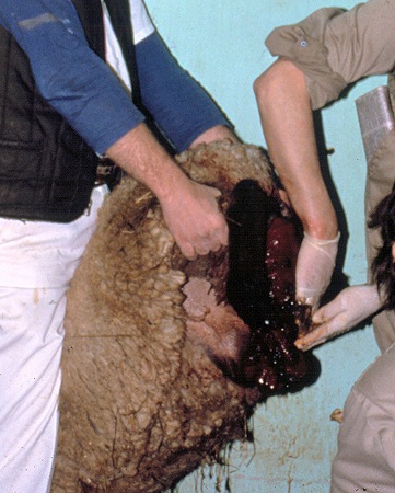
Traumatized Prolapsed Uterus.
This ewe's prolapsed uterus is at risk of trauma from the wire panel. Note the meconium accumulated beneath the lamb's tail as the ewe has been distracted by the discomfort of the prolapse and has not attended properly to its lamb.
Smith MC (2010)
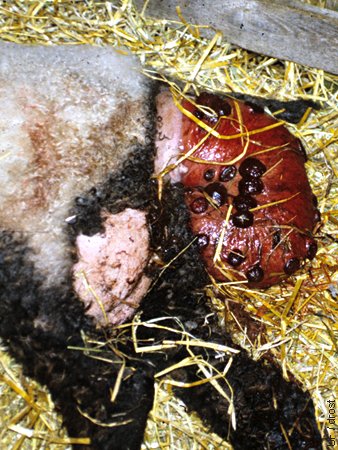
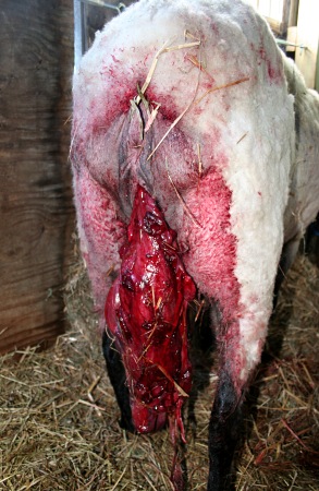
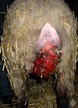
Prolapsed Uterus - Close Up.
Partially prolapsed uterus showing evidence of trauma and contamination. Wool needed to be clipped from the perineum before replacement of the prolapse to facilitate cleaning of the uterus.
Smith MC (2010)
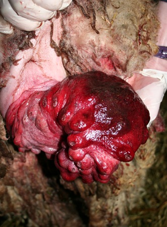
Cleaned Up Uterine Prolapse.
Fairly fresh prolapse of the previously gravid uterine horn. The fetal membranes are gone. The maternal caruncles are swollen.
Smith MC (2010)
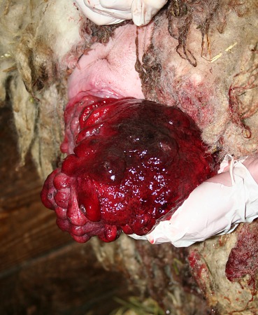
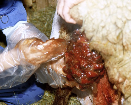
Prolapsed Uterus Replacement.
A caudal epidural has been administered and the prolapsed uterus and the surrounding area have been cleansed. The prolapsed mass is ready for replacement. Note the caruncles on the left and bruising on top.
Smith MC (2010)
Positioning for Replacement.
To facilitate replacement of the prolapsed uterus the ewe is placed over a hay bale with her head down to allow gravity to facilitate the uterus moving forward during replacement under epidural anesthesia.
Smith MC (2010)
Total Prolapse of the Uterus.
Total prolapse of the uterus with a few remnants of the fetal membranes still attached. The Suffolk ewe had a short show dock which may have predisposed it to the prolapse. Hypocalcemia is another possible contributing factor.
Smith MC (2010)
Suffolk Ewe with Prolapsed Uterus.
Suffolk with live newborn lamb and a prolapsed uterus.
Blough E (2013)
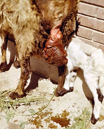
Suffolk Ewe with Prolapsed Uterus.
Suffolk with live newborn lamb and a prolapsed uterus. This was likely the result of hypocalcemia.
Blough E (2013)
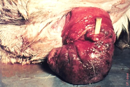
Prolapsed Uterus.
Severe bruising of this prolapsed uterus indicates that the prolapse occurred a number of hours, perhaps as long as a day, ago. A piece of wood is inserted into the lumen of the non-everted, nongravid horn, for identification.
Utrecht (1976)
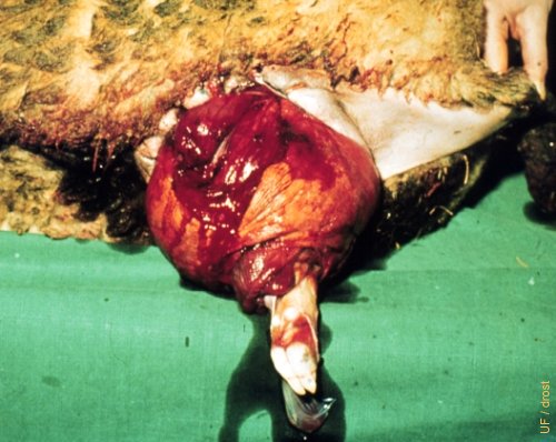
Perforated Prolapsed Uterus.
A fetal foot has perforated the prolapsed uterus.
Utrecht (1976)
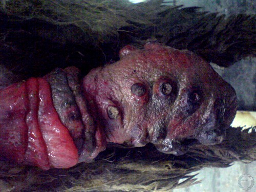
Chronic Uterine Prolapse.
Total chronic uterine prolapse. The tissues are indurated and dessicated. Replacement is not possible. Amputation is indicated.
Mogheiseh A (2013)
Chronic Prolapse of the Uterus.
Chronic prolapse of the uterus of several days duration. The endometrium of both horns had sloughed. The uterus was successfully amputated. During the amputation the uterine arteries should be ligated.
Wright JM (1973)
Total Uterine Prolapse.
Total uterine prolapse in a fat-tailed ewe after the delivery of a live lamb. The prolapsed mass has been cleansed before the picture was taken. Several cup-shaped caruncles are clearly visible.
El Naggar MA (2005)
Uterine Prolapse.
Total eversion after the delivery of a live lamb. The prolapsed mass has been cleansed before the picture was taken. Several cup-shaped caruncles are clearly visible.
Shipley C (2006)
Prolapse Replacement.
Under epidural anesthesia and cleaning of the prolapsed mass the uterus is slowly reinserted by raising the uterus up horizontally.
Shipley C (2006)
Prolapse Replacement.
Elevating the hindquarters of the ewe aids in replacement of the prolapsed uterus with the help of gravity.
Shipley C (2006)
Prolapse Replacement.
Replacement of the prolapsed uterus should be started with the tip of the prolapsed mass.
Shipley C (2006)