The Visual Guide to
Ovine Reproduction
Reproductive Technology: Ultrasonography
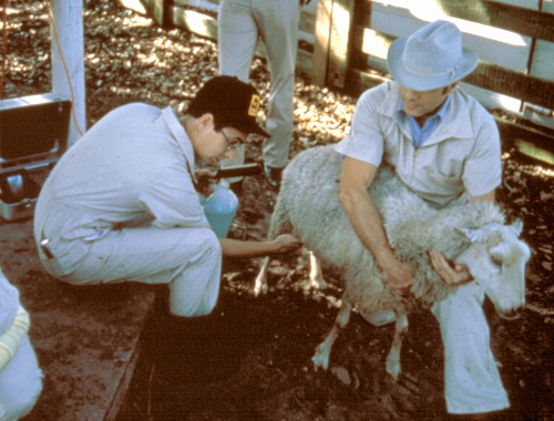
Transabdominal Ultrasound.
The ewe may be placed on her rump while the transducer scans the lower abdomen for the presence of a fetus or fetuses and fetal fluids.
Drost M (1980)
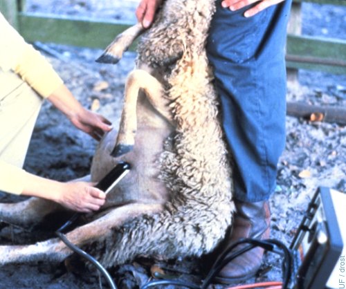
Transabdominal Ultrasound.
With the ewe standing, the ultrasound transducer (probe) is placed in the woolless / hairless inguinal area and aimed towards the uterus.
Smith MC (2006)
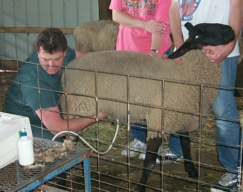
Transabdominal Ultrasound.
A coupling agent is applied to the woolless inguinal area prior to the placement of the transducer.
Drost M (1995)
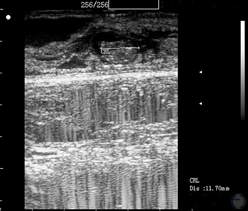
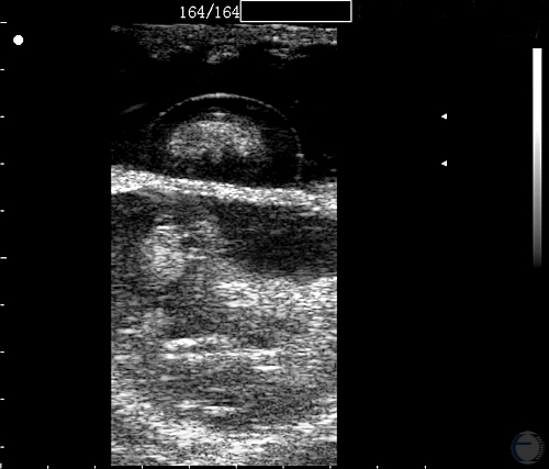
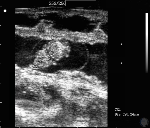
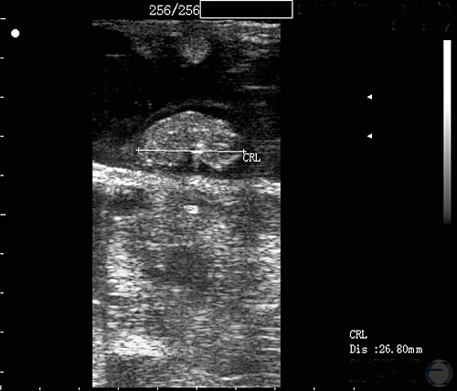
Ultrasonogram at 24 Days.
Crown rump length at 24 days of gestation.
Mogheiseh A (2013)
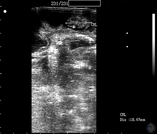
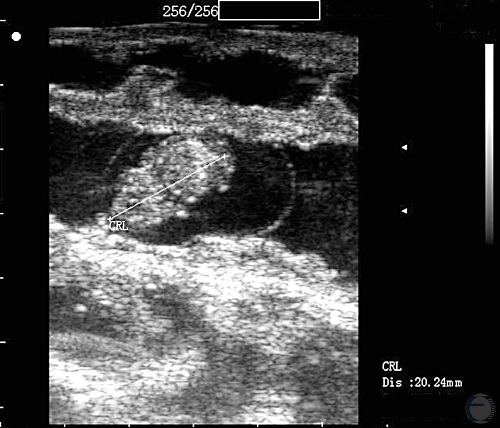
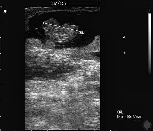
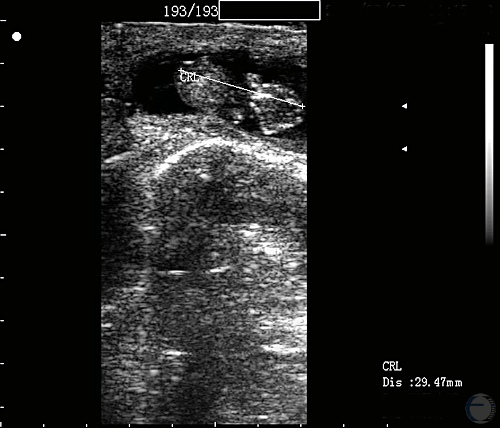
Ultrasonogram at 25 Days.
Crown rump length at 25 days of gestation.
Mogheiseh A (2013)
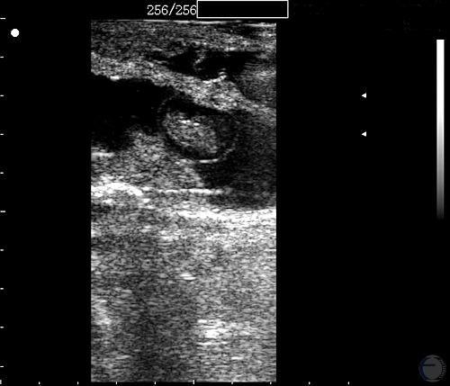
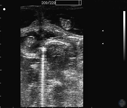
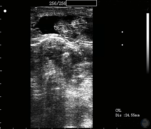
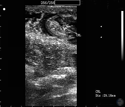
Ultrasonogram at 28 Days.
Crown Rump length at 28 days of gestation.
Mogheiseh A (2013)
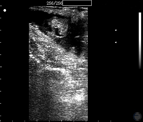
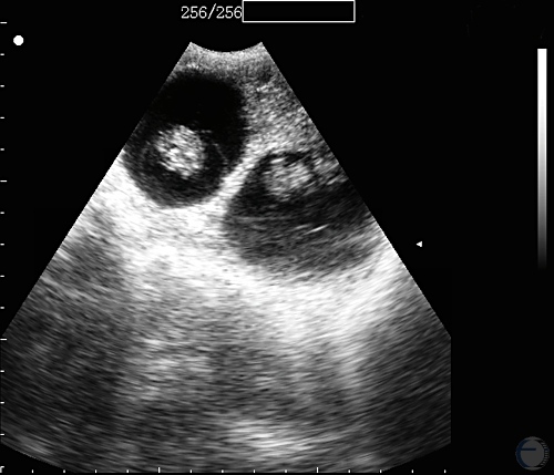
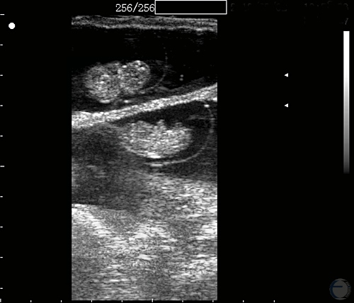
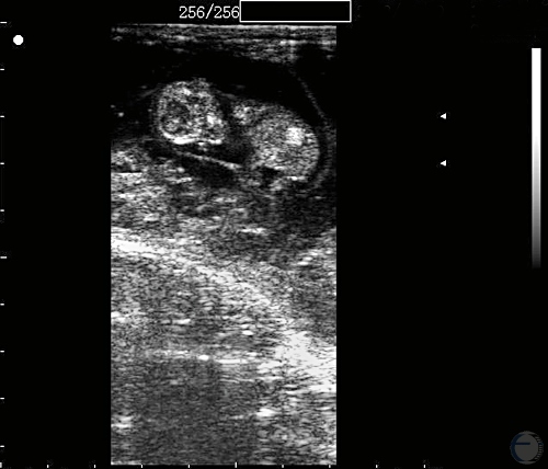
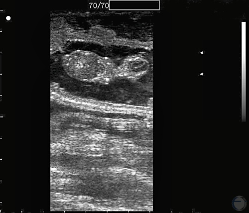
Ultrasonogram at 30 Days - Twins.
Twins at 30 days of gestation.
Mogheiseh A (2013)
Ultrasonogram at 30 Days - Twins.
Twins at 30 days of gestation.
Mogheiseh A (2013)
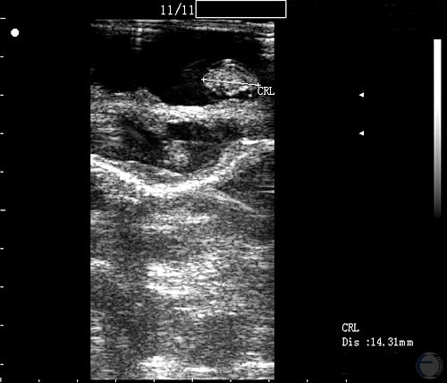
Ultrasonogram at 32 Days.
Crown rump length at 32 days of gestation.
Mogheiseh A (2013)
Ultrasonogram at 33 Days.
Crown rump length at 33 days of gestation.
Mogheiseh A (2013)
Ultrasonogram at 34 Days.
Crown rump length at 34 days of gestation.
Mogheiseh A (2013)
Ultrasonogram at 34 Days.
Crown rump length at 34 days of gestation.
Mogheiseh A (2013)
Ultrasonogram at 35 Days.
Crown rump length at 35 days of gestation.
Mogheiseh A (2013)
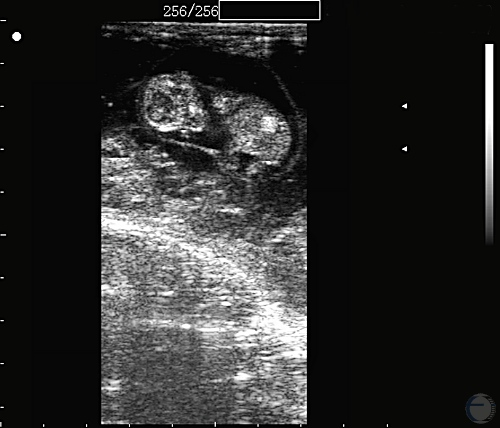
Ultrasonogram of 35 Days - Twins.
Twins at 35 days of gestation.
Mogheiseh A (2013)
Ultrasonogram at 36 Days.
Crown rump length at 36 days of gestation.
Mogheiseh A (2013)
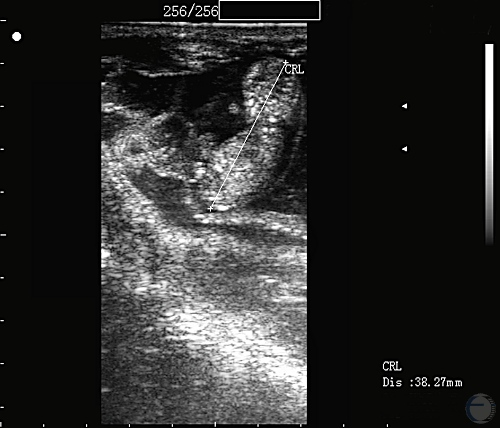
Ultrasonogram at 37 Days.
Crown rump length at 37 days of gestation.
Mogheiseh A (2013)
Ultrasonogram at 37 Days.
Crown rump length at 37 days of gestation.
Mogheiseh A (2013)
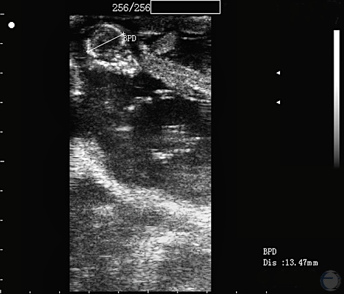
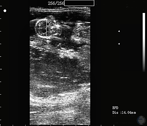
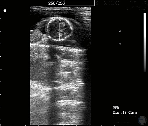
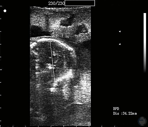
Ultrasonogram at 38 Days.
Fetal skull width at 38 days of gestation.
Mogheiseh A (2013)
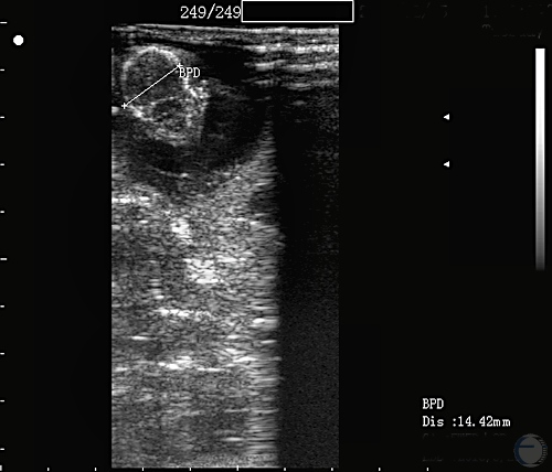
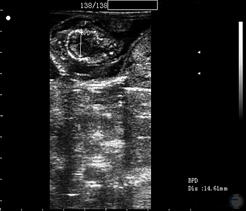
Ultrasonogram at 40 Days.
Fetal skull width at 40 days of gestation.
Mogheiseh A (2013)
Ultrasonogram at 40 Days.
Fetal head and body at 40 days of gestation.
Mogheiseh A (2013)
Ultrasonogram at 40 Days.
Fetal head and body at 40 days of gestation.
Mogheiseh A (2013)
Ultrasonogram at 40 Days.
Fetal skull width at 40 days of gestation.
Mogheiseh A (2013)
Ultrasonogram at 40 Days.
Crown rump length at 40 days of gestation.
Mogheiseh A (2013)
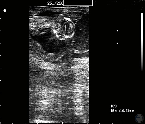
Ultrasonogram at 43 Days.
Fetal skull width at 43 days of gestation.
Mogheiseh A (2013)
Ultrasonogram at 44 Days.
Fetal skull width at 44 days of gestation.
Mogheiseh A (2013)
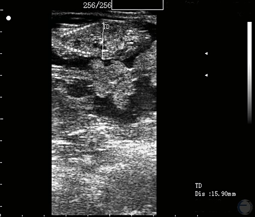
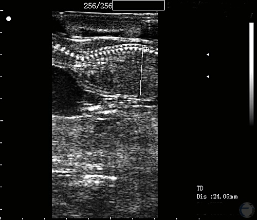
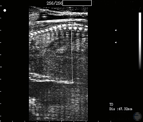
Ultrasonogram at 52 Days.
Thoracic diameter at 52 days of gestation.
Mogheiseh A (2013)
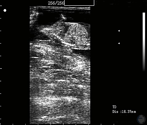
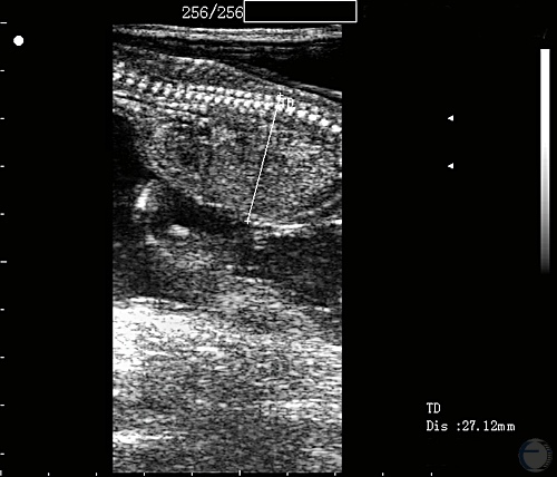
Ultrasonogram at 54 Days.
Thoracic diameter at 54 days of gestation.
Mogheiseh A (2013)
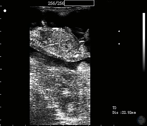
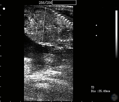
Ultrasonogram at 57 Days.
Thoracic diameter at 57 days of gestation.
Mogheiseh A (2013)
Ultrasonogram at 59 Days.
Thoracic diameter at 59 days of gestation.
Mogheiseh A (2013)
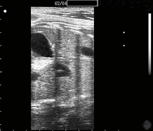
Ultrasonogram at 62 Days.
Thoracic diameter at 62 days of gestation.
Mogheiseh A (2013)
Ultrasonogram at 68 Days.
Thoracic diameter at 68 days of gestation.
Mogheiseh A (2013)
Ultrasonogram at 79 Days.
Thoracic diameter at 79 days of gestation.
Mogheiseh A (2013)
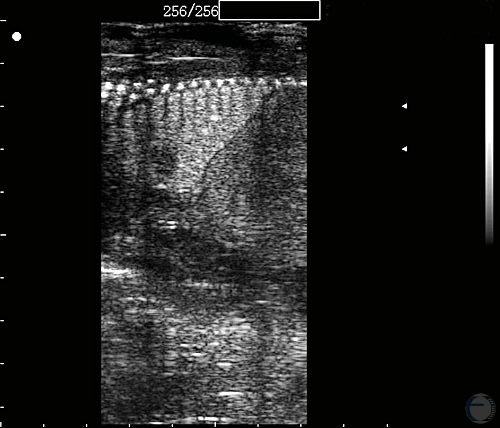
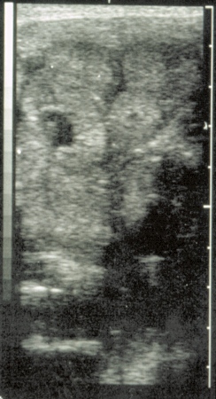
Fetal Lung and Liver at 80 Days.
Fetal lung and liver at 80 days of gestation.
Mogheiseh A (2013)
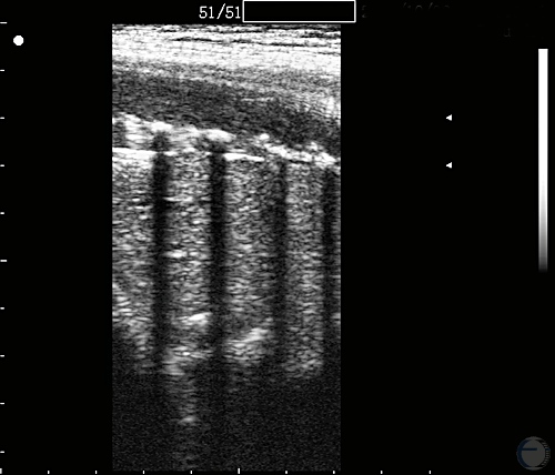
Ultrasonogram Fetal Ribs at 140 Days.
Fetal ribs at 140 days of gestation.
Mogheiseh A (2013)
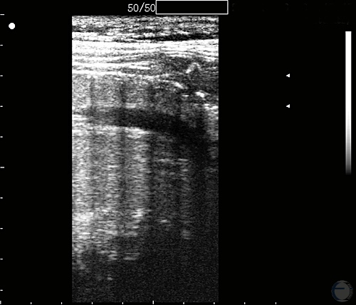
Fetal Thorax and Aorta at 140 Days.
Fetal thorax and aorta at 140 days of gestation.
Mogheiseh A (2013)
Fetal Lung and Abomasum at 140 Days.
Fetal thorax and abomasum at 140 days of gestation.
Mogheiseh A (2013)
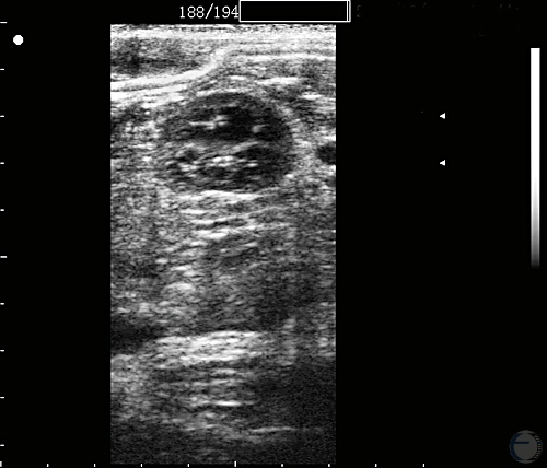
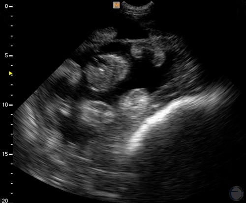
Ultrasonogram Fetal Kidney at 140 Days.
Fetal kidney at 140 days of gestation.
Mogheiseh A (2013)
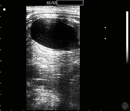
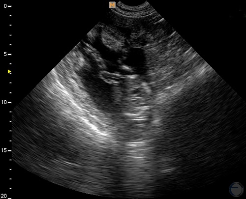
Fetal Urinary Bladder at 140 Days.
Fetal urinary bladder at 140 days of gestation.
Mogheiseh A (2013)
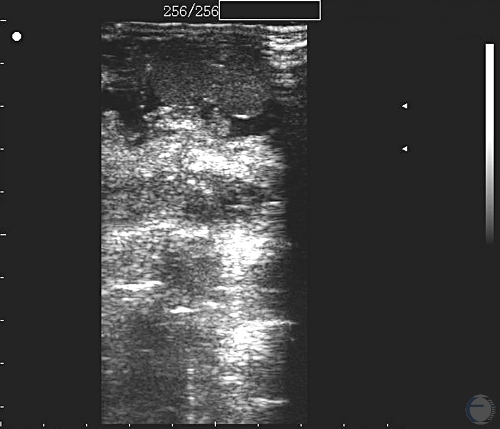
Ultrasonogram Fetal Kidney at 80 Days.
Fetal kidney at 120 days of gestation.
Mogheiseh A (2013)
Ultrasonogram of Placentomes.
Large placentomes during the period of gestation.
Pugh DG (2007)
Ultrasonogram of Placentomes.
Large cup-shaped placentomes during the third trimester in the ewe.
Villarroel A (2013)
Umbilical Cord.
Clear view of the umbilical cord at 60 days of gestation (cm scale).
Villarroel A (2013)
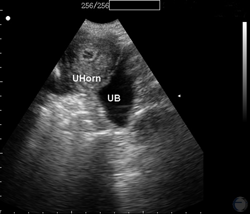
Postabortion Uterine Involution.
Cross-section of the uterine horn and urinary bladder.
Mogheiseh A (2013)
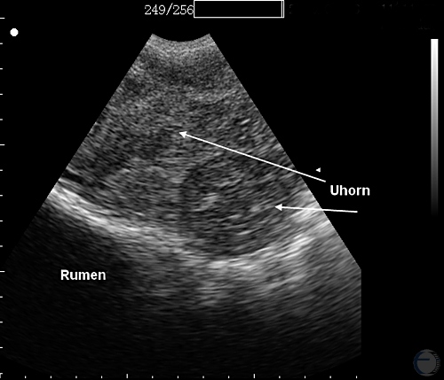
Postabortion Uterine Involution.
Cross-section of the uterine horns. Upper left: previously nongravid uterine horn. Lower right: previously gravid uterine horn. Note the fluid filled rumen off to the lower left.
Mogheiseh A (2013)
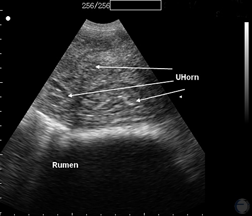
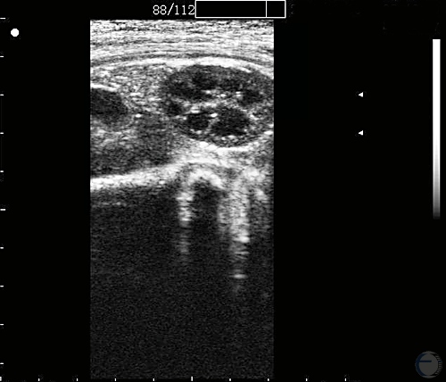
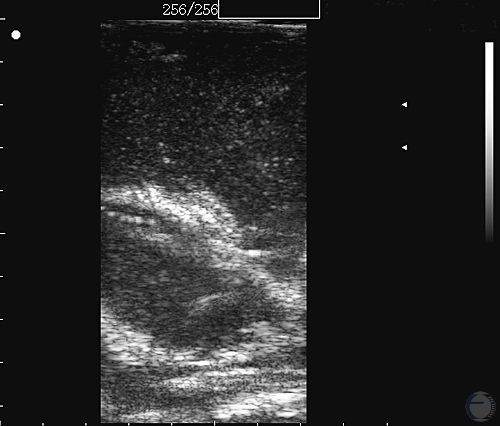
Postabortion Uterine Involution.
Several cross-sections of the uterine horns. The size of the diameter varies between the previously gravid and nongravid horns. Note the fluid filled rumen at the bottom of the images.
Mogheiseh A (2013)
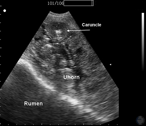
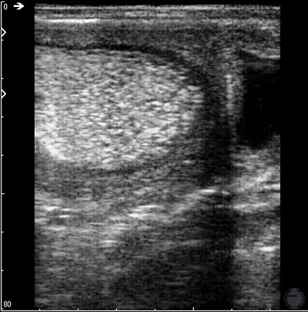
Postabortion Uterine Involution.
Involuting caruncles post abortion. Rumen is at the lower left.
Mogheiseh A (2013)