The Visual Guide to
Equine Reproduction
Reproductive Technology: Ultrasonography
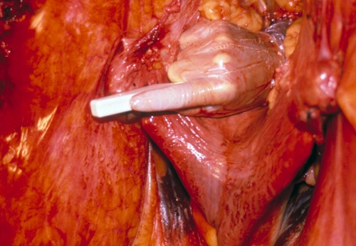
Placement of the Transducer.
Demonstration of the placement of the transducer per rectum to scan the right ovary and the uterine horn of the mare. The rectum has been removed from the specimen for ease of orientation.
Shipley C (2006)
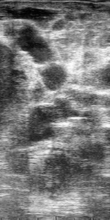
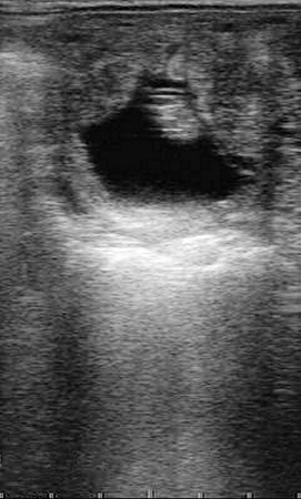
Granulosa Cell Tumor.
Ovary of a mare with an granulosa cell tumor. On palpation the right ovary was very large and very firm, while the left ovary was small and inactive. Ultrasonography revealed this multilocular structure which measured approximately 11 cm in diameter.
King AC (2008)
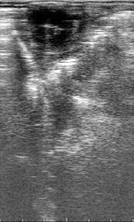
GCT - Contralateral Ovary.
Ovary of a mare with a granulosa cell tumor. On palpation the right ovary was very large and very firm, while the left ovary was small and inactive. Ultrasonography of the left ovary revealed a small, inactive ovary with no follicles developing beyond ~ 5 mm. GCTs secrete large quantities of inhibin, which blocks follicular development on the contralateral ovary.
King AC (2008)
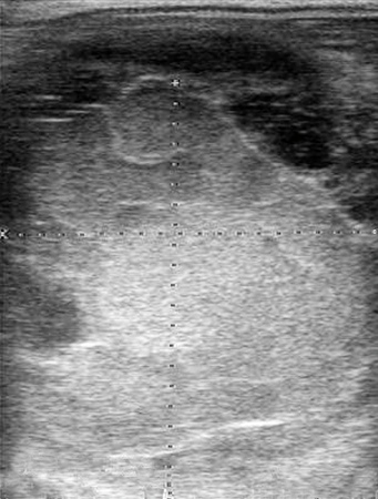
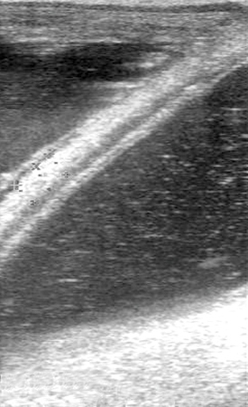
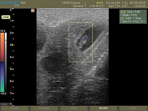
Ovarian Hematoma.
Ultrasonographic image of a large ovarian hematoma. It developed slowly over a period of several days. Ovarian tumors usually develop luteal tissue that does not respond to prostaglandin leading to anestrus.
King AC (2008)
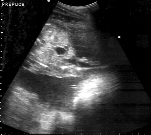
Day 120 Male Fetus.
Male fetus at 120 days of gestation. The large, pendulous prepuce can be seen directly behind the area where the umbilical cord (represented by the distinct, round, anechoic area) meets the fetal abdomen.
King AC (2008)
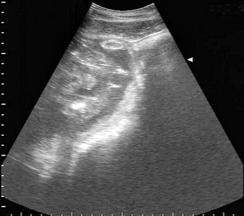
Combined Utero-Placental Thickness.
Measurement of the combined utero-placental thickness in a mid-gestation mare. The bladder is seen on the right hand side of the image, the fetal fluids on the left. A large vessel that runs along the cranioventral surface of the uterus is used to delineate the ventral edge of the uterus, and separate it from the wall of the bladder. Three measurements of the CTUP are taken. Note that the amnion seen within the fetal fluids is not included in the measurement. This technique can be useful in the diagnosis and evaluation of mares with ascending placentitis.
King AC (2008)
Day 120 Female Fetus.
Female fetus at 120 days of gestation. The teats of the mammary gland become visible after Day 118. In this image, two halves of the mammary gland can be seen along the midline with two very small, hyperechoic teats at their lateral edges.
King AC (2008)
Normal Day 40 Embryo.
Ultrasonographic image of a normal, healthy, day 40 developing embryo.
King AC (2008)
Normal Day 30 Pregnancy.
Ultrasonographic image of a normal, healthy, day 30 developing embryo.
Zambrano-Varon J (2012)
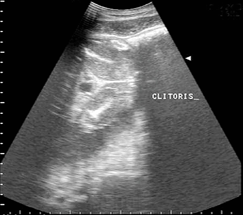
Hyperechoic Clitoris.
Day 120 female fetus. At this stage the clitoris appears as a hyperechoic, trilobed structure that can be seen on the midline. The distinct, round, anechoic area is where the umbilicus joins the fetal abdomen.
King AC (2008)