The Visual Guide to
Equine Reproduction
- Parturition
- Presentation
- Injuries to the Foal
- Cesarean Section
- Fetotomy
- Twinning
- Prolapsed Uterus
- Prolapsed Rectum
Obstetrics: Cesarean Section
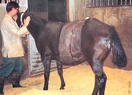
Mare after C-section.
This mare had a cesarean section because of an old healed pelvic fracture which decreased the functional diameter of her birth canal. Incision in the left paralumbar fossa.
Drost M (1970)
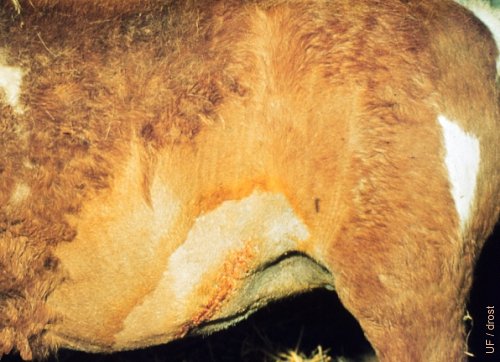
Ventro-lateral Incision.
A ventrolateral approach was used to deliver a dead abnormal foal by cesarean section. The birth canal was too small to perform a fetotomy in this Shetland pony mare.
Utrecht (1976)
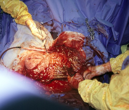
Whipstitch of the Uterine Edge.
The fetal membranes should be pulled away from the incision line in the uterus to avoid suturing them to the uterine wall.
Pozor MA (2009)
Uterine Closure.
Closure of the uterine incision (following c-section) with the Utrecht suture pattern, a continuous infolding pattern, to seal the uterine wall and minimize formation of adhesions.
Pozor MA (2009)
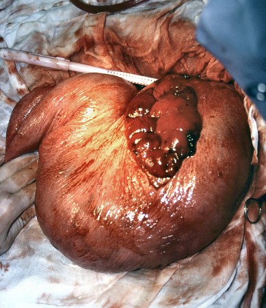
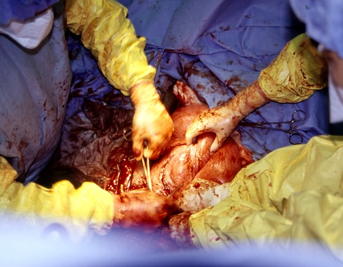
Uterine Tear.
Uterine tear at the base of the horn and in the dorsal aspect of the horn. It was repaired 2 days post partum. The uterus has partially involuted.
Pozor MA (2009)
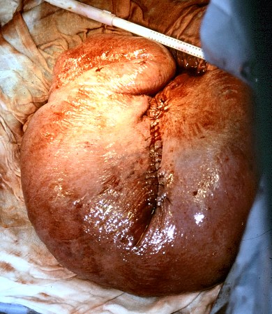
Uterine Tear Repair.
The uterine tear sustained during a dystocia was repaired 2 days post partum. Notice the extensive bruising.
Pozor MA (2009)