The Visual Guide to
Equine Reproduction
Reproductive Technology: Embryos
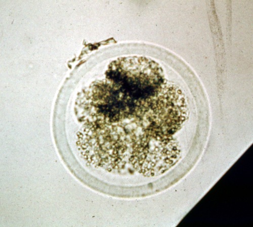
Expanding Blastocyst.
The zona pellucida is becoming thinner due to the expansion of the blastocyst. The inner cell mass is a small circumscribed dark area. Trophoblast cells can be seen around the translucent blastocele.
Ball BA (1987)
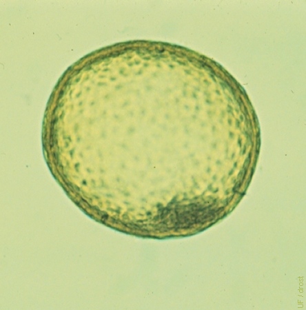
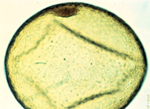
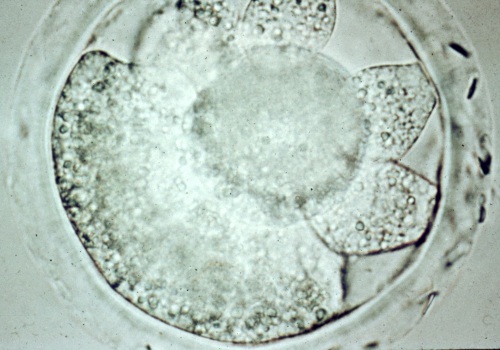
Day 7, Expanded Blastocyst.
The zona pellucida has become very thin. The diameter of the embryo has also noticeably increased. The embryo sometimes takes on the appearance of a golf ball. Day 7.
Ball BA (1987)
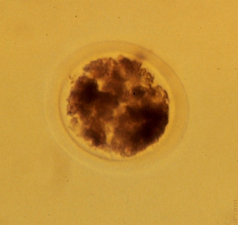
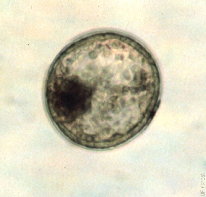
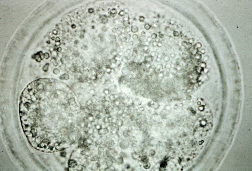
Unfertilized Ovum.
Unfertilized ovum (UFO) collected on Day 7. Note the fragmentation of the cytoplasm.
Ball BA (1987)
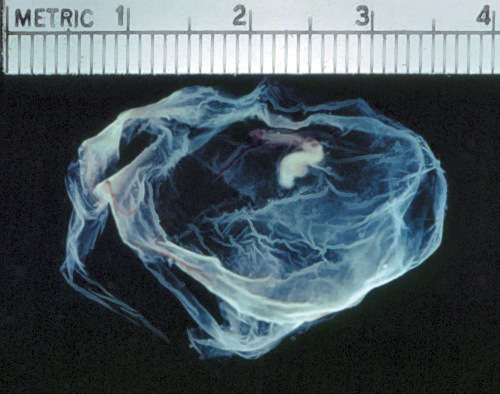
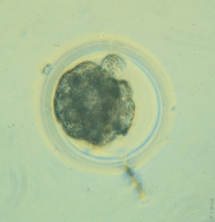
Day 20 Embryo.
Day 20 embryo surrounded by the trophoblast. The embryo itself is 5 mm long.
Bergin WC (1973)
Degenerating Embryo.
Early 6-cell embryo in the process of degenerating. One blastomere at 10 o'clock is still intact.
Bergin WC (1973)
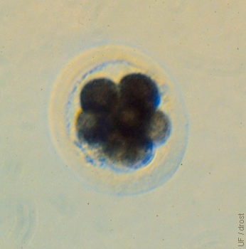
Degenerating 8-cell Embryo.
Degenerating 8-cell embryo. The blastomeres have taken on a granular appearance.
Bergin WC (1973)
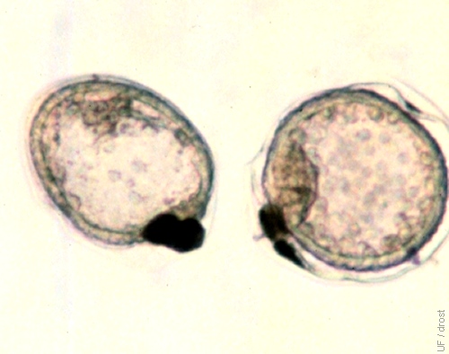
Early Degenerating Embryo.
Six- to eight-cell degenerating embryo. One large original 2-cell blastomere is still intact. Spermatozoa are present in the zona pellucida, on the right.
Bergin WC (1973)