The Visual Guide to
Equine Reproduction
Placenta: Abnormal Placenta
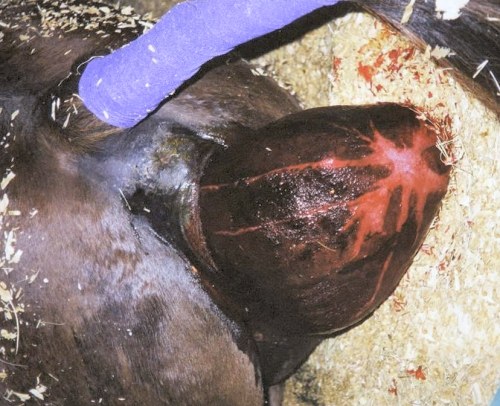
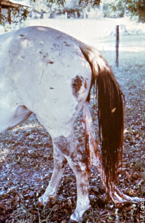
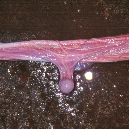
Premature Placental Separation.
The chorioallantois may separate from the endometrium before birth. The fetus frequently dies from asphyxia or will be weak at birth. The separated chorioallantois may present at the vulva without rupturing, leaving the cervical start intact.
Hopper RM (2007)
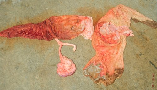
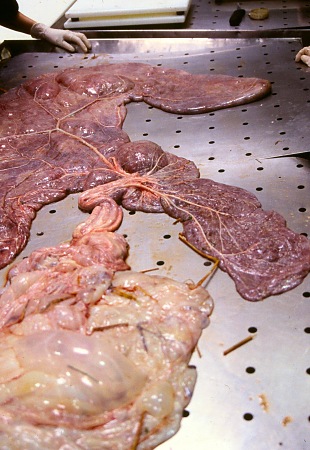
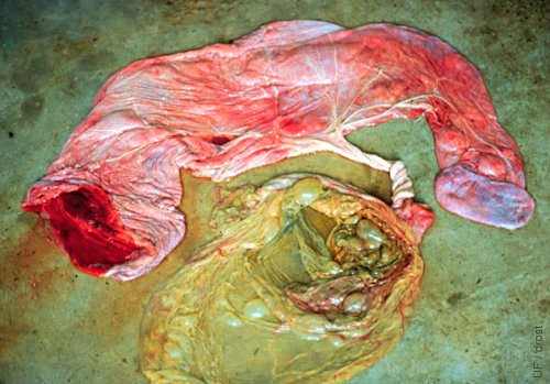
Twin Placentas.
These two sets of fetal membranes belonged to twins. One of the twins was in the process of mummification.
LeBlanc MM (1982)
Expulsion of the Placenta.
The placenta is normally expelled within 3 to 8 hours after delivery of the foal. Mares should be treated within 12 to 24 hours to avoid septicemia, toxemia, and possible postparturient laminitis.
Roberts SJ (1972)
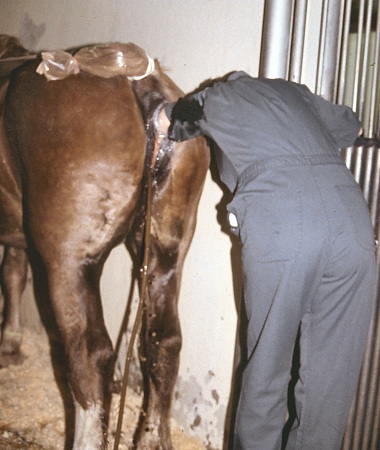
Post Dystocia Lavage.
After dystocia mares should be routinely lavaged to prevent the development of postpartum metritis. The tail is wrapped in a palpation sleeve.
Pozor MA (2009)
Retained Placenta.
The placenta (actually the fetal membranes) are normally spontaneously delivered within 3 to 6 hours. They are considered retained when not expelled by 8 hours after delivery of the foal.
Asbury AC (1977)
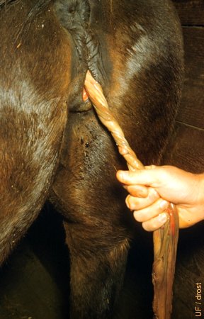
Retained Placenta.
The fetal membranes are twisted to facilitate identification of the membranes from the endometrium, and in some cases to aid in removing them with gently traction. Manual removal generally is not recommended.
Roberts SJ (1972)
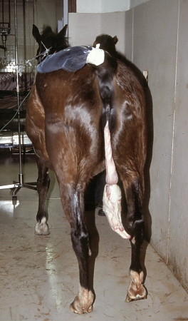
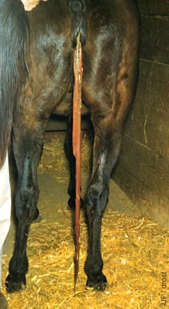
Retained Fetal Membranes.
The membranes have been tied in a knot to keep the mare from stepping on it. Cleanliness is essential.
Pozor MA (2009)
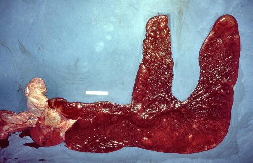
Fetal Membranes.
Fetal membranes. Chorionic side out. Laid out for evaluation in the lazy F position.
Pozor MA (2009)
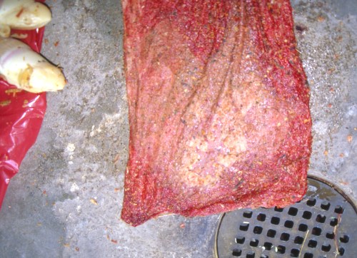
Edema of the Tip of the Placenta.
Typical edema of the tip of the fetal membranes of the pregnant horn.
Pozor MA (2009)
Membranes Spread Out.
Allantoic side out. It is important to ascertain that the fetal membranes are intact. Atypical attachment of the umbilical vessels.
Pozor MA (2009)
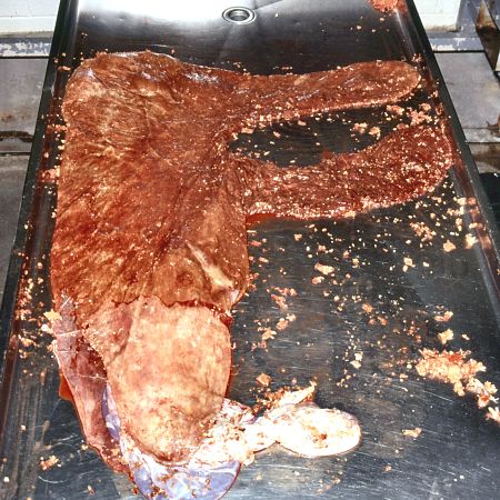
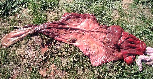
Ascending Placentitis.
Fetal membranes laid out in the Lazy F position. Cervical star at beginning of the large avillous area. [the flocculent appearing debris is bedding shavings].
Pozor MA (2009)
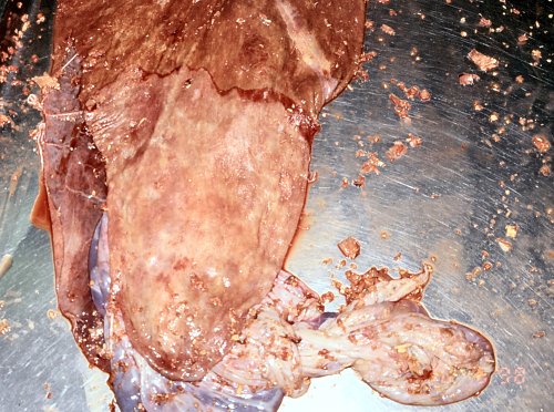
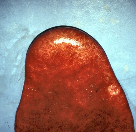
Tip of the Infected Membranes.
Unusually large avillous portion of the fetal membranes near the cervical star.
Pozor MA (2009)
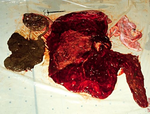
Necrotic Placenta.
Comparison of a necrotic placenta of a macerated fetus with that of its normal twin.
Roberts SJ (1972)
Necrotic Amnion.
The chorioallantois has been turned inside out. The amniotic membrane below is necrotic.
LeBlanc MM (1982)
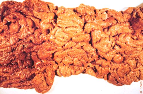
Mycotic Placentitis.
Thickened allantochorion. The diagnosis of fungal infection may be confirmed by making an impression smear and finding hyphae.
McEntee K (1972)
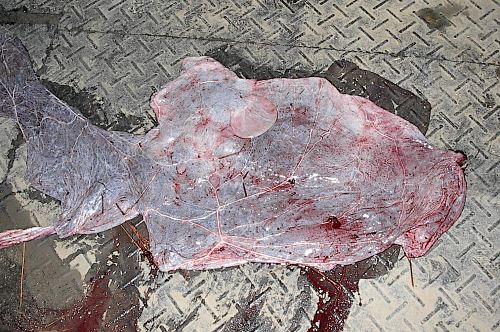
Avillous Chorioallantois.
Most of the surface of the chorioallantois is avillous. The pluriparous mare, a 3/4 Cleveland Bay, had been bred to a purebred Cleveland Bay stallion. She delivered a live foal at 333 days. There was no twin. This was her fourth foal.
Norman S (2010)
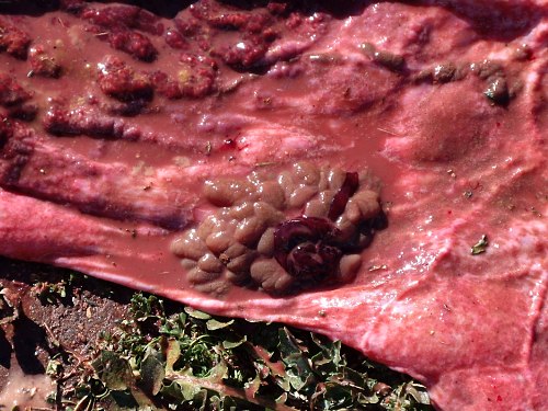
Hypertrophied Chorionic Villi.
Large fist size mass of hypertrophied chorionic villi in an avillous area of a placenta at term. The pluriparous mare had a live singleton foal.
Norman S (2010)
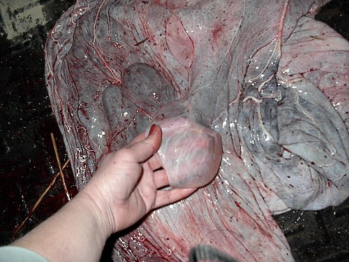
Large Allantoic Cyst.
Single, flaccid allantoic cyst. Incidental finding at normal term.
Campbell J (2007)
Allantoic Cyst.
Flaccid allantoic cyst. Incidental postpartum finding.
Campbell J (2007)
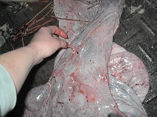
Peduncluated Allantoic Cyst.
Pedunculated allantoic cyst observed after a normal delivery at term.
Campbell J (2007)
Yolk Sac Remnant.
Small remnant of the yolk sac along the umbilical cord. Incidental finding. The occurrence is uncommon.
Pozor MA (2009)
Avillous Fetal Membranes.
Large avillous area which is close to cervical star. The small granular particles are bedding.
Pozor MA (2009)
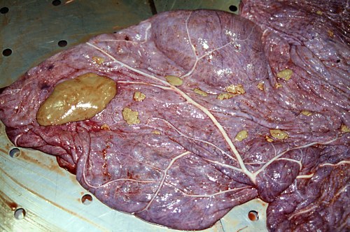
Hippomane.
Hippomane which is also called an allantoic calculus. To owners it sometimes looks like a fetal tongue.
Pozor MA (2009)