The Visual Guide to
Equine Reproduction
Postpartum Care: Trauma to Birth Canal
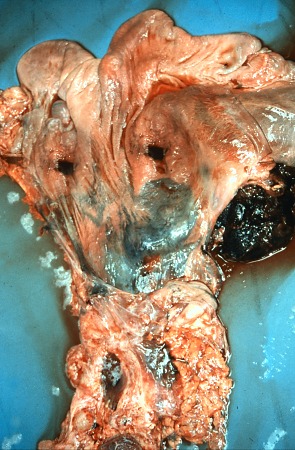
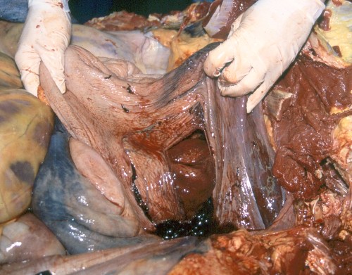
Ruptured Hematoma in Broad Ligament.
Haematoma in the broad ligament. Occasional complication during dystocia or at times during normal parturition. They are usual fatal when ruptured. Without rupture the mare may survive.
Pozor MA (2009)
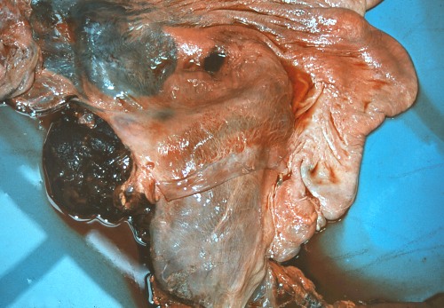
Close Up of Ruptured Hematoma.
Close up of a ruptured hematoma in the broad ligament. Occasional complication during dystocia or, at times, during normal parturition.
Pozor MA (2009)
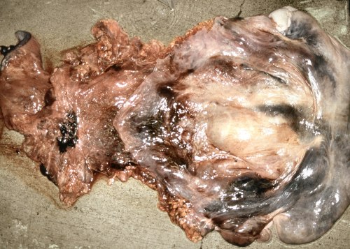
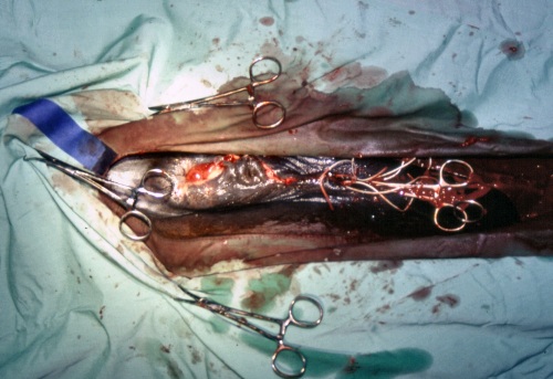
Necrotic Uterus after Torsion.
Chronic uterine torsion accompanied by signs of colic for 1 week which led to uterine rupture and necrosis.
Pozor MA (2009)
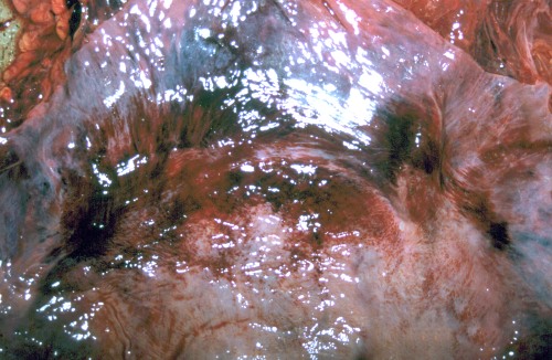
Bruised Uterus - Close Up.
Circular band of bruising of the uterus at the location of a chronic torsion as seen during the postmortem examination.
Pozor MA (2009)
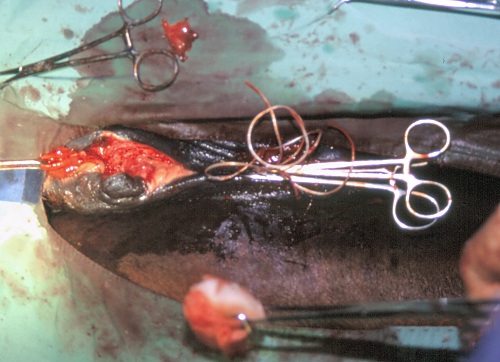
Uterine Tear.
Uterine tear sustained during corrective rotation of the uterine torsion.
Pozor MA (2009)
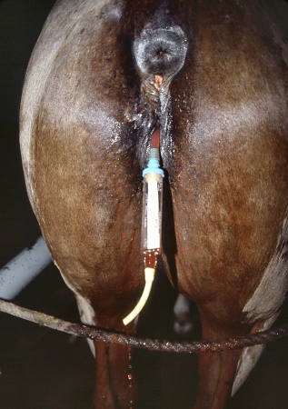
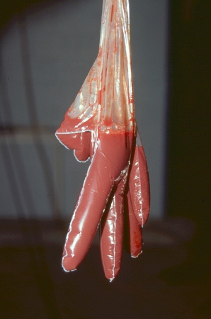
Vaginal Hematoma.
Vaginal hematoma due to trauma during a dystocia. A balloon catheter has been placed in the pocket of the hematoma to drain and lavage it.
Pozor MA (2009)
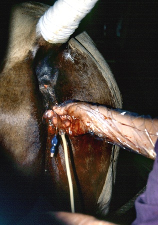
Balloon Catheter Placement.
A balloon catheter, like a Foley catheter, has been place in the crater produced by a ruptured vaginal hematoma.
Pozor MA (2009)
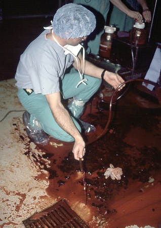
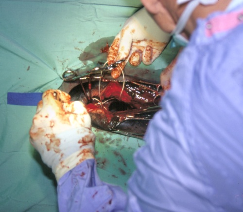
Lavage of the Peritoneal Cavity.
Blood and blood clots are lavaged from the abdominal cavity of a mare. The hemorrhage was the result of a uterine tear sustained during a dystocia.
Pozor MA (2009)
Lavaged Fluid.
Blood lavaged from a vaginal hematoma which was sustained during a dystocia.
Pozor MA (2009)
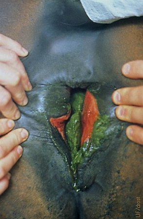
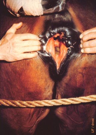
Third-degree Perineal Laceration.
In a third-degree perineal laceration the dorsal commissure of the labia is torn as well as the perineal body and the anal sphincter. In the vernacular, it is called a Gillflirter.
Utrecht (1976)
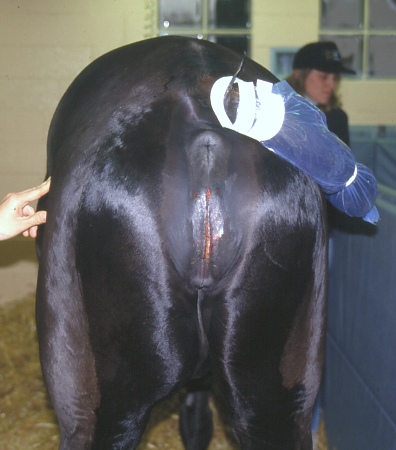
Third-degree Perineal Laceration.
The torn anal sphincter, perineal body and dorsal commissure of the vulva have healed. The mare was maintained on green grass to keep the consistency of the feces soft to expedite healing and awaiting reconstruction.
Utrecht (1976)
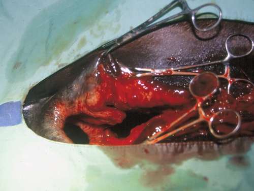
Perineal Laceration Surgery.
In a 3rd degree perineal laceration the dorsal commissure of the vulva is split, the perineum is torn and the anal sphincter is severed. Here, the wounds have been debrided and cleansed in preparation for surgical repair.
Pozor MA (2009)
Perineal Laceration Surgery.
The first step in surgical repair of a 3rd degree perineal laceration is realignment of the anal sphincter muscle.
Pozor MA (2009)
Perineal Laceration Surgery.
Step 3 is to close the labiae as in a Caslick's procedure leave adequate space at the ventral commissure for urination.
Pozor MA (2009)
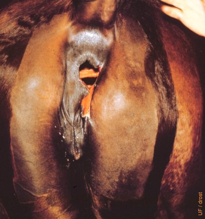
Vulvar Edema after Dystocia.
Extensive edema of both vulvar lips after an assisted delivery to relieve a dystocia.
Pozor MA (2009)
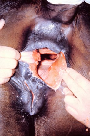
Recto-vaginal Fistula.
The anal sphincter and perineal body are ripped apart. The vulva is intact. R-V fistulas can also have an intact anal sphincter and merely have a hole between the roof of the vestibule and the floor of the rectum.
Utrecht (1975)
Recto-vaginal Fistula.
The anal sphincter and perineal body are torn apart. The vulva is intact. R-V fistulas can also have an intact anal sphincter and merely have a hole (fistula) between the roof of the vagina and the floor of the rectum.
Utrecht (1975)
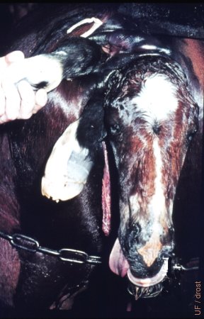
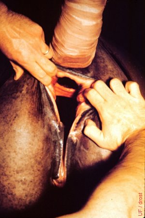
Recto-vaginal Perforation.
The right forelimb of the foal has perforated the rectal floor and can be seen to exit via the anus. The anal sphincter is intact. The head and the other forelimb are extended through the vulva.
Utrecht (1976)
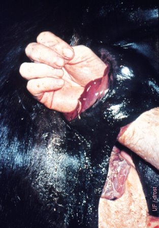
Recto-vaginal Perforation.
After assisted delivery of the foal, insertion of the hand and arm through the rectovaginal fistula demonstrates its extent. The vulva is intact. A portion of the placenta can be seen.
Utrecht (1976)
Gillflirter.
The rectum and the vestibule became a continuous cavity, a "cloaca". This is a third-degree perineal laceration; associated with a forceful delivery in the mare.
Utrecht (1975)