The Visual Guide to
Caprine Reproduction
Reproductive Technology: Ultrasonography
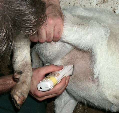
Clipping the Inguinal Region.
The lower right flank is clipped in preparation for ultrasonography to allow better contact of the probe to evaluate the pregnancy status of the doe.
Smith MC (2006)
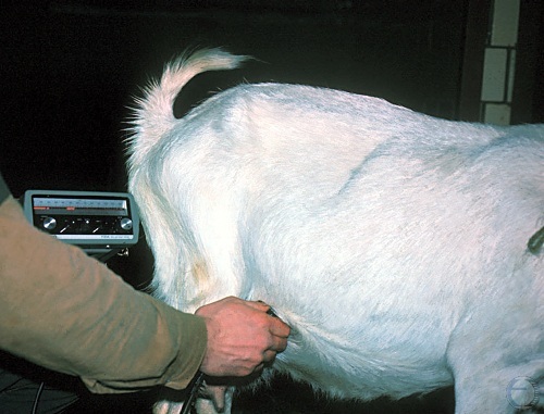
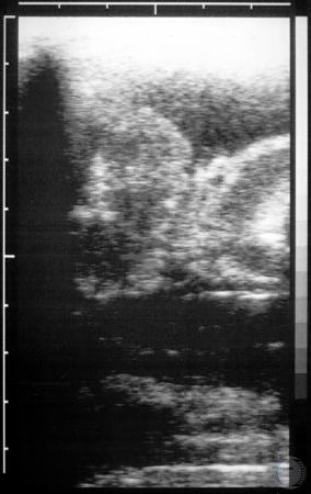
Ultrasonography - Standing.
Pregnancy diagnosis via the lower right rear flank with a 5 MHz sector scanner, with the doe in standing position.
Smith MC (2006)
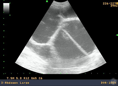
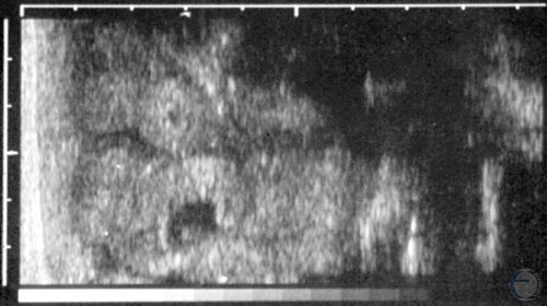
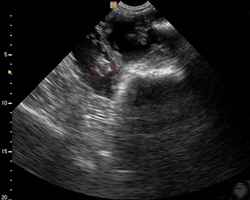
Hydrometra.
Sector scanner image of echolucent fluid accumulation in the uterus of a doe with hydrometra. Trabeculae are the result of reflections.
Smith MC (2006)
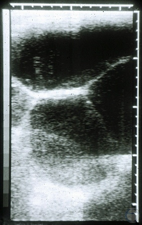
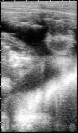
Hydrometra Fluid.
Ultrasonographic image of fluid accumulation in a doe with hydrometra. The image was made with a linear probe. The white material in the lower compartment mixed with the fluid after ballottement of the abdomen, and then settled out over time.
Smith MC (2006)
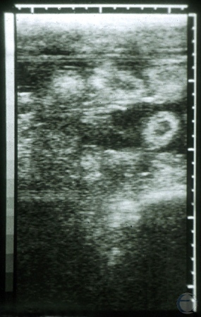
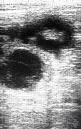
Placentomes.
The doughnut shaped structures are cup shaped placentomes of a doe in her second or third trimester of pregnancy. In sheep, the placentomes reach maximum size at 90 days of gestation.
Smith MC (2006)
Ultrasonogram at 2 Months.
Normal gestation at about two months in a Nubian goat. Amniotic fluid is present in the center surrounding part of the fetus. Placentomes off to the left. 5mHz linear array.
Pugh DG (2007)
Ultrasonogram in a Pygmy Goat.
Normal pregnancy at 35 days of gestation in a Pygmy goat with a tight compact abdomen. Tiny fetus surrounded by dark amniotic fluid.
Pugh DG (2007)
Ultrasonogram at 2 Months.
Normal gestation at about two months in a Nubian goat. Amniotic fluid is present in the center surrounding part of the fetus. 5mHz linear array.
Pugh DG (2007)
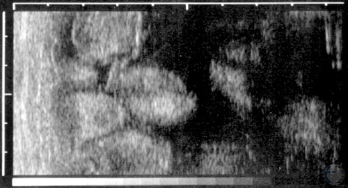
Ultrasonogram of Placentomes.
Large placentomes during the third trimester. Caruncular size reaches its maximum around 3 months of gestation. The scale along the top and the left side is in centimeters.
Pugh DG (2007)
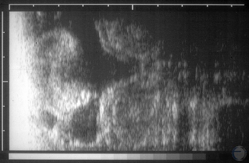
Third Trimester Placentomes.
Large placentomes during the third trimester.
Pugh DG (2007)
Ultrasonogram.
Large placentomes in a pregnant nanny. Placentomes reach their full size around three months of gestation. The scale along the top and the left side is in centimeters.
Pugh DG (2007)
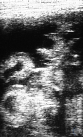
Pregnancy Diagnosis.
A fetus and small placentomes are visible outlined by black uterine fluid in this transabdominal ultrasound.
Smith MC (2010)
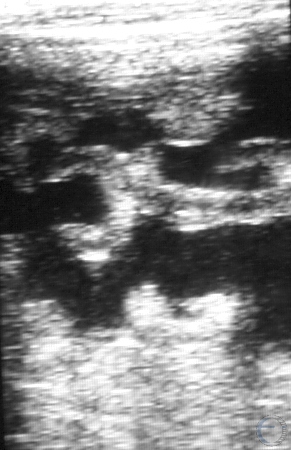
Pregnancy Diagnosis.
Donut- and cup-shaped placentomes are visible in this transabdominal ultrasound. The placentomes attain maximum size at about 90 days of gestation.
Smith MC (2010)
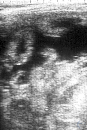
Pregnancy Diagnosis.
A prominent cup- or C-shaped placentome is visible on this transabdominal ultrasound. Placentomes are proof of pregnancy.
Smith MC (2010)
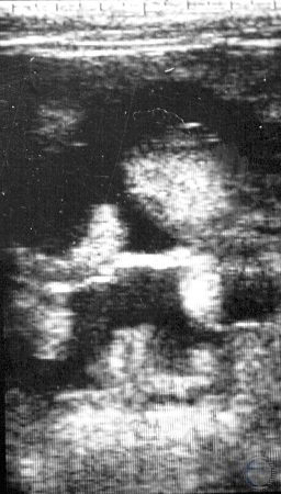
Pregnancy Diagnosis.
Depending on the plane of transection by the ultrasound beam, placentomes appear to be donut- or cup-shaped.
Smith MC (2010)
Pregnancy Diagnosis.
Placentomes visible in transabdominal ultrasound of a pregnant goat using a 5 MHz linear probe. Caruncular size reaches its maximum around 3 months of gestation. The scale across the top is in centimeters.
Smith MC (2011)
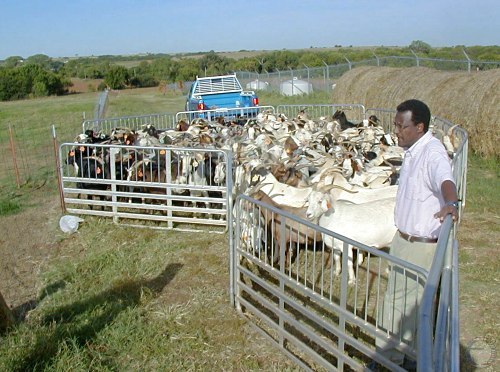
Goats Ready for Ultrasound.
Round up of female goats for pregnancy examination with ultrasound.
Prado TM (2007)
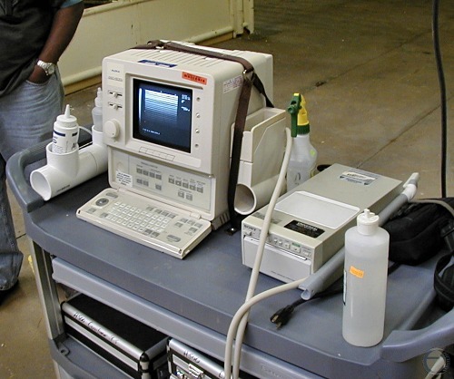
Ultrasound Unit.
Standard non-portable unit with a transducer at the end of a rectal probe.
Prado TM (2007)
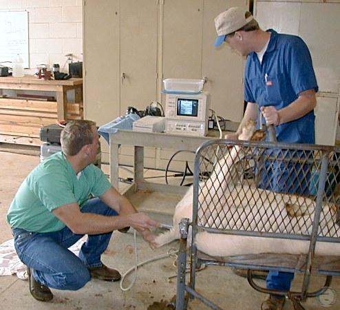
Positioning of the Patient.
Patient in dorsal recumbency. It is helpful to keep the goats off feed for 12 hours prior to the examination.
Prado TM (2007)
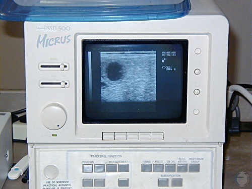
Ultrasound Unit Image.
Fetal fluids and presence of the embryo at the 9 0'clock location.
Prado TM (2007)