The Visual Guide to
Caprine Reproduction
Accidents of Gestation: Prolapsed Vagina
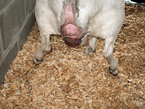
Prolapsed Vagina.
The doe is straining. The exposed vaginal mucosa is dried out and irritated.
Smith MC (2006)
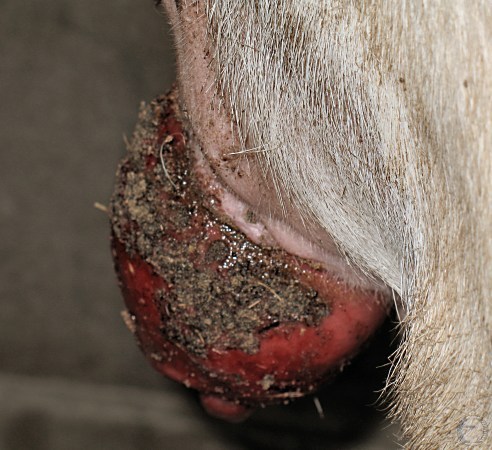
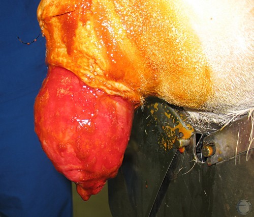
Lateral View of Prolapse.
Close-up lateral view of a traumatized and infected prolapsed vagina.
Smith MC (2006)
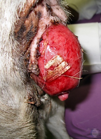
Cervico-Vaginal Prolapse.
Recurrent prolapse of the vagina. The external cervical os protrudes.
Breed M (2006)
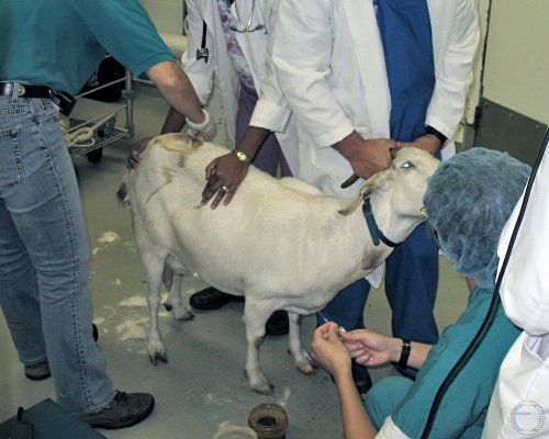
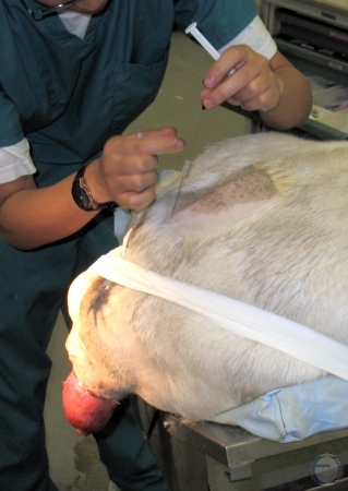
High Epidural Anesthesia.
Surgical preparation of the tail head prior to the administration of lidocaine to induce high epidural anesthesia, via the lumbosacral space.
Breed M (2006)
Lumbosacral Anesthesia.
Placement of the needle in the lumbosacral space, with the doe in sternal recumbency.
Breed M (2006)
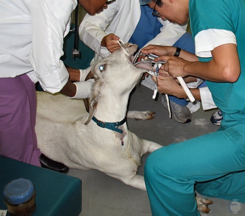
Endotracheal Intubation.
Endotracheal intubation of a pre-anesthetized doe. A goat can be somewhat tricky to intubate because it cannot open its mouth very wide to permit visualization of the rima glottis.
Breed M (2006)
Surgical Preparation.
The doe is placed in sternal recumbency and the operative area is clipped, scrubbed and disinfected.
Breed M (2006)
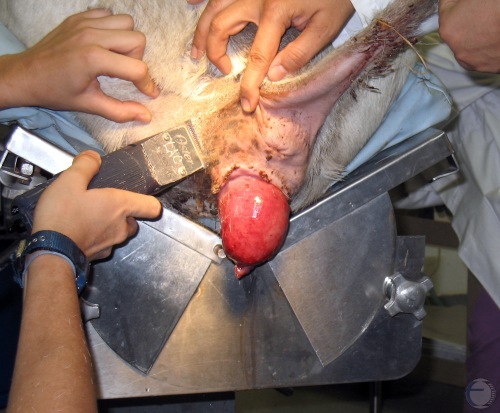
Ready for Surgery.
The prolapsed vagina and surrounding area have been prepared and disinfected.
Breed M (2006)
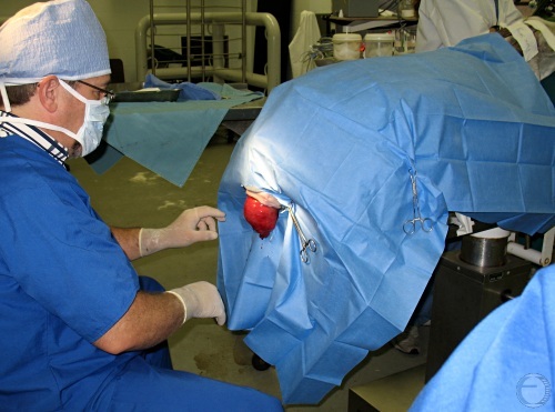
Draped for Surgery.
The doe is in dorsal recumbency, anesthetized, surgically prepared, and ready for corrective surgery.
Breed M (2006)
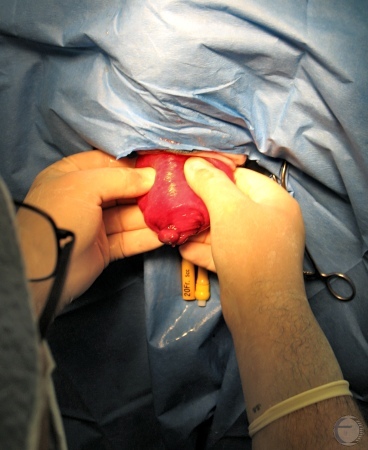
Exam of the Prolapsed Vagina.
Palpation of the prolapsed vagina and examination of the contents.
Breed M (2006)
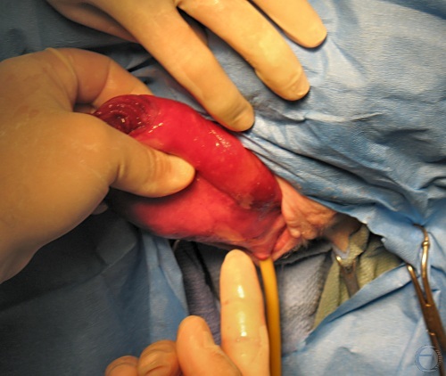
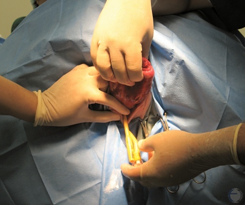
Location of the Urethral Orifice.
The external urethral opening was located underneath the prolapsed mass, and the bladder was catheterized with a Foley catheter.
Breed M (2006)
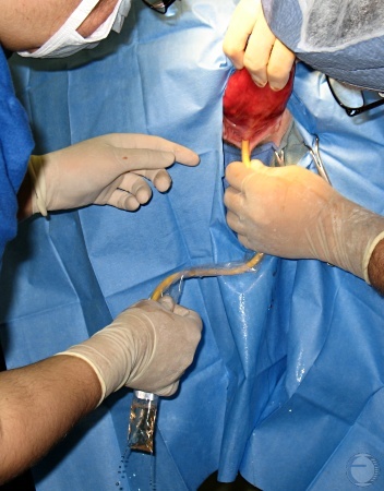
Catheterization of the Bladder.
A sterile 20-French gauge Foley catheter is inserted into the urethra.
Breed M (2006)
Drainage of the Bladder.
Urine flows freely via a balloon catheter.
Breed M (2006)
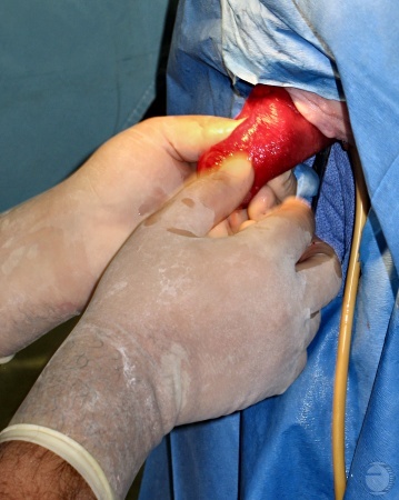
Positioning of the Prolapse.
The empty prolapsed vagina is positioned for an incision.
Breed M (2006)
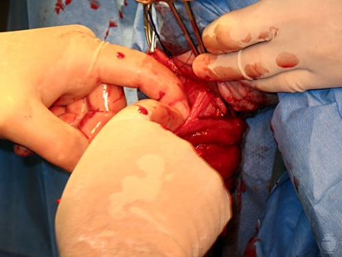
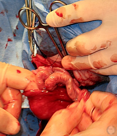
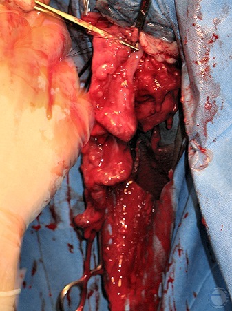
Exploration.
The contents of the prolapsed mass are being examined.
Breed M (2006)
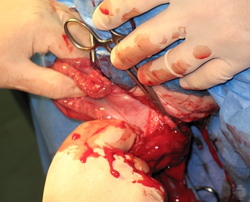
Exploration.
The uterus is identified within the incised prolapsed mass.
Breed M (2006)
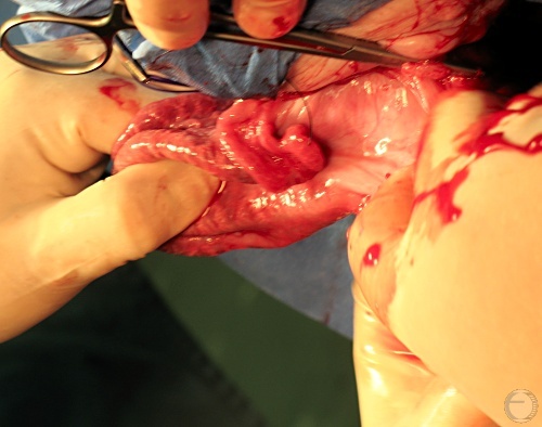
Exteriorizing the Uterus.
The uterus is pulled out through the prolapsed vagina.
Breed M (2006)
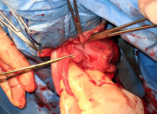
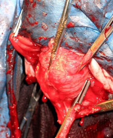
Entire Uterus Exposed.
Locating the blood supply in the broad ligament.
Breed M (2006)
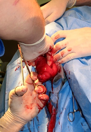
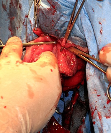
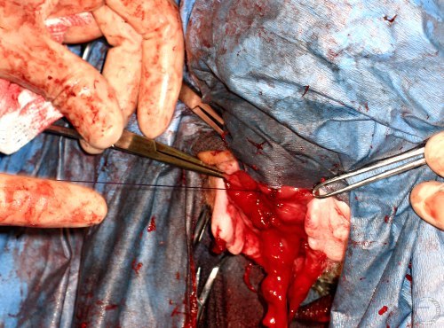
Clamping the Broad Ligament.
The broad ligament and the uterine blood vessels are being clamped.
Breed M (2006)
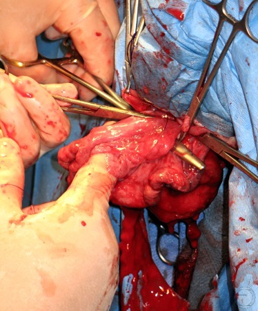
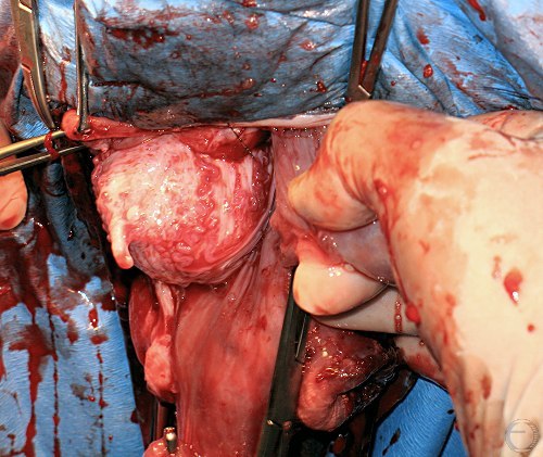
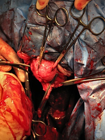
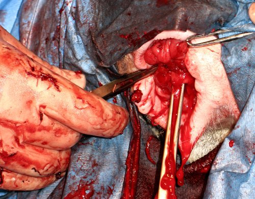
Ready to Amputate.
The isolated uterus is now ready for amputation.
Breed M (2006)
Hemostasis.
The bilateral uterine blood supply has been clamped and ligated.
Breed M (2006)
Exposure of the Bladder.
Exposure of the bladder via the incision in the prolapsed vagina.
Breed M (2006)
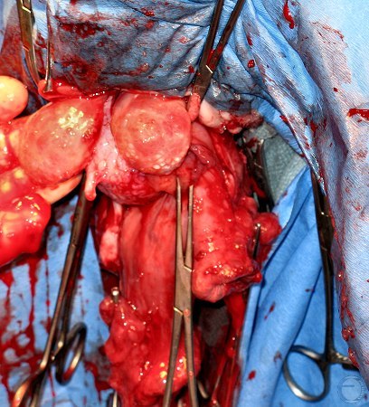
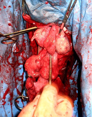
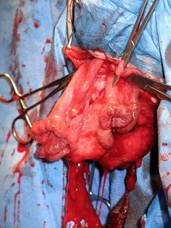
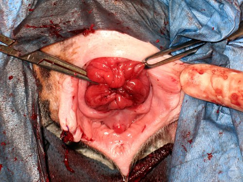
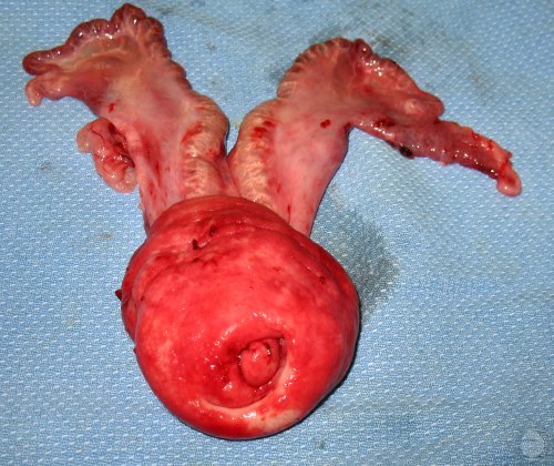
Uterus and Ovaries.
The complete uterus and both ovaries, and the bladder, are exposed via the incision in the prolapsed mass.
Breed M (2006)
Ready to Ligate.
The uterus and the prolapsed cervico-vaginal mass have been removed. The bladder is ready to be returned to the abdomen after the various blood vessels have been ligated.
Breed M (2006)
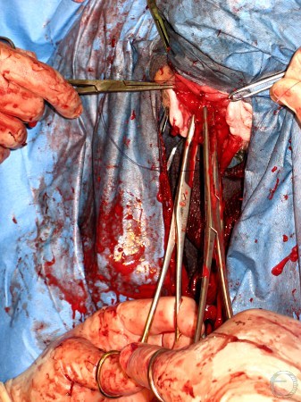
Ligation of the Stump.
Ligation of the bilateral uterine arteries, potentially significant bleeders.
Breed M (2006)
Ready to Release Stump.
The stump is now ready to be returned to the remaining portion of the vagina.
Breed M (2006)
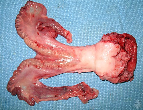
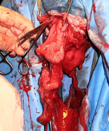
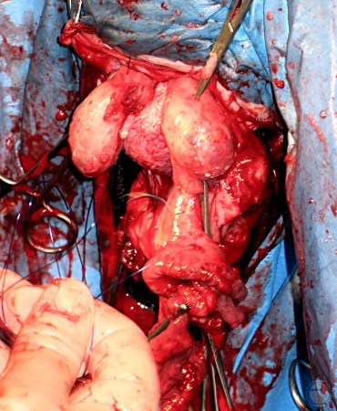
Removed Cervicovaginal Mass.
External cervical os, prolapsed portion of the vagina, and the uterus were all removed in one piece.
Breed M (2006)
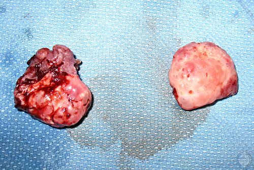
Removed Ovaries.
Both ovaries contained multiple cystic follicles ranging in size from 2 to 20 mm in diameter. The ovarian stroma was markedly congested and edematous.
Breed M (2006)
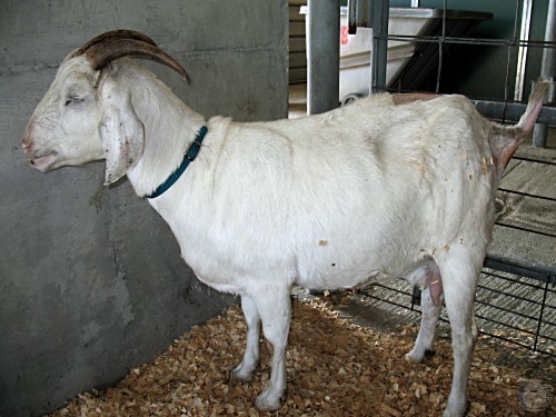
Patient after Operation.
The 6-year old, 75 kg, pluriparous Nubian crossbred doe after the surgery.
Breed M (2006)
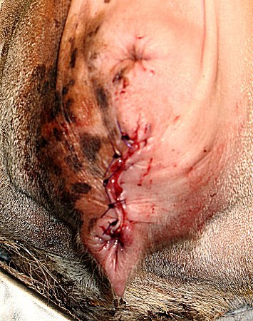
Caslick Closure.
The vulva was closed with a Caslick operation. The ventral tip of the vulva was left open to permit urination.
Breed M (2006)