The Visual Guide to
Caprine Reproduction
Male Reproductive System: Testis and Epididymis
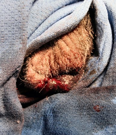
Sperm Granuloma.
Sperm granuloma in the head of the left epididymis. Extraductal spermatozoa are very irritating and immunogenic, which leads to the formation of granulomata. The buck was genetically polled.
Smith MC (2006)

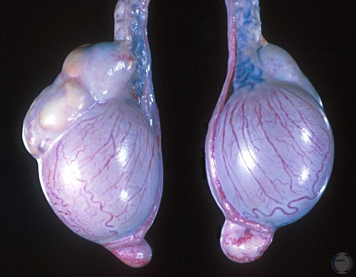
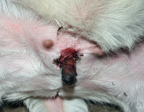
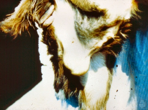
Cross Section of Granuloma.
The testis and the head of the epididymis have been transsected to demonstrate the normalcy of the testis and the formation of a sperm granuloma in the head of the epididymis.
Smith MC (2006)
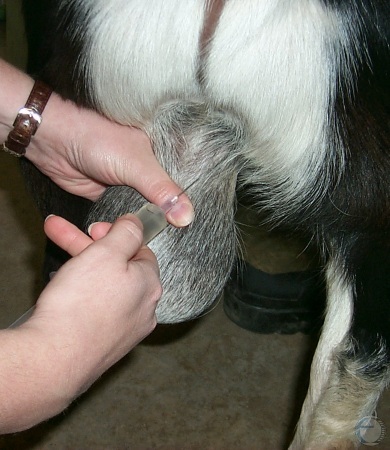
Local Anesthesia.
Each spermatic cord is injected with 1% Lidocaine to produce local anesthesia prior to castration. The total amount of lidocaine used should probably not exceed 5 mg/kg, to avoid convulsions from lidocaine toxicity.
Smith MC (2006)
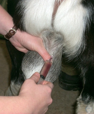
Blood from Spermatic Vessels.
Prior to injection of the Lidocaine the anesthetist should aspirate to check for the presence of blood which would indicate that the needle is in a blood vessel. If blood is obtained the needle should be relocated prior to injection.
Smith MC (2006)
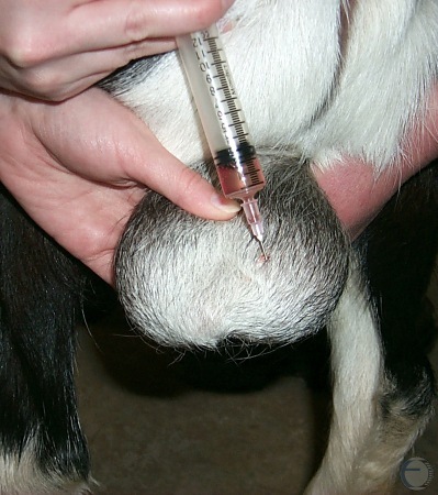
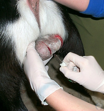
Local Infiltration.
Local infiltration of the incision site at the bottom of the scrotum, prior to castration.
Smith MC (2006)
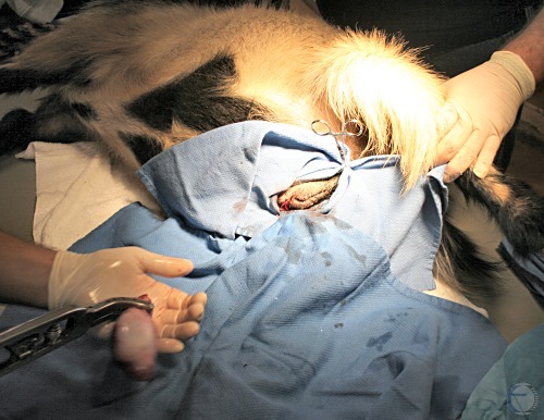
Surgical Castration.
Two longitudinal incisions are made on the bottom of the scrotum.
Smith MC (2006)
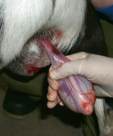
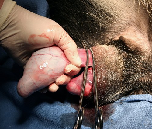
Isolated Testicle.
The testis, still within the tunics, has been freed from the scrotum, and the spermatic cord is ready to be clamped. Hemorrhage in the cord is the result of lidocaine injection.
Smith MC (2006)
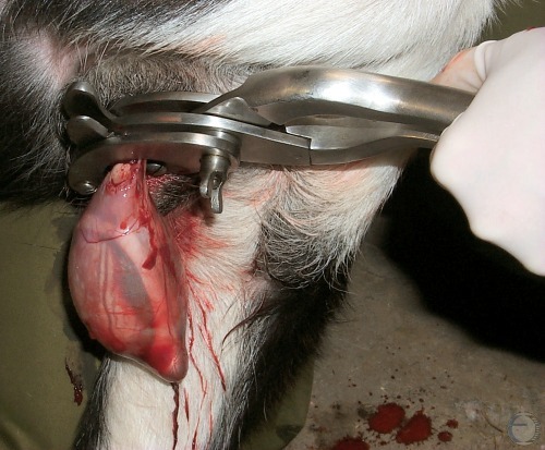
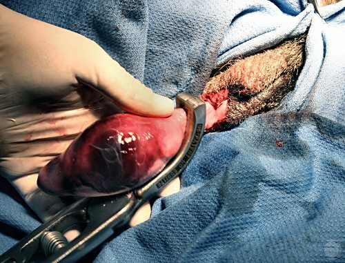
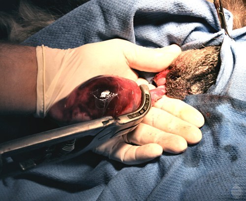
Emasculator Applied.
The jaws of the emasculator are placed on the spermatic cord with the "nut towards the nut" (the wing nut of the instrument towards the testicle). This orientation ensures that the proximal end of the cord is crushed and the distal end is severed, to prevent bleeding. Because of the large size of caprine testes, an equine emasculator is needed.
Smith MC (2006)
Preparation for Castration.
Positioning of the clean scrotum prior to castration.
Smith MC (2010)
Ligation of the Blood Supply.
Two clamps are placed on the spermatic cord to control hemorrhage during castration.
Smith MC (2010)
Application of Henderson Tool.
The Henderson tool is placed on the spermatic cord in preparation for removal of the testicle.
Smith MC (2010)
Close-up of Henderson Tool.
Close-up of the Henderson tool placed on the spermatic cord. The testicle and cord have been crushed and then twisted multiple times by the drill action of the tool until the tissues separate.
Smith MC (2010)
Henderson Tool Castration - Testicle Separated.
Rotating the Henderson tool has detached the testicle with no hemorrhage in this castration.
Smith MC (2010)
Scrotal Wound after Henderson Tool Castration.
No hemorrhage occurs with this castration technique.
Smith MC (2010)
Elastrator Band Failure.
The elastrator band used to castrate this buckling failed before the blood supply to the deeper tissues of the scrotum was completely interrupted, leaving viable tissue that needed to be surgically removed.
Smith MC (2010)
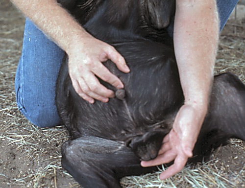
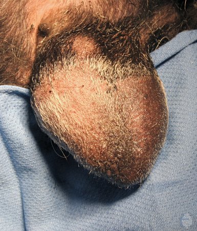
Testicular Hypoplasia.
The testes are small for the size and age of the buck. The consistency is soft.
Pugh DG (2007)
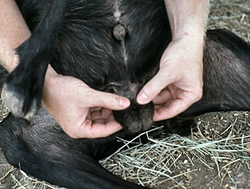
Testicular Hypoplasia.
Small, soft, symmetrical testes, too small for the size of the buck, indicating bilateral hypoplasia.
Pugh DG (2007)
Epididymitis.
Both testes are necrotic after Burdizzo castration at too advanced an age for complete resorption to occur. Blood supply to the epididymis survived the two crimps, which are visible in the spermatic cords.
Smith MC (2006)