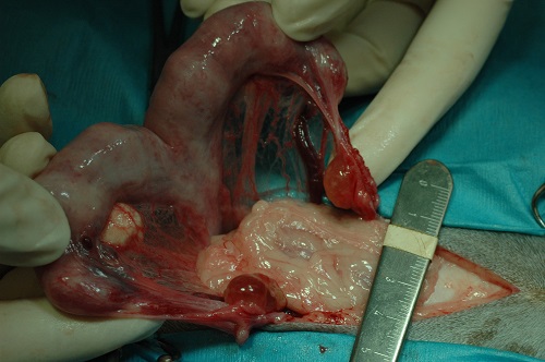
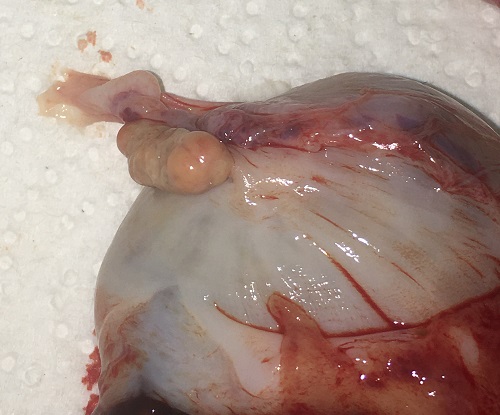
Ovarian Cysts.
This is a case of an enlarged thickened uterus and bilateral ovarian cysts, observed at the time of an ovariohysterectomy.
Johnson AK (2021)
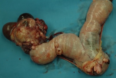
Granulosa Cell Tumor.
This female cat was diagnosed with a granulosa cell tumor. Although rare in cats, this ovarian neoplasia was associated with multiple other nodules throughout the uterus and a thickening of the endometrium.
Johnson AK (2021)
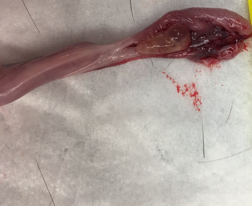
Normal Feline Ovary with Follicles.
The feline ovary is not located within a bursa like the canine. It is easily seen at the tip of each horn.
Johnson AK (2021)
Normal ovary with follicles showing follicular development.
The feline ovary is not located within a bursa like the canine. It is easily seen at the tip of each horn. During estrus, multiple small (2-5 mm) follicles will be present on both ovaries. Cats are induced ovulators. When mature follicles like these are present and a queen is mated LH is released and ovulation occurs within 48 hours later. It requires multiple mating episodes to induce ovulation.
Johnson AK (2021)
Normal Corpora Lutea.
Normal corpora lutea in a pregnant queen. There are usually 2-5 corpora lutea on each ovary and they range 3-5 mm in diameter.
Johnson AK (2021)
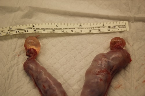
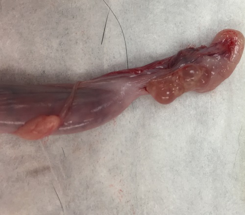
Follicular ovarian cysts located on both ovaries.
Follicular cysts are enlarged follicles that are not fertile. They often occur in conjunction with other pathology and they cause infertility. This queen had a concurrent pyometra (notice the enlarged/thickened uterine horns).
Johnson AK (2021)
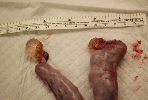
A large cystic follicle is present on both ovaries.
Also present at the base of each cyst on each ovary are multiple CL from her last ovulatory cycle (small dark yellow colored nodules). The uterus is enlarged due to pyometra.
Johnson AK (2021)