Postcervical Fold.
The post-cervical vaginal fold may obstruct the view of the external cervical os of the cervix during vaginoscopy. It may also interfere with the placement of a catheter into the cervix.
Shille VM (1980)
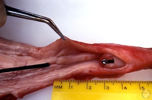
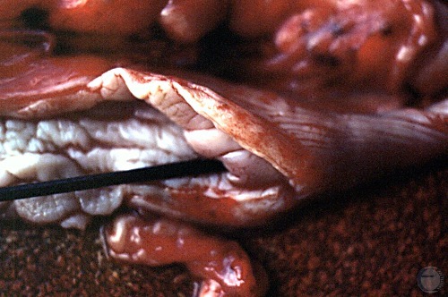
Cervix Traversed.
A small back rod has been inserted through the cervical canal from the uterine lumen into the fornix.
Shille VM (1980)
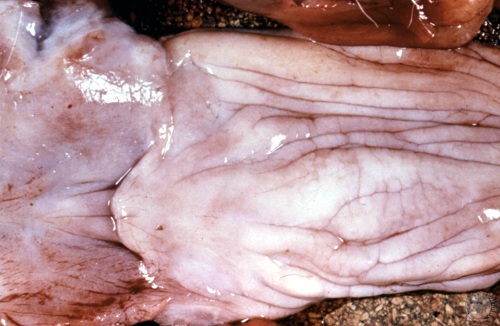
Vestibulovaginal Junction.
Junction of the vagina and the vestibule in the area of the hymen.
Shille VM (1980)
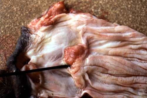
Transmissible Venereal Tumor.
A Transmissible Venereal Tumor (TVT) is located at the vestibulovaginal junction near the urethral meatus.
Shille VM (1980)
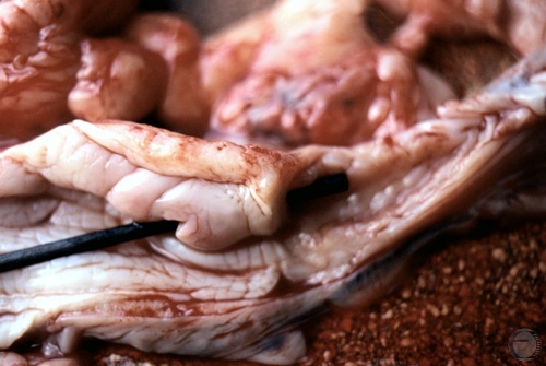
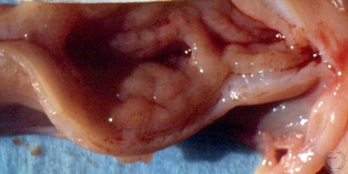
External Cervical Os.
External cervical os. Note the pronounced longitudinal cervical fold.
Shille VM (1980)
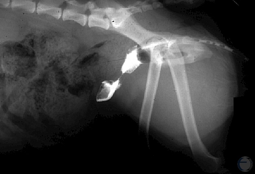
Constricted Cervix.
Infusion of a radio-opaque solution into the anterio vagina demonstrates a severely constricted cervical lumen.
Verstegen J (2009)
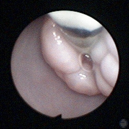
View of the Cervix by Vaginal Endoscopy.
View of the cervical os during a transcervical intrauterine insemination in the dog.
Verstegen J (2009)