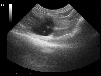
Ovarian Cysts.
Ultrasound image of the right ovary containing multiple ovarian cysts shown by the black, anechoic circular structures surrounded by gray soft tissue.
Johnson AK (2021)
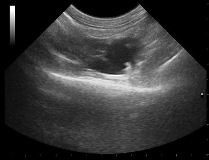
Ovarian Cysts.
Ultrasound image of the left ovary containing multiple ovarian cysts shown by the black, anechoic circular structures surrounded by gray soft tissue.
Johnson AK (2021)
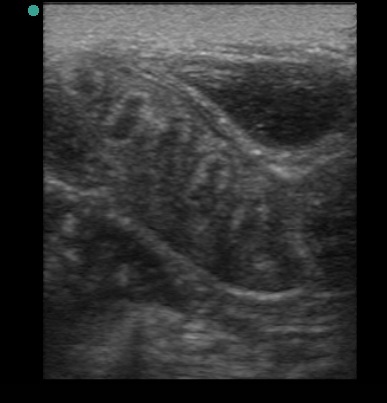
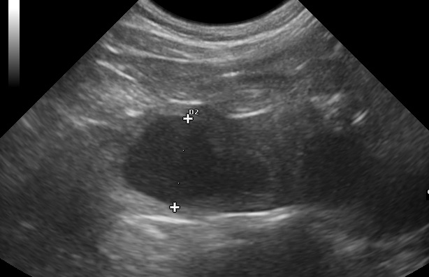
Cystic endometrial hyperplasia.
Ultrasound image of a 5-year old queen that presented for infertility. Ultrasound showed moderate cystic endometrial hyperplasia (CEH) with prominent endometrial folds (light and dark striping throughout the uterus).
Johnson AK (2021)
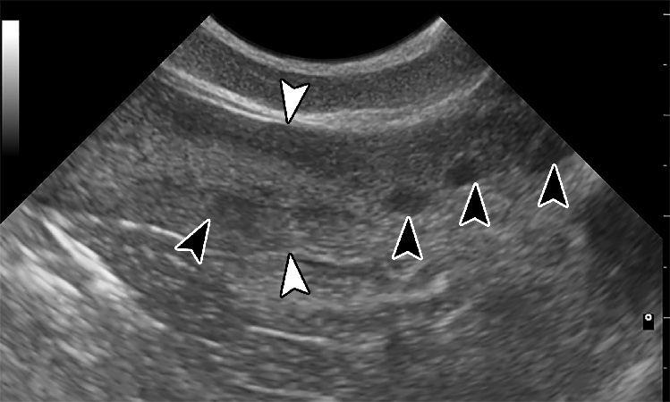
Queen with Cystic endometrial hyperplasia.
Ultrasonographic image of a queen with cystic endometrial hyperplasia (CEH). The uterus is marked with white arrows. The endometrium is thickened and multiple small cysts are visualized with in the lumen (gray arrows).
Johnson AK (2021)
Ultrasound image of the uterus of a queen with pyometra.
The loops of distended uterus measured 1.36cm. Two other distended loops can be seen to the right of the largest loop.
Johnson AK (2021)