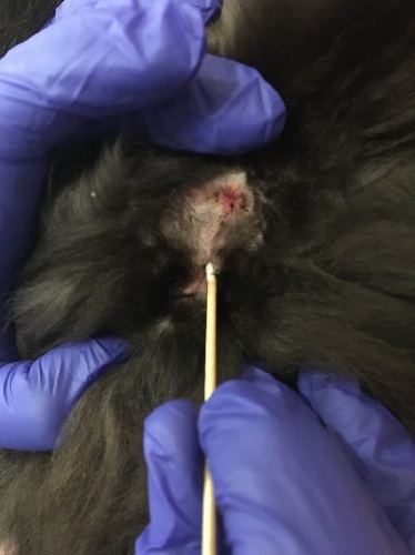
Vaginal cytology in a queen.
Vaginal cytology in a queen. Place a sterile moistened cotton applicator into the vulva to the level of the cotton. Rotate the cotton to the left and right to exfoliate cells. Roll the cotton tip onto a slide and stain for analysis.
Johnson AK (2021)
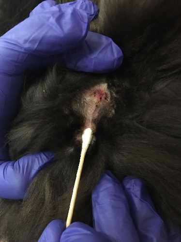
Vaginal cytology in a queen.
Vaginal cytology in a queen. Place a sterile moistened cotton applicator into the vulva to the level of the cotton. Rotate the cotton to the left and right to exfoliate cells. Roll the cotton tip onto a slide and stain for analysis.
Johnson AK (2021)
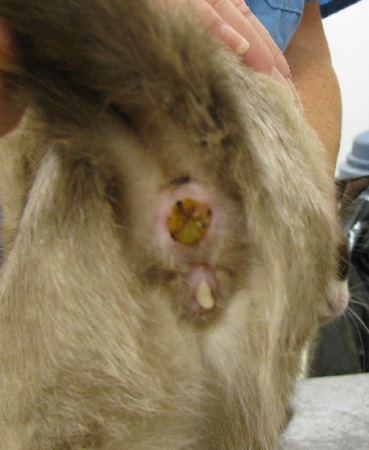
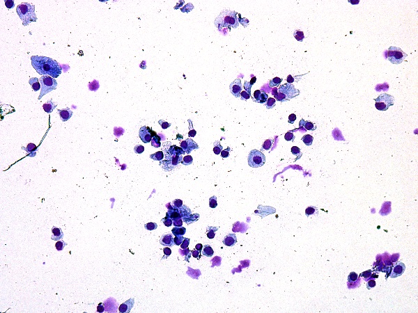
Vaginal discharge.
Vaginal discharge in a queen with an open pyometra.
Johnson AK (2021)
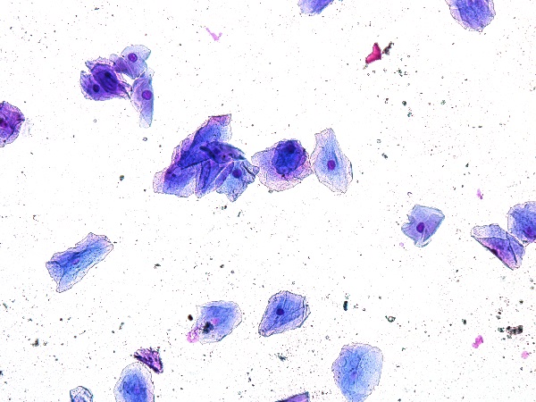
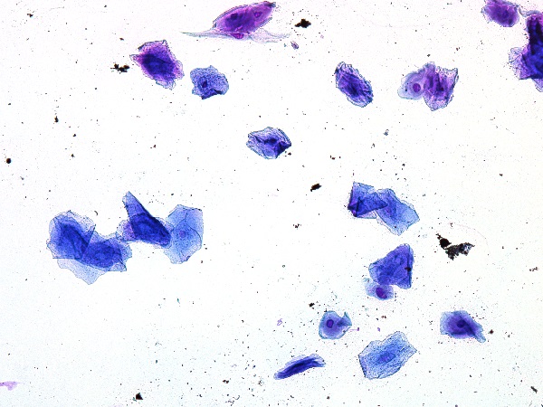
Vaginal cytology.
Vaginal cytology of a queen in estrus. Greater than 50% of the cells are cornified (95% in this image). Many retain their nucleus, but the nucleus is small and pyknotic. In the queen, red blood cells and white blood cells are not typically seen on vaginal cytology.
Johnson AK (2021)
Vaginal cytology.
Vaginal cytology of a queen in mid estrus. This queen has mostly cornified epithelial cells and most lack their nucleus.
Johnson AK (2021)
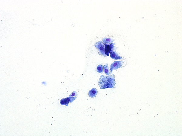
Vaginal cytology.
Vaginal cytology of a queen in interestrus. Note the cornified cell intermixed with intermediate and parabasal cells. The percentage of cornified cells would be less than 50%.
Johnson AK (2021)
Vaginal cytology.
Vaginal cytology of a queen in diestrus. This cytology is characterized by small intermediate and parabasal cells.
Johnson AK (2021)