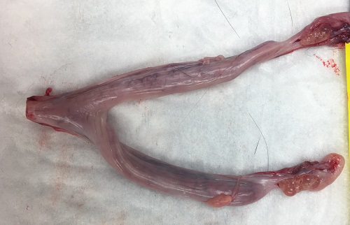
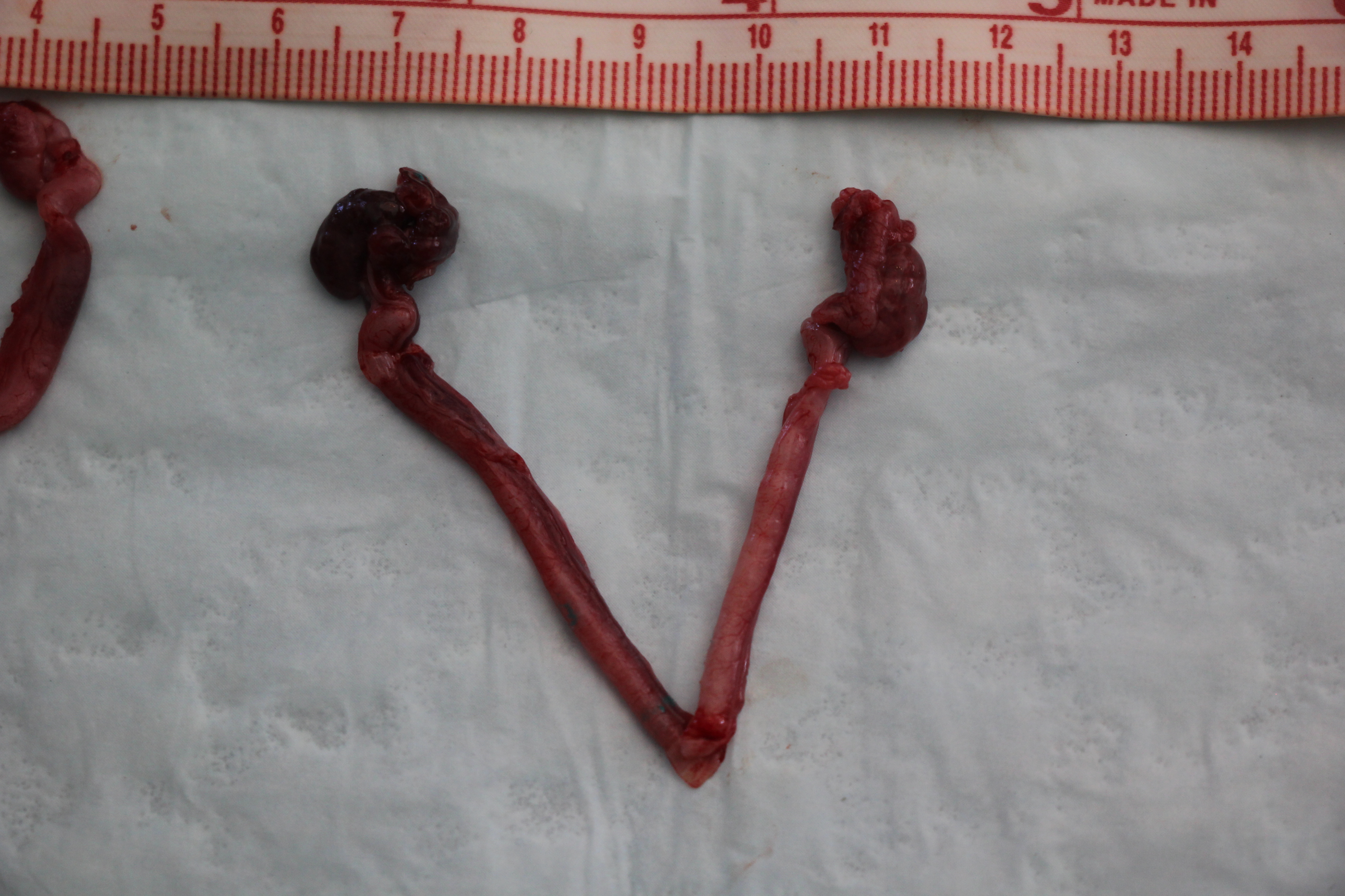
Normal bicornuate uterus in a multiparous cat after ovariohysterectomy.
Multiple follicles are present on both ovaries indicating the queen was in estrus.
Johnson AK (2021)
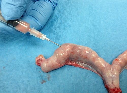
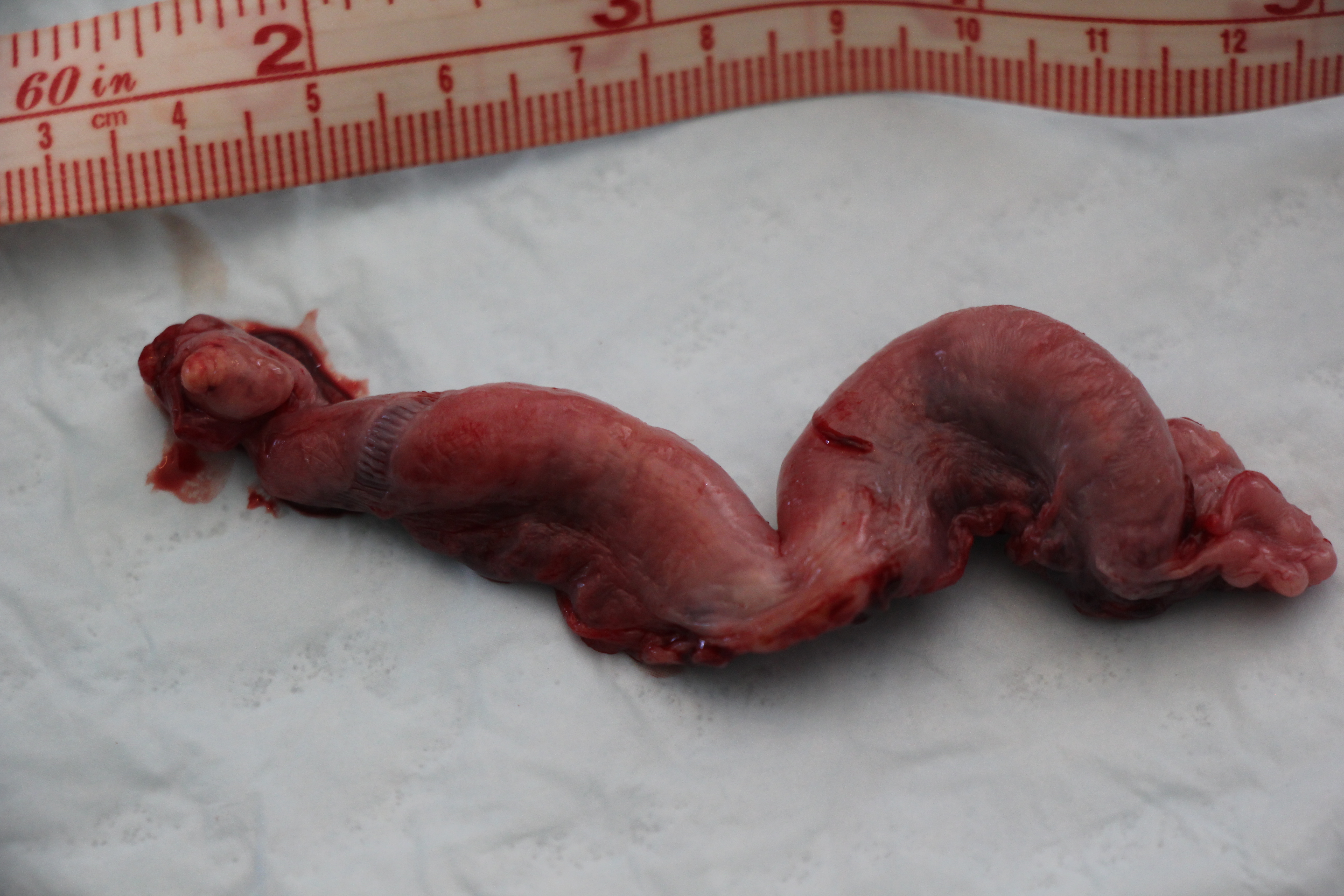
Pyometra in a queen.
The uterus is segmented and not uniformly enlarged throughout which is typical in the queen. Purulent material is being aspirated from the lumen. The queen presented with a vaginal discharge but was otherwise not ill.
Johnson AK (2021)
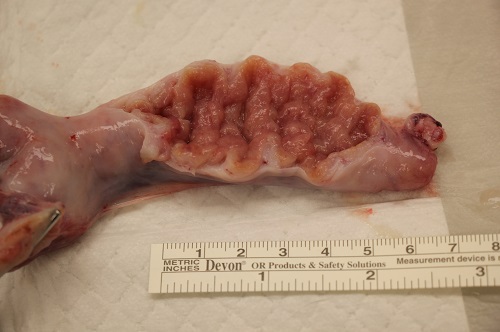
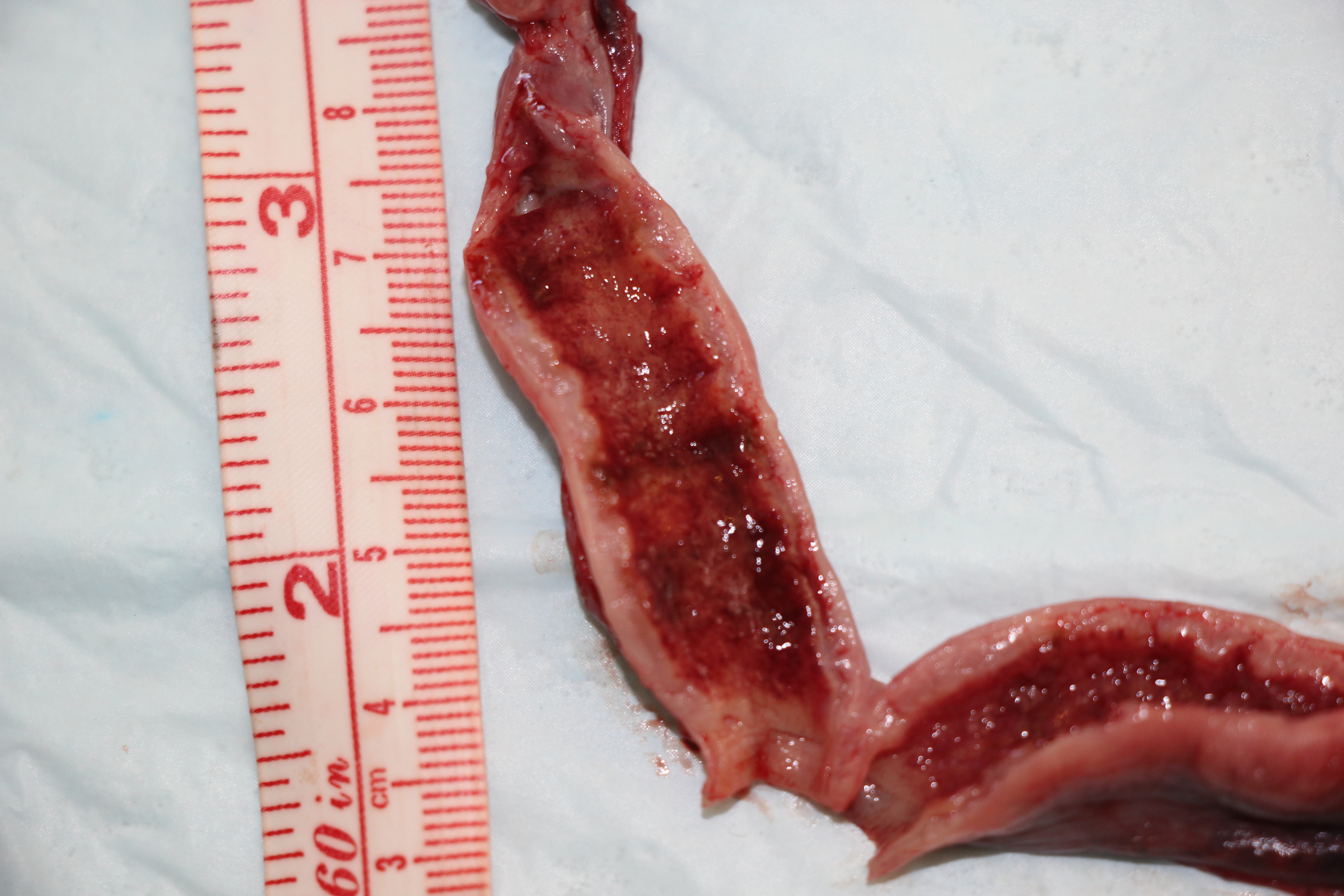
A uterus in a pluriparous queen.
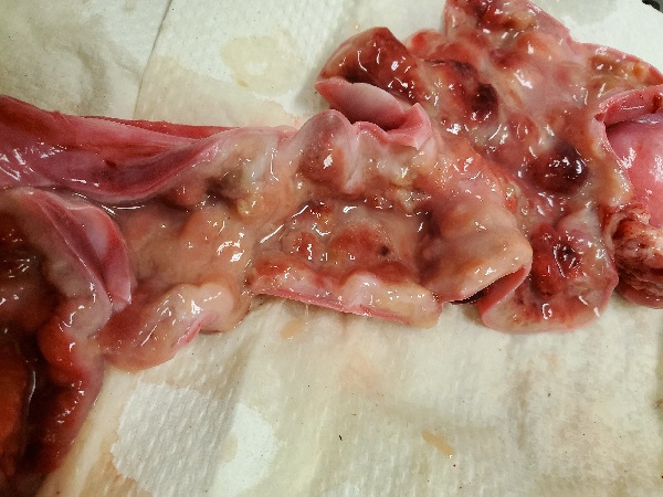
Endometrial hyperplasia. The endometrial folds are thickened and prominent causing enlargement of the uterus.
Johnson AK (2021)
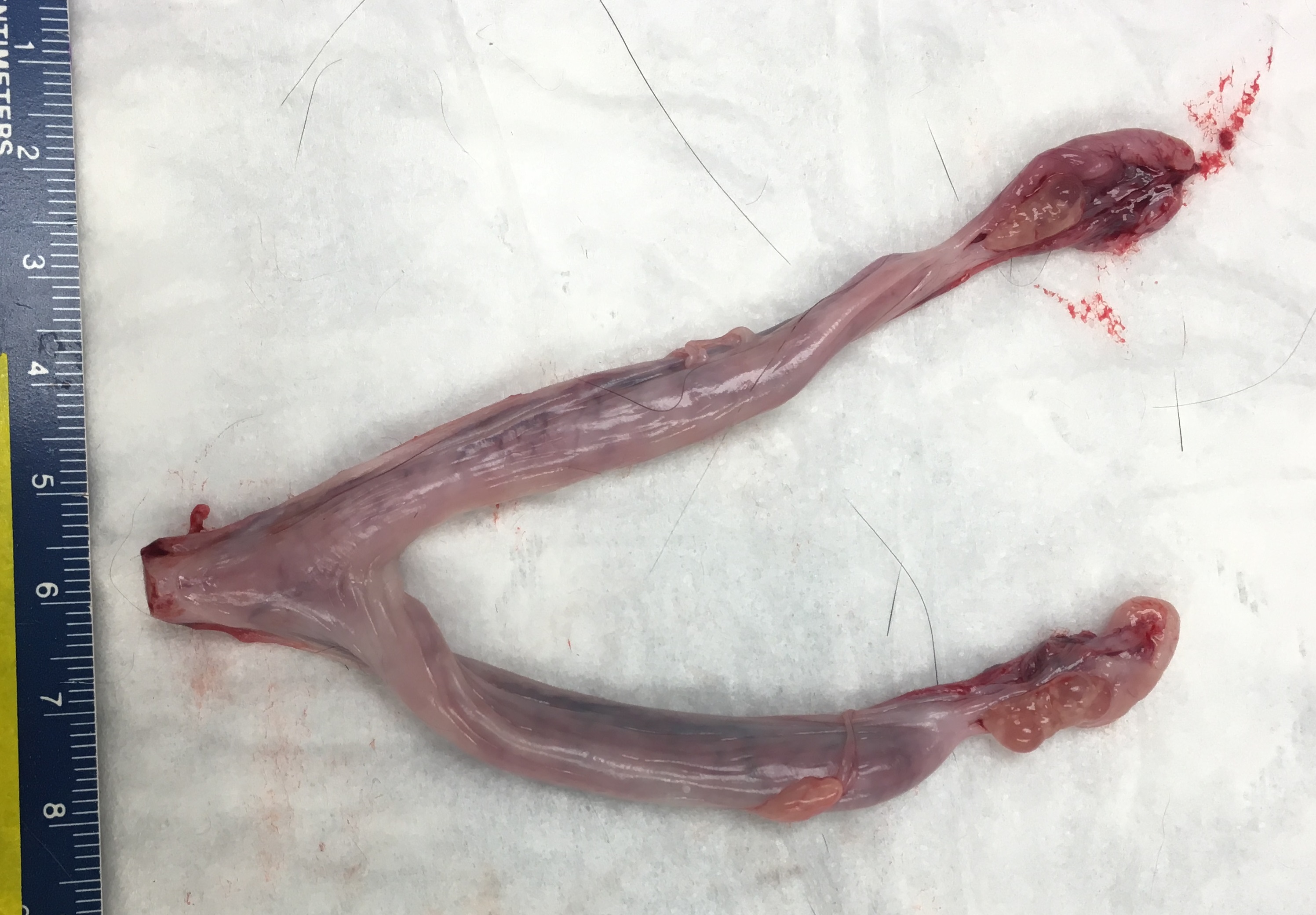
Normal Uterus
Normal adult uterus during estrus. Bicornuate tract, tract was transected at level of cervix when ovariohysterectomy was performed.
Johnson AK (2021)
Normal Uterus.
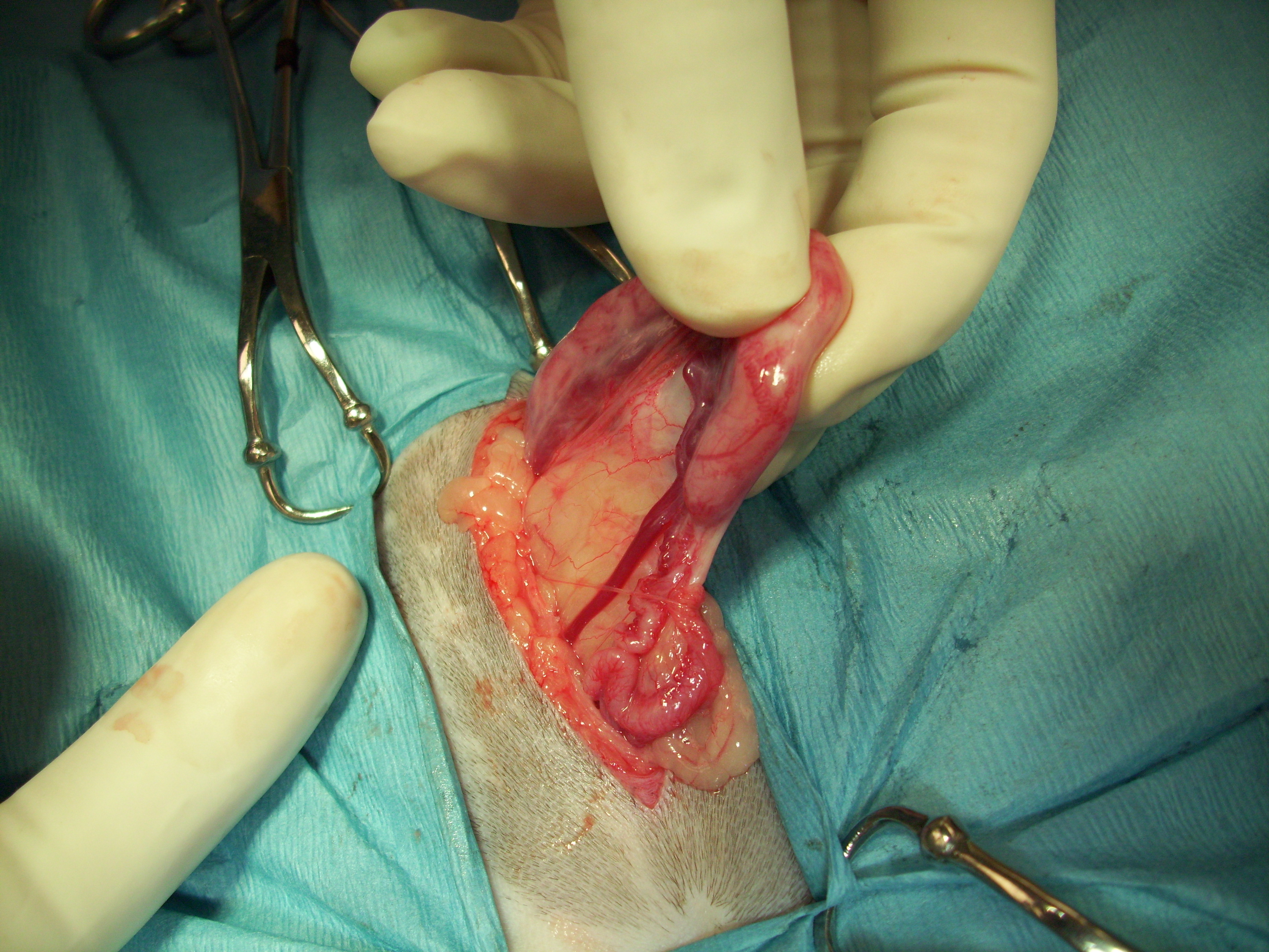
Normal uterus and ovary showing mesovarium connecting uterus to ovary and broad ligament connecting the ovary to the body wall.
Johnson AK (2021)
Normal Uterus.
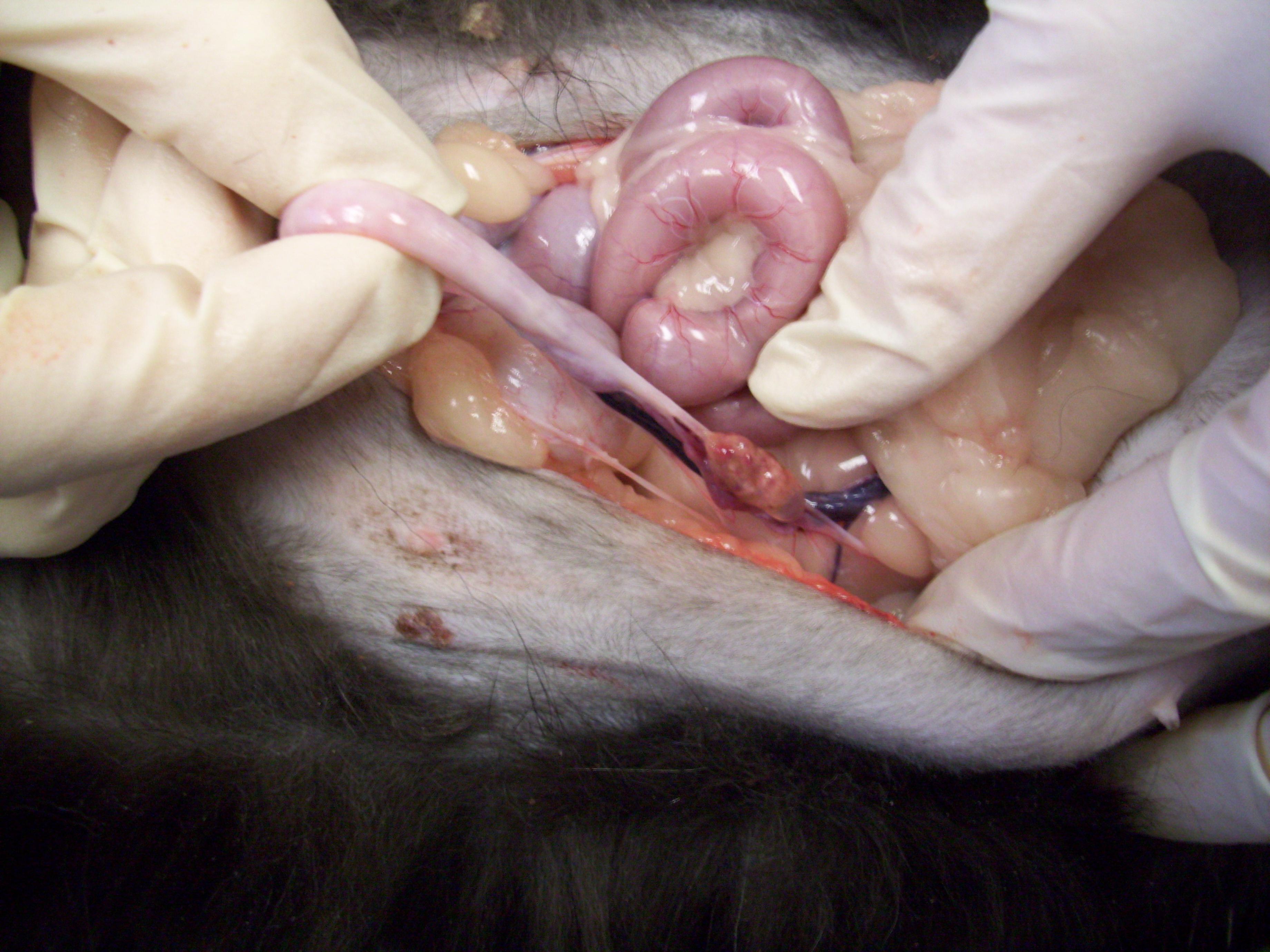
Normal adult uterus in estrus (to right of the picture). Oviduct with prominent Uterine Artery beneath the oviduct.
Johnson AK (2021)
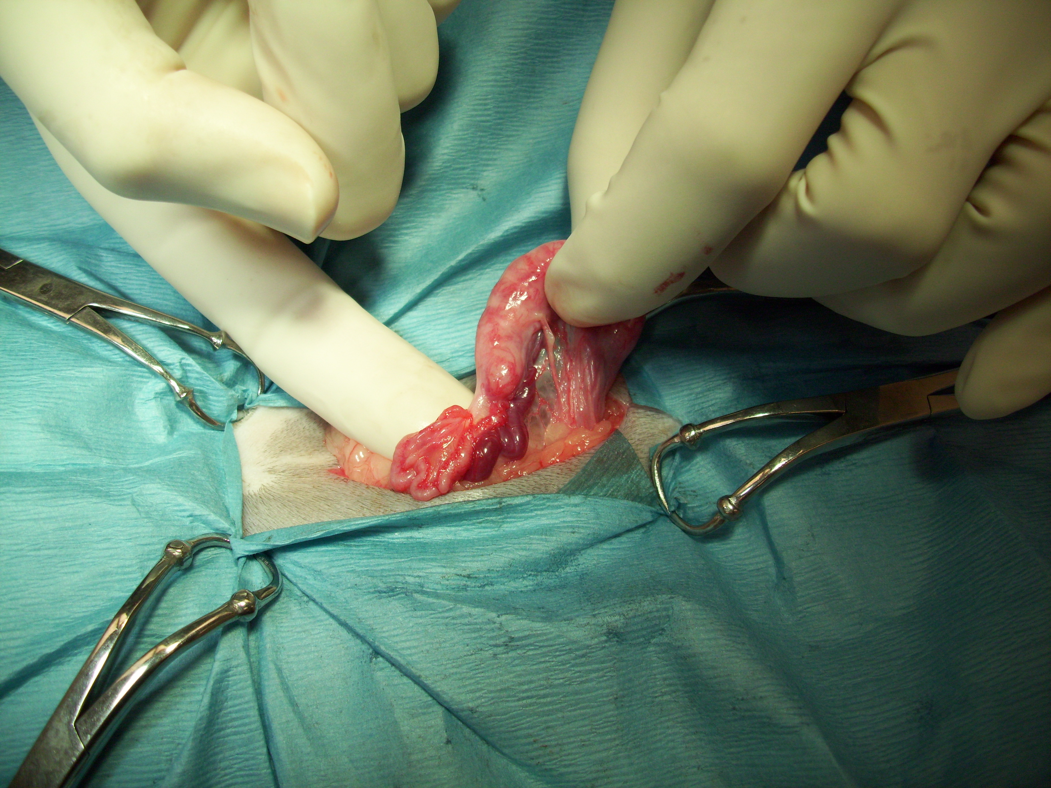
Uterine Horn.
Uterine horn (in hand) at surgery, Ovarian artery seen coursing ventrally below oviduct and uterine horn. Ovary not shown.
Johnson AK (2021)
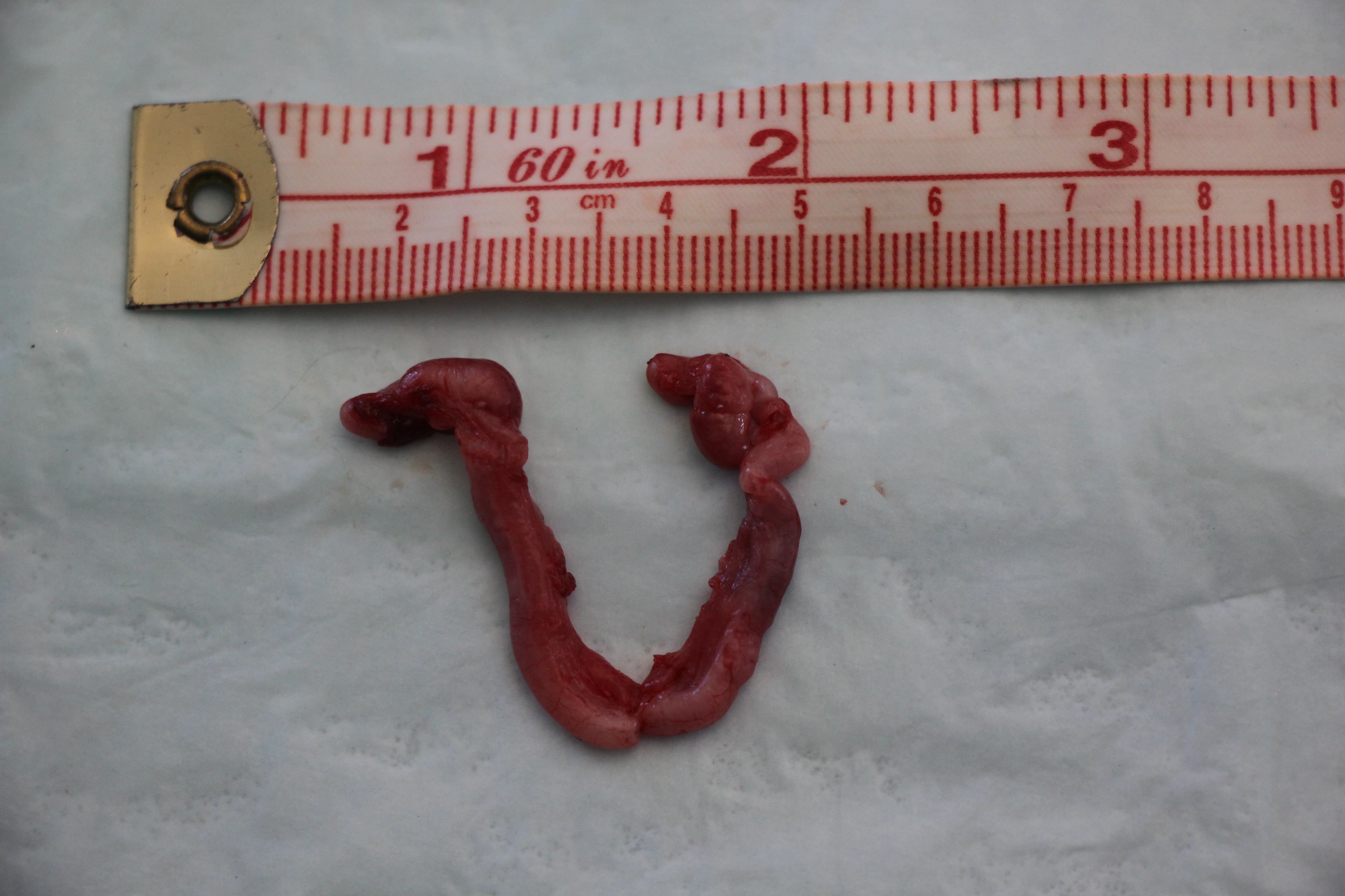
Normal primiparous (immature) uterus from a 2-month old kitten.
Normal immature uterus and ovaries from a 2 month old kitten.
Johnson AK (2021)
Primiparous Uterus
Normal immature uterus and ovaries from a 4-5 month old kitten.
Johnson AK (2021)
Primiparous Uterus
Endometrial surface of the same uterus showing endometrial folds.
Johnson AK (2021)
Postpartum Uterus
Involuting postpartum uterus following ovariohysterectomy.
Johnson AK (2021)
Postpartum Uterus
Endometrial surface of a late postpartum uterus. Note the remnants of the placental sites (dark stripes).
Johnson AK (2021)
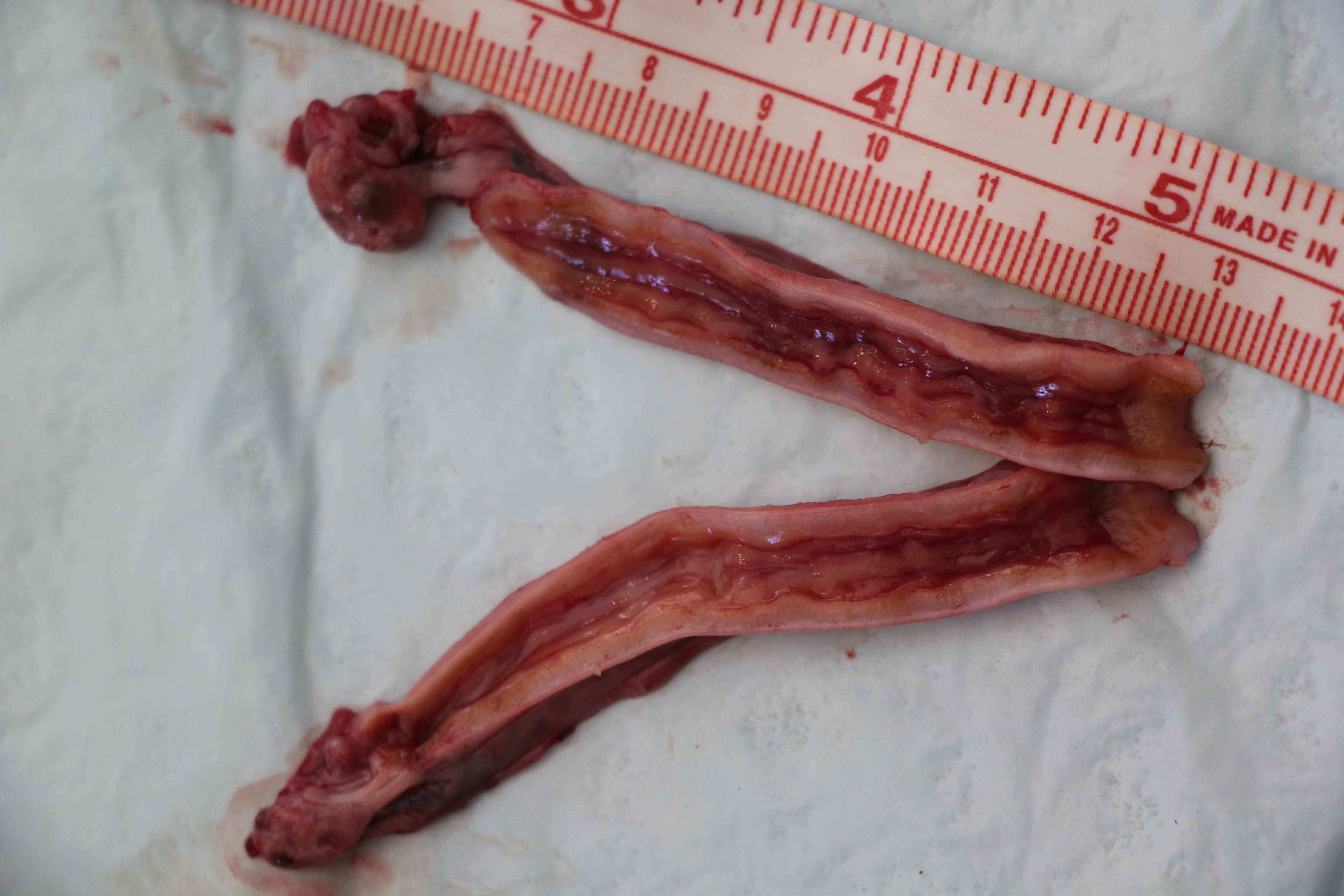
Surgical treatment of a pyometra
Cut section of uterus following surgical treatment of a pyometra. Cystic endometrium is noted throughout the endometrial surface.
Johnson AK (2021)
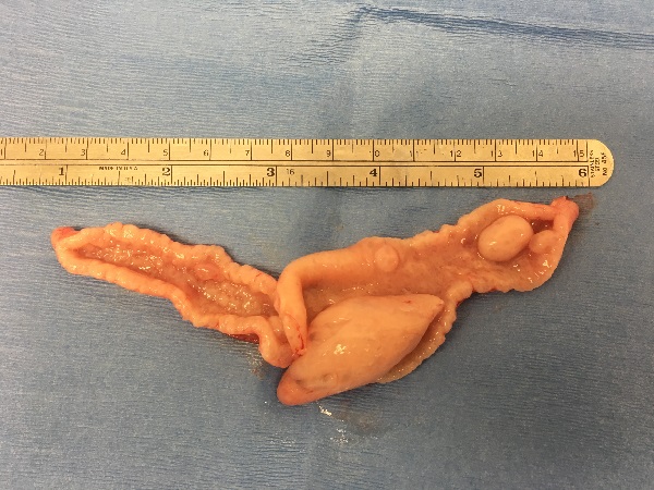
Uterine polyps
A uterus removed from a subfertile queen that was diagnosed with a uterine mass by ultrasound. Several endometrial polyps are visible protruding into the uterine lumen. On histopathology, the masses were confirmed to be cystic endometrial hyperplasia.
Johnson AK (2021)
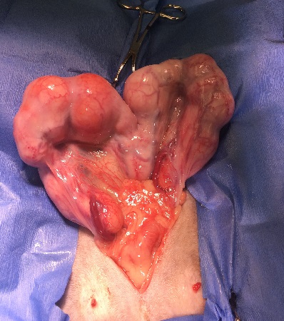
A queen with pyometra at surgery.
A queen with pyometra at surgery. The fluid filled uterus is not uniformly filled, rather segmental as is typical in the cat.
Johnson AK (2021)
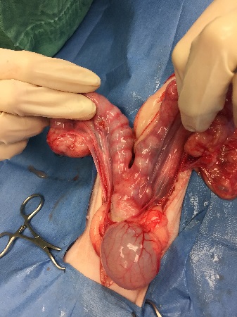
Queen during surgery with suspected pyometras
An 11-year-old queen presented for bloody vaginal discharge and suspected pyometra. During surgery, the uterus contained multiple masses confirmed to be fibrosarcoma via histopathology.
Johnson AK (2021)
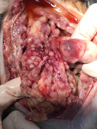
Queen with metastasis in the abdomen
The same cat in the previous picture at necropsy showing the degree of metastasis within the abdomen.
Johnson AK (2021)