Proestrous Uterus.
Normal uterus from an animal in proestrus. Old, pale corpora lutea and hyperemic follicles are present on the ovaries.
Evans LE (2009)
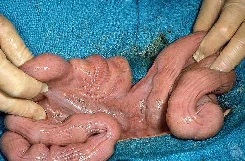
Estrous Uterus.
Regressing corpora lutea, large follicles. Sow had been in estrus for 24 hours. Notice the normal tortuous cervix which has been opened up in this specimen. The empty bladder is off to the right.
Roberts SJ (1973)
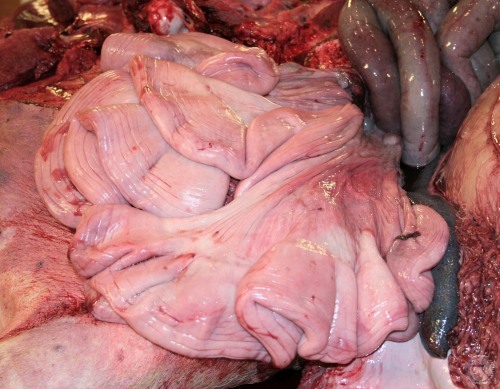
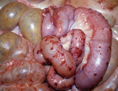
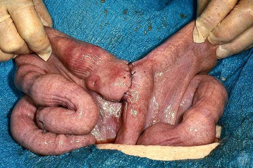
Firm Estrus Uterus.
Uterus of a sow in heat. Some of the large follicles are hyperemic. There are some corpora albicantia of the previous cycle. The fimbriae have been laid out.
Evans LE (2009)
Diestrous Uterus.
Uterus and ovaries 6 to 7 days after ovulation. Mature corpora lutea. Vascularization of the uterine horns is nicely illustrated by the prominent blood vessels in the broad ligament.
Evans LE (2009)
Midcycle Uterus.
Uterus and ovaries of a sow in diestrus. Mature corpora lutea are present.
Evans LE (2009)
Anestrous Tract of a Gilt.
Anestrous, nulliparous tract. Ovaries have small follicles but no CLs. Note the mesonephric cyst in the fimbrae of the left ovary (an embryonic remnant of the male duct system).
Evans LE (2009)
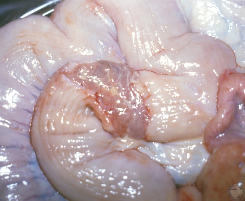
Involuting Uterus.
Normal involuting uterus of a sow with gastric torsion at 3 days after farrowing.
Smith MC (2011)
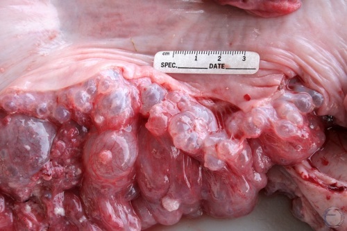
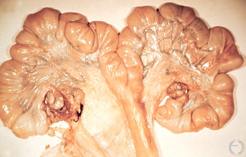
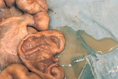
Endometrial Hyperplasia.
Endometrial hyperplasia in an 8-year old sow with cystic ovaries.
Smith MC (2011)
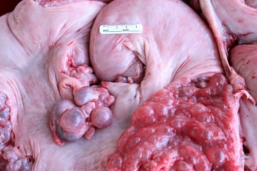
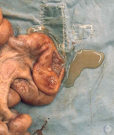
Endometrial Hyperplasia.
Endometrial hyperplasia in an 8-year old sow with cystic ovaries.
Smith MC (2011)
Peritonitis in a Sow.
Signs of adhesions between the uterus and the intestines. Cause was probably uterine infection.
Shipley C (2006)
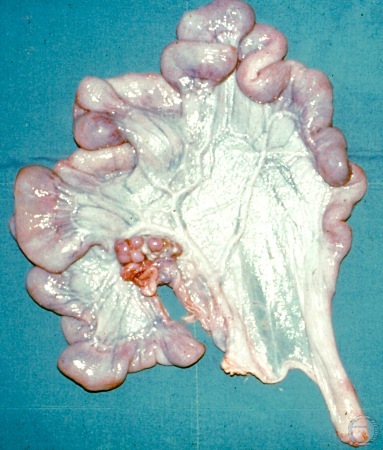
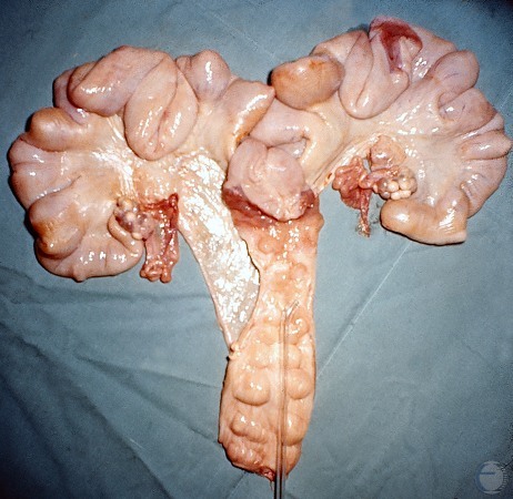
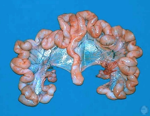
Uterus Unicornis.
Uterus unicornis in a gilt. The right ovary is also missing. It is common for the contralateral ovary to be present even in the absence of the horn.
Evans LE (2009)
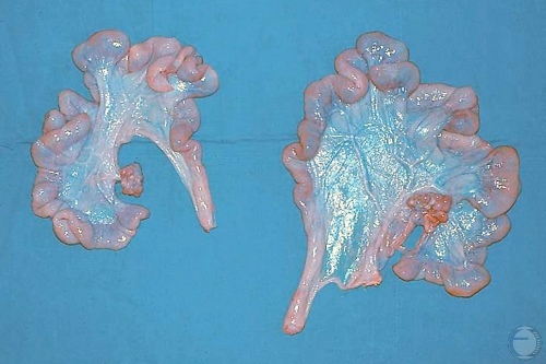
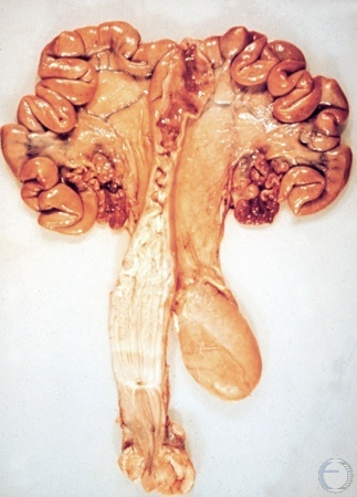
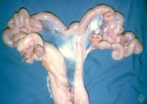
Uterus Unicornis.
Uterus unicornis in two gilts. The contralateral ovary is also missing in each case. These tracts were slaughter house specimens hence the abdomens were not searched for the missing ovary.
Evans LE (2009)
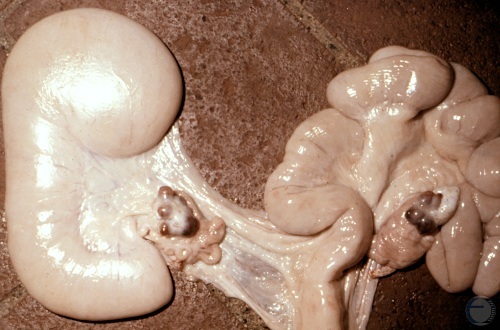
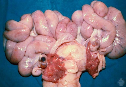
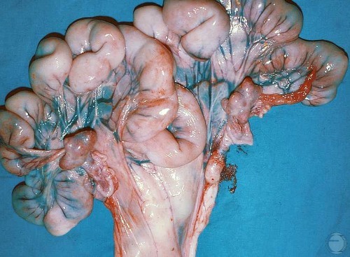
Segmental Hypoplasia.
Congenital defect. The base of the left horn is missing causing the distal portion to distend with uterine secretions. Conception on the contralateral side is possible. Normal corpora lutea are present on both ovaries.
Larsen RE (1979)
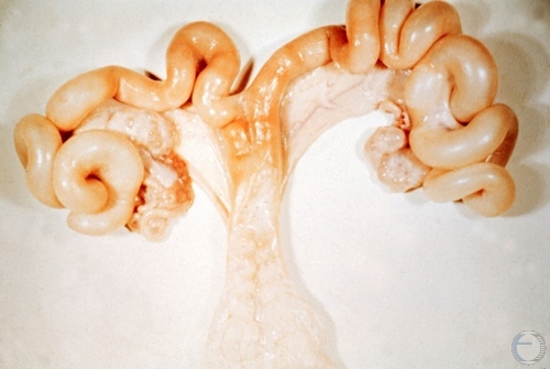
Segmental Hypoplasia.
Segmental hypoplasia in a 13-month old gilt that was a repeat breeder with a history of recycling with discharges. The blocked nonpregnant horn caused the recycling by failure to give a pregnancy signal to it corresponding ovary. The nonpregnant horn releases prostaglandins into general circulation and lyses the CLs on both ovaries. Each horn needs 1 to 2 fetuses to maintain a pregnancy.
Evans LE (2009)
Segmental Hypoplasia Corrected.
Segmental hypoplasia in a 13-month old gilt that was a repeat breeder with a history of recycling with discharges. Hypoplasia was surgically corrected. The gilt subsequently bred successfully and farrowed normal pigs after surgery.
Evans LE (2009)
Pyometra.
Post-breeding pyometra. No obvious odor. Most likely the result of contamination at breeding by environmental organisms (Strep. Staph. E. coli) or from the boars prepuce.
Evans LE (2009)
Pyometra.
Post-breeding pyometra. No obvious odor. Most likely the result of contamination at breeding by environmental organisms (Strep. Staph. E. coli) or from the boars prepuce.
Evans LE (2009)