The Visual Guide to
Porcine Reproduction
Teratology: Congenital Anomalies
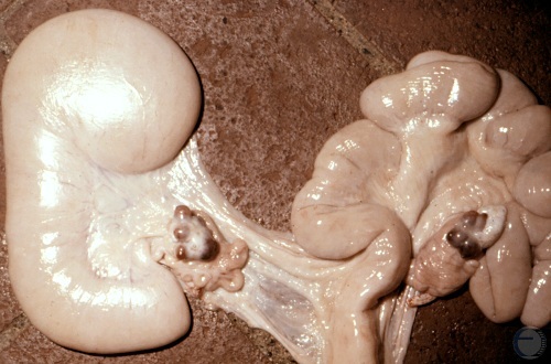
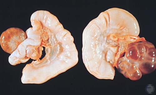
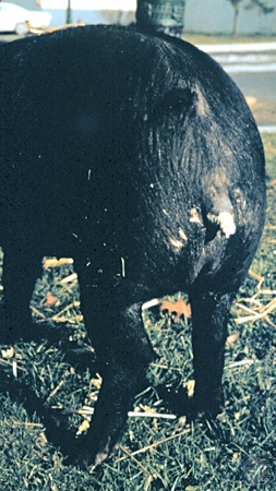
Segmental Aplasia.
Congenital defect. The base of the left horn is missing causing the distal portion to distend with uterine secretions. Conception on the contralateral side is possible. Normal corpora lutea are present on both ovaries.
Larsen RE (1979)
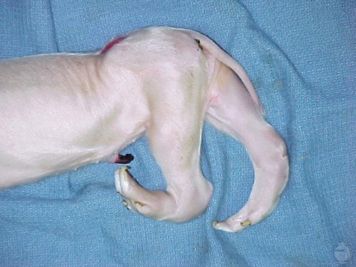
Perosomus Elumbis.
Perosomus elumbis is a congenital defect characterized by lack of vertebrae and spinal cord caudal to the thoracic region.
McEntee K (1973)
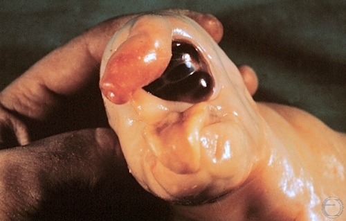
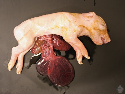
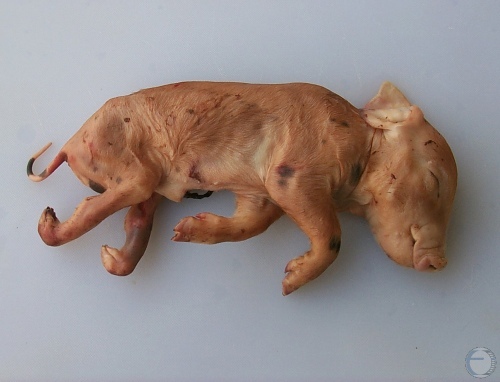
Severe Cyclops.
Extreme expression of cyclopia. The fetus has a distinct proboscis while major portions of the mouth are absent.
Roberts SJ (1973)
True Hermaphrodite.
The uterine horn on the left is accompanied by a testis and a mesonephric duct, the uterine horn on the right is flanked by an ovary with cystic follicles.
Roberts SJ (1973)
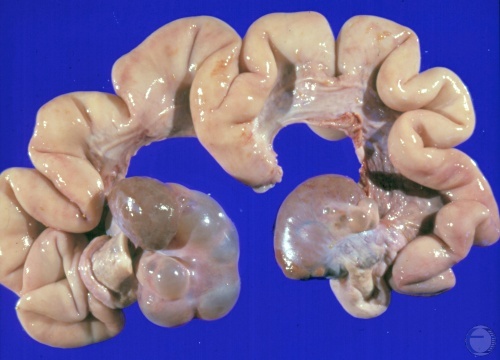
True Hermaphrodite.
The left gonad is a combination of testicular and ovarian tissue. The right gonad consists primarily of testicular tissue. Both ovotestes are accompanied by a uterus.
Shipley C (2006)
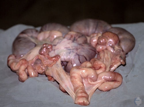
Ovotestis.
Gravid uterus with six fetuses. The ovaries contain several corpora lutea. The right gonad is an ovotestis which also shows one or two corpora lutea. This congenital defect can be compatible with fertility, as confirmed by the pregnant status.
Evans LE (2009)
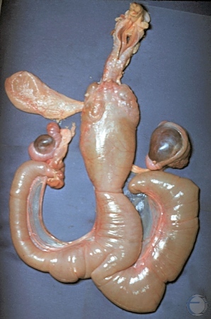
Male Pseudohermaphrodite.
The rudimentary fluid-filled uterus is accompanied by male gonads. At the upper left is an empty bladder.
McEntee K (1973)
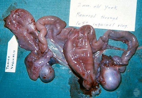
Male Pseudohermaphrodite.
This rudimentary fluid-filled uterus is accompanied by male gonads. At the upper left is the tunica vaginalis. The tract was removed via the left inguinal ring of a 2 month old Yorkshire pig.
Evans LE (2009)
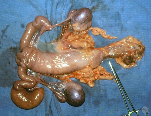
Male Pseudohermaphrodite.
The rudimentary fluid-filled uterus is accompanied by male gonads. The cervical area appears to be constricted.
Shipley C (2006)
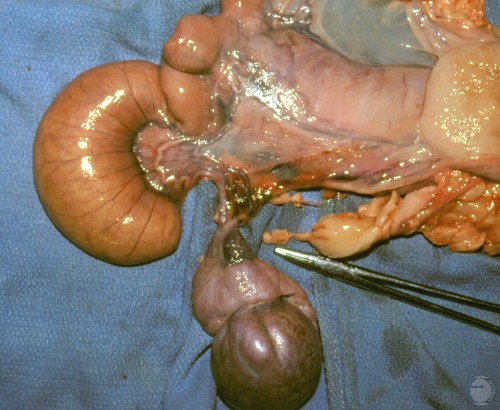
Male Pseudohermaphrodite.
The left testis is connected to the fluid-filled rudimentary uterus in the location of the ovary.
Shipley C (2006)
Male Pseudohermaphrodite.
The right testis is connected to the fluid-filled rudimentary uterus in the location of the ovary.
Shipley C (2006)
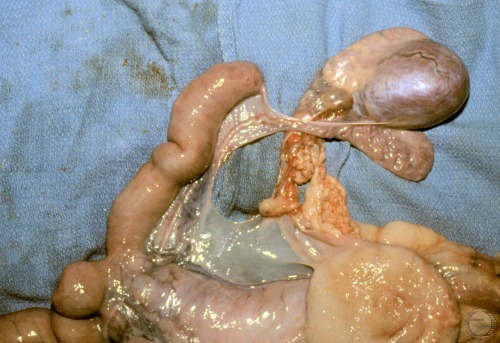
Male Pseudohermaphrodite.
The male gonads are located in anatomical location of the ovaries. The round structure at the base of the uterus is the bladder.
Shipley C (2006)
Hermaphrodite.
The piglet on the left has a hermaphroditic vulva; note the fishhook tip of the enlarged clitoris. The piglet on the right is a normal female litter mate for comparison.
Roberts SJ (1973)
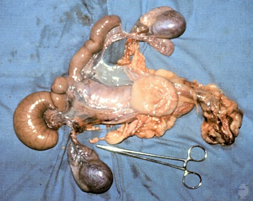
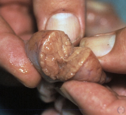
Pregnant Hermaphrodite.
Tract of a hermaphroditic gilt. Testicular structures on the right, ovary on the left, plus a pregnant horn. This has only been seen in swine. The testicular tissue on the right, opposite to a normal ovary on the left, was likely not functional.
Evans LE (2009)
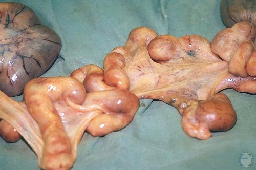
Testis of a Pregnant Hermaphrodite.
Close up cross section of a testicular structure from a true hermaphrodite. The tract of a hermaphrodite gilt showed a nonfunctional testicular structure on the right, a functional ovary on the left, plus a pregnant horn.
Evans LE (2009)
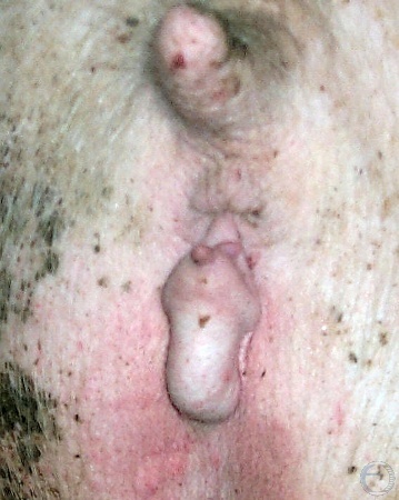
Intersex Vulva.
Male pseudohermaphrodite. Intra-abdominal testes. Notice the prominent clitoris.
Smith MC (2011)
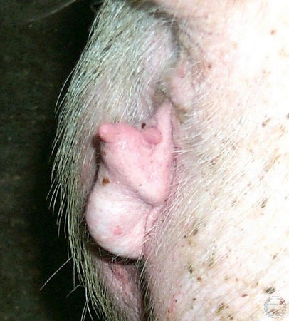
Intersex Vulva - Lateral View.
Male pseudohermaphrodite. Intra-abdominal testes. Lateral view of the vulva. Notice the prominent clitoris at the dorsal commissure.
Smith MC (2011)
Vulvar Hypoplasia.
Very small labiae in a male pseudohermaphrodite. There is evidence of clitoral enlargement. Intra-abdominal testes.
Shipley C (2006)
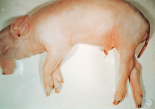
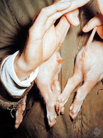
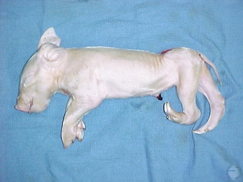
Epitheliogenesis Imperfecta.
Epitheliogenesis imperfecta is a condition whereby the skin fails to form. This condition is the result of a recessive lethal or sub-lethal defect.
Drost M (1974)
Epitheliogenesis Imperfecta.
Epitheliogenesis imperfecta is a condition whereby the skin fails to form. This condition is the result of a recessive lethal or sub-lethal defect.
Drost M (1974)
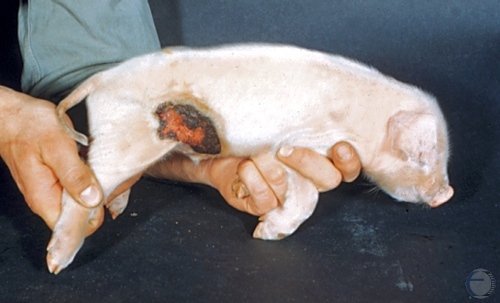
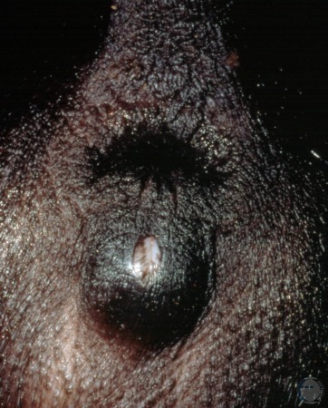
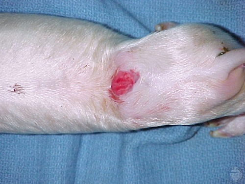
Small Epitheliogenesis Imperfecta.
Small healing area of epitheliogenesis imperfecta.
Roberts SJ (1973)
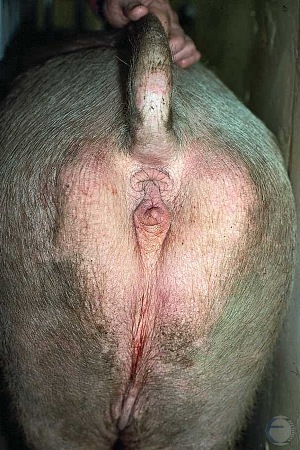
Atresia Ani.
Atresia ani and a rectovaginal fistula leading to the formation of a vulvar cloaca which allowed the animal to defecate.
Utrecht (1976)
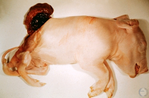
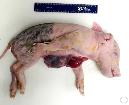
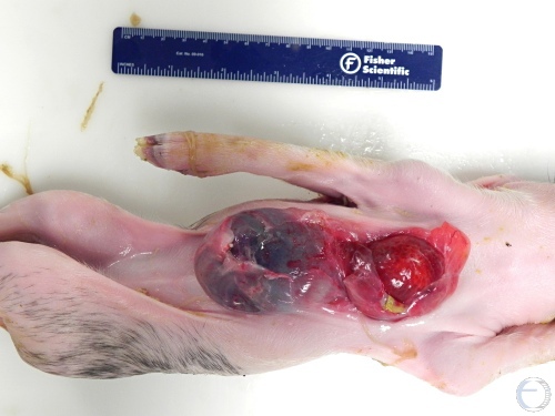
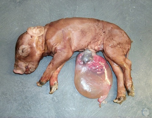
Abdominoschisis.
Piglet born with a wide open abdominal wall. Resembled a massive open umbilical hernia.
Smith MC (2010)
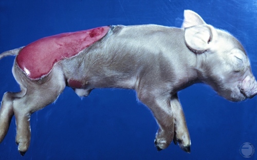
Sternal Fissure - Lateral View.
Piglet born with a split ventral sternum. Atelectic lung exposed. Umbilical cord hematoma.
Smith MC (2011)
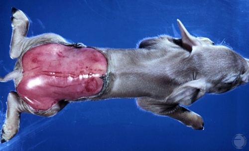
Sternal Fissure - Ventral View.
Piglet born with a split ventral sternum. Atelectic lung exposed. Umbilical cord hematoma.
Smith MC (2011)
Umbilical Hernia.
Piglet born with an open umbilical hernia whereby the abdominal are exposed and visible.
Smith MC (2011)
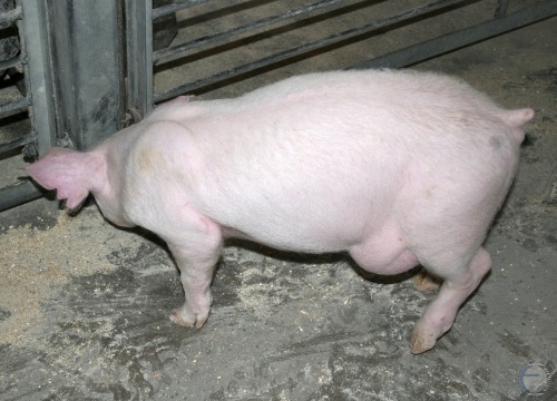
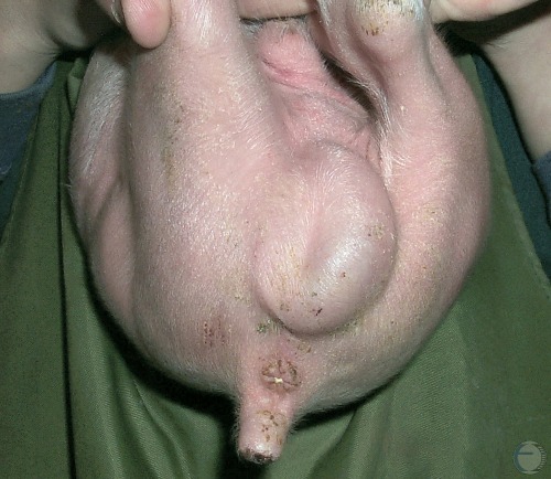
Inguinal Hernia.
Inguinal hernia in a boar in dorsoventral position.
Smith MC (2011)
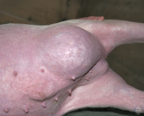
Inguinal Hernia.
Close up of a left inguinal hernia in a boar in right lateral recumbency.
Smith MC (2011)
Arthrogryposis.
Neonate with congenitally retroflexed hind limbs.
Shipley C (2006)
Arthrogryposis.
Spinal deformity as part of congenital arthrogryposis in a neonate.
Shipley C (2006)