The Visual Guide to
Porcine Reproduction
Abortion: Infectious Causes
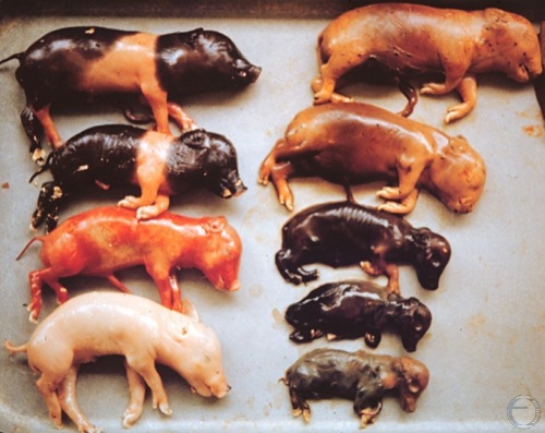
Mummification.
Largely mummified litter believed to be caused by Parvo virus (but not confirmed in this case).
Roberts SJ (1973)
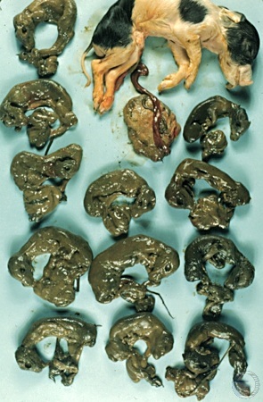
Mummification / Uterus.
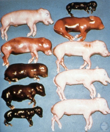
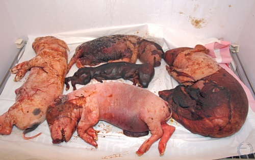
Some fetuses died and started to mummify earlier than others, based on their relative sizes.
Larsen RE (1979)
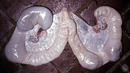
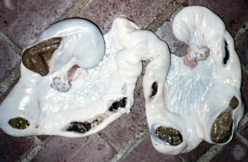
Small Mummies.
Multiple firm masses of different sizes could be palpated within the uterus.
Larsen RE (1979)
Leptospirosis.
The sow had a high titer for Leptospira spp. Some or all of the fetuses may have been infected and died due to a leptospiral septicemia. Mummification had started in some fetuses.
McEntee K (1973)
Leptospirosis or Parvo Virus.
Some of the fetuses may have been infected and died due to a leptospiral septicemia or a Parvo virus infection. Mummification is in various stages.
Shipley C (2006)
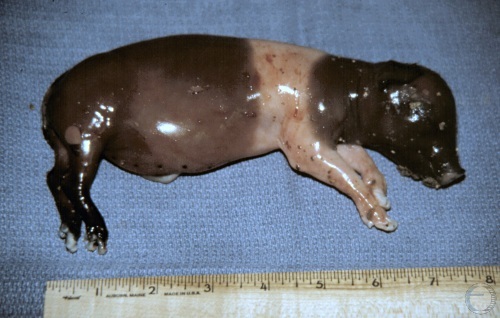
Late Fetal Death.
Dead fetus with abdominal distension and in early stage of mummification [note sunken-in eye socket] due to Parvo virus infection.
Shipley C (2006)
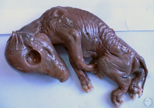
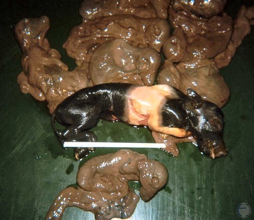
Mummified Fetus.
Porcine mummy believed to be associated with a respiratory syndrome.
Prado TM (2007)
SMEDI Virus Abortion.
Porcine fetuses likely infected with an enterovirus [SMEDI: Stillborn, Mummification, Embryonic Death, Infertility].
Smith MC (2011)
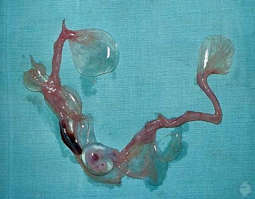
Parvo Virus.
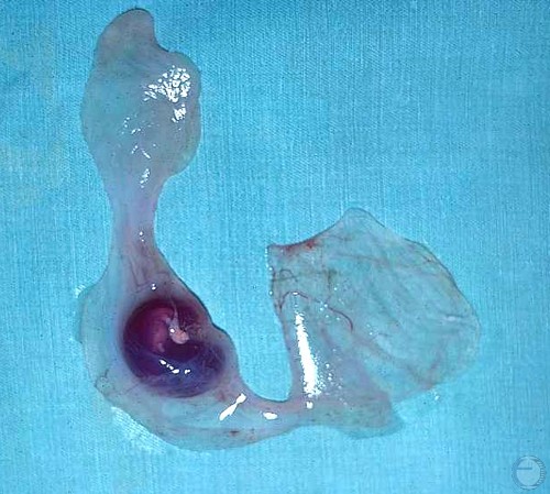
24 day embryo in a blood tinged amniotic vesicle. Parvo virus infection caused embryonic death.
Evans LE (2009)
Parvo Virus.
This is an example of a mid-trimester Parvo virus infection. Fetuses have died at different ages and have mummified.
Evans LE (2009)
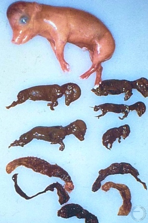
Parvo Virus.
Multiple mummies plus one fetus that survived for a while longer, the result of Parvo virus infection.
Evans LE (2009)
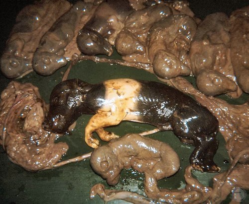
Parvo Virus.
Multiple mummies plus one fetus that succumbed later, as the result of Parvo virus infection.
Evans LE (2009)
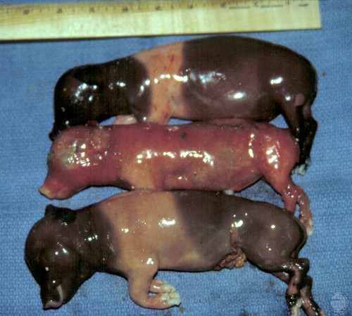
Pseudorabies.
Abortion due to pseudorabies. The membranes are intact and appear to be autolysed.
Evans LE (2009)
Pseudorabies.
Abortion due to pseudorabies. The membranes are intact and appear to be autolytic.
Evans LE (2009)
Pseudorabies.
Abortion due to pseudorabies. All fetuses were about the same age at the time of death.
Evans LE (2009)
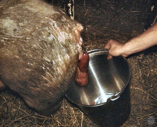
24 Day Embryo.
Vascular damage prior to abortion. Embryo still intact. Etiology: Parvo virus.
Evans LE (2009)
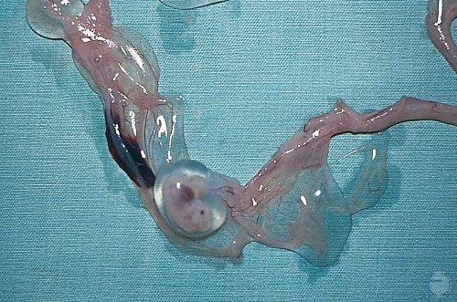
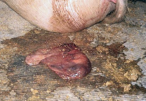
24 Day Embryo - Close Up.
Vascular damage in the chorioallantois prior to abortion. Embryo still intact. Etiology: Parvo virus.
Evans LE (2009)
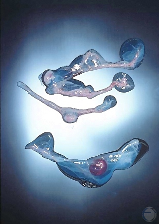
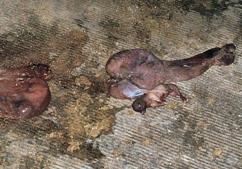
Degenerating Embryos.
Three degenerating embryos at slightly different ages. Etiology: Parvo virus.
Evans LE (2009)
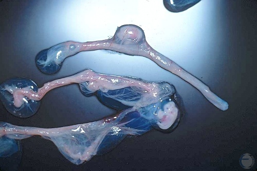
Degenerating Embryos.
Two 24 to 26 day embryos from the uterus with a damaged feto-placental unit. Cause not determined. There is evidence of hemorrhage in the chorionic vessels of the upper embryo.
Evans LE (2009)