The Visual Guide to
Porcine Reproduction
Male Reproductive System: Penis and Prepuce
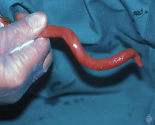
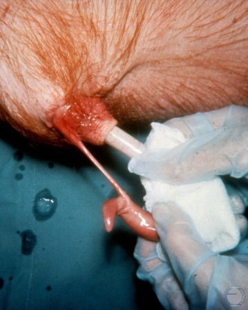
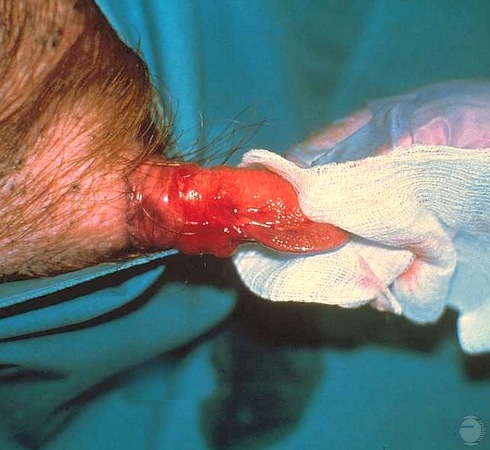
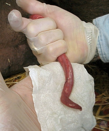
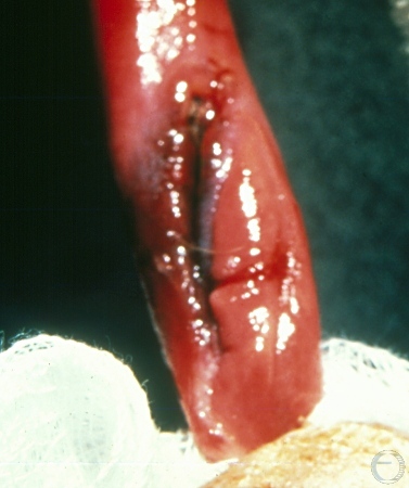
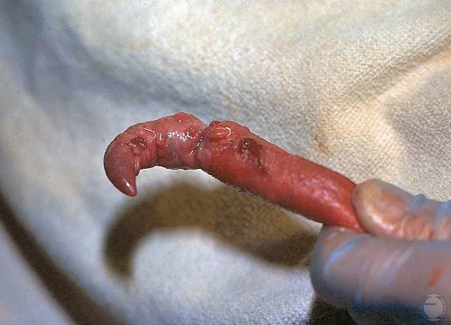
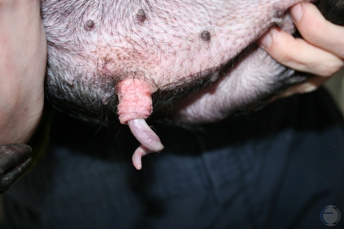
Persistent Frenulum.
The frenulum is a band of tissue that extends from the ventral tip of the glans penis to the prepuce. Separation of the penis from the sheath normally occurs spontaneously at 4 to 6 months of age in the boar. When the frenulum persists there is frequently a small artery present in the center of the tissue band comprising the frenulum. Persistence of the frenulum causes curvature of the extended penis.
Roberts SJ (1973)
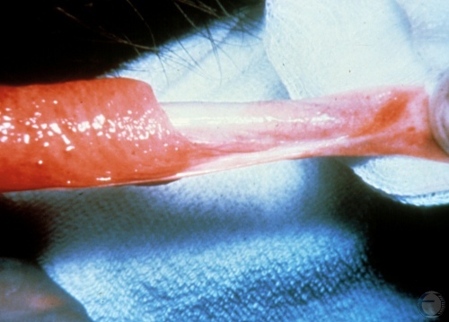
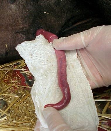
Frenulum.
The frenulum is a band of tissue that extends from the ventral tip of the glans penis to the prepuce. Separation of the penis from the sheath normally occurs spontaneously at 4 to 6 months of age in the boar. When the frenulum persists there is frequently a small artery present in the center of the tissue band comprising the frenulum. Persistence of the frenulum prevents the penis from full extending.
Evans LE (2009)
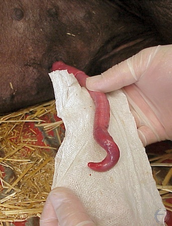
Frenulum.
The frenulum is a band of tissue that extends from the ventral tip of the glans penis to the prepuce. Separation of the penis from the sheath normally occurs spontaneously at 3-4 months of age in the boar. When the frenulum persists there is frequently a small artery present in the center of the tissue band comprising the frenulum. Persistence of the frenulum prevents the penis from fully extending.
Evans LE (2009)
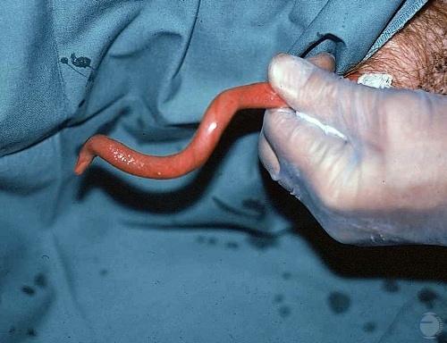
Persistent Frenulum.
The frenulum is a band of tissue that extends from the ventral tip of the glans penis to the prepuce. Separation of the penis from the sheath normally occurs spontaneously at 3-4 months of age in the boar. When the frenulum persists there is frequently a small artery present in the center of the tissue band comprising the frenulum. Persistence of the frenulum prevents the penis from fully extending.
Shipley C (2006)
Hypoplasia of the Penis.
Incomplete erection, making intromission impossible.
Evans LE (2009)
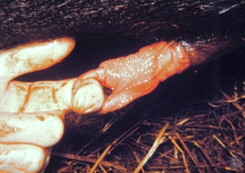
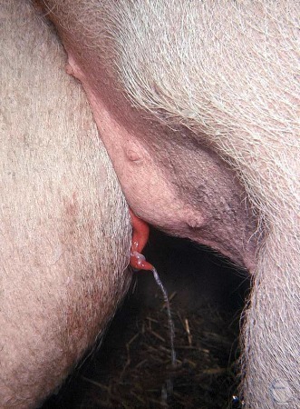
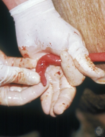
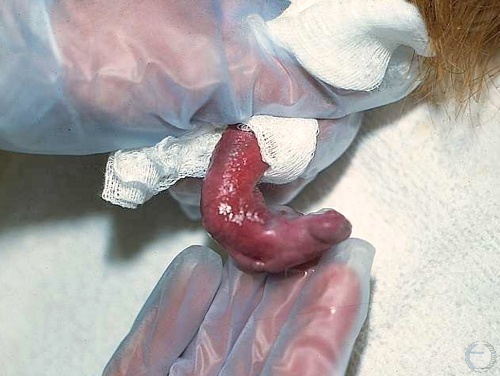
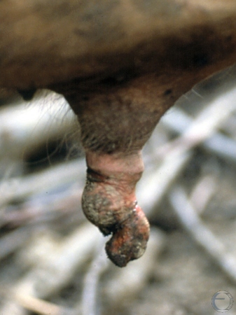
Prepuberal Penile Hypoplasia.
Hypoplasia of the penis. Adherence of the penis to the prepuce in a 6 month old boar. Bleeding is due to trauma while extracting the penis from the prepuce.
Evans LE (2009)
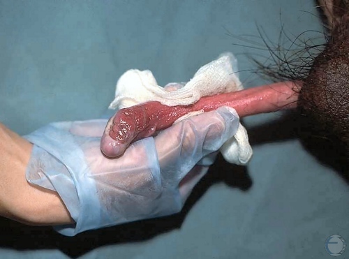
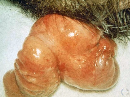
Penile Hypoplasia.
Hypoplasia of the penis. An underdeveloped spiral glans penis which leads to erectile dysfunction.
Shipley C (2006)
Penile Hypoplasia.
Hypoplasia of the penis. Underdeveloped flaccid spiral glans penis.
Shipley C (2006)
Penile Hypoplasia.
Hypoplasia of the penis. Underdeveloped flaccid spiral glans penis.
Shipley C (2006)
Erectile Dysfunction.
Erectile dysfunction in a 7 month old virgin boar. The young boar had difficulty extending his penis (phimosis). Note the lack of a tight clockwise spiral tip. This is a heritable condition.
Evans LE (2009)
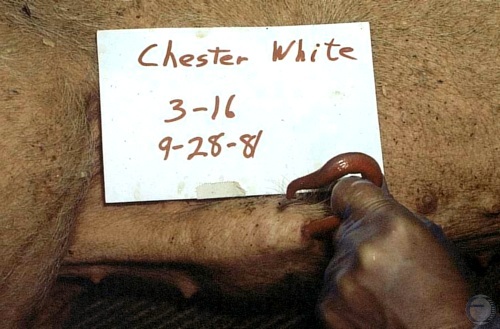
Hypoplasia and Erectile Dysfunction.
Hypoplasia and erectile dysfunction in a young Chester White boar.
Evans LE (2009)
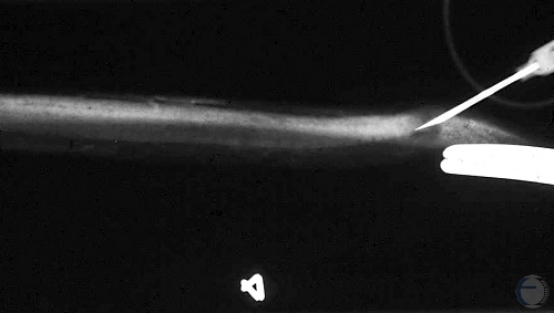
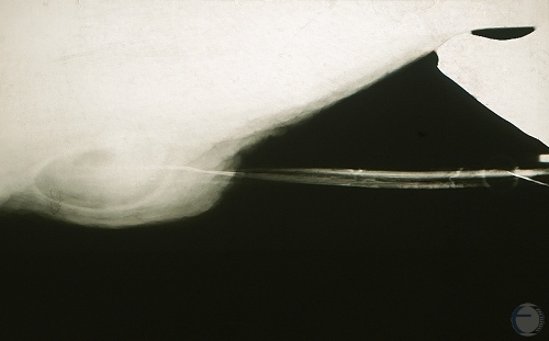
Diagnosis of Erectile Dysfunction.
Radiography and the use of a contrast medium illustrate the dye escaping near the tip of the penis and demonstrating erectile dysfunction.
Evans LE (2009)
Cavernosography.
Radiography of erectile dysfunction using contrast radiography. The contrast medium escaped near the tip of the penis in the diagnosis of erectile dysfunction in this boar.
Evans LE (2009)
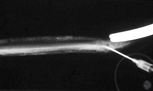
Cavernosogram.
Radiograph of erectile dysfunction, or caversonography. Several seconds after the injection of the contrast medium it can be seen in the surface vessels of the penis. Procedure was used to aid in the diagnosis of erectile dysfunction in this boar.
Evans LE (2009)
Cavernosogram.
Radiograph of erectile dysfunction, or caversonography. Several seconds after the injection of the contrast medium it can be seen in the surface vessels of the penis. This procedure was used to aid in the diagnosis of erectile dysfunction in this boar.
Evans LE (2009)
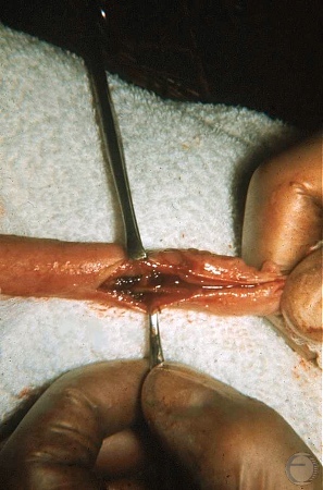
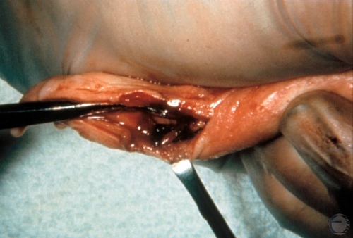
Urethral Hemorrhage.
Surgical exposure of the urethra near the tip of the penis. Urethral bleeding was caused by an old bite wound. Externally, the penis has healed but urethral ulceration into the corpus spongiosum persists.
Evans LE (2009)
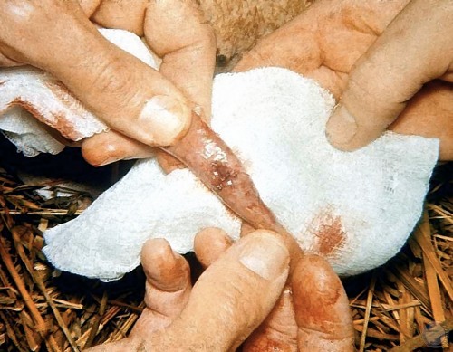
Penile Laceration.
Laceration of the spiral tip of the glans penis. Profuse hemorrhage due to the vascular nature of the tissues.
Shipley C (2006)
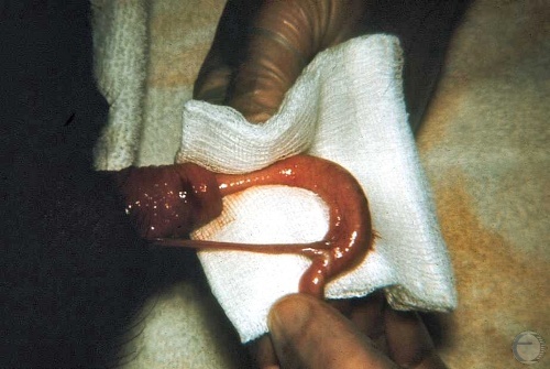
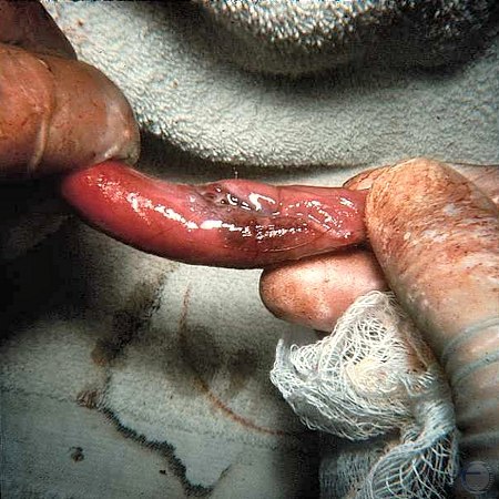
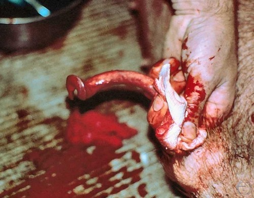
Penile Laceration.
Laceration of the spiral tip of the glans penis along the ventral surface.
Shipley C (2006)
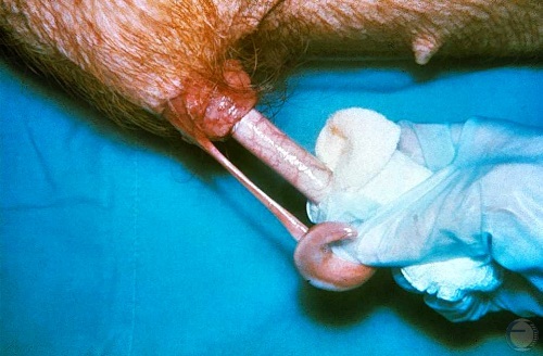
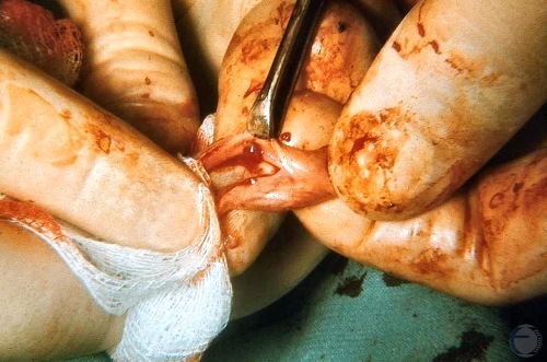
Penile Laceration - Close Up.
Close up of the laceration of the spiral tip of the glans penis along its ventral aspect and into the urethra.
Shipley C (2006)
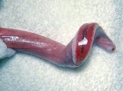
Penile Laceration.
Laceration of the spiral tip of the glans penis along its ventral aspect.
Shipley C (2006)
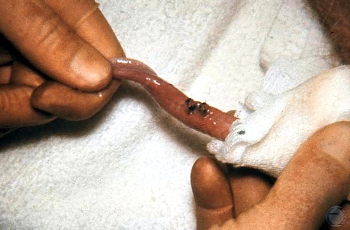
Penile Laceration.
Spread of the laceration to assess its depth into the urethra.
Shipley C (2006)
Bite Wound into the Urethra.
Bite wound, 7 to 8 days old, into the urethra near the tip of the penis.
Evans LE (2009)
Bite Wound.
Bite wound on the penis in the process of healing. The boar may be unable to breed naturally due to the damaged penis and inability to lock into the cervix.
Evans LE (2009)
Healed Bite Wound.
Healed bite wound to the penis which is free from adhesions.
Evans LE (2009)
Bite Wound with Amputation.
Bite wound with amputation of the glans penis. Four out of six of the boars in the breeding herd sustained bite wounds (usually) inflicted by an intervening sow.
Evans LE (2009)
Bite Wound into Urethra.
Nearly healed bite wound into the urethra. Semen escaped here. The boar was still fertile.
Evans LE (2009)
Acute Hemorrhage during Breeding.
Acute hemorrhage at erection in a virgin boar due to urethral bleeding.
Evans LE (2009)
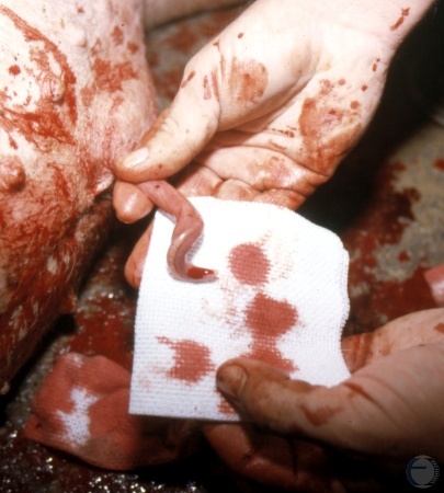
Vascular Urethral Polyp.
Acute hemorrhage at erection in a virgin boar. Upon opening of the urethra there was a vascular polyp from the corpus cavernosum urethrae (spongiosum). The boar bled upon erection resulting in hemospermia and infertility.
Evans LE (2009)
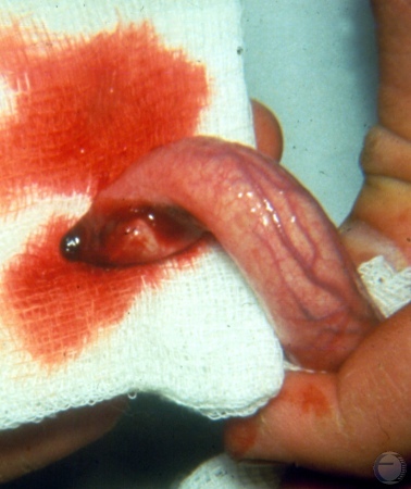
Removal of Urethral Polyp.
Acute hemorrhage at erection due to a urethral polyp in a virgin boar. The polyp was surgically removed and the incision was closed.
Evans LE (2009)
Intromission Problem.
Trauma sustained during breeding. The boar had difficulties with intromission and abraded the surface of the penis on the sow's bristles. The ejaculate contained blood due to the erosion into the erectile tissues.
Evans LE (2009)
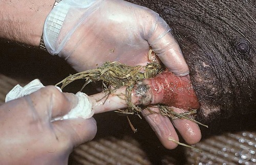
Phimosis.
The penis was trapped in the prepuce by foreign (hay bedding) material, which has also caused irritation, inflammation, and infection, leading to a preputial discharge. The boar was unable to retract the penis, leaving it exposed.
Evans LE (2009)
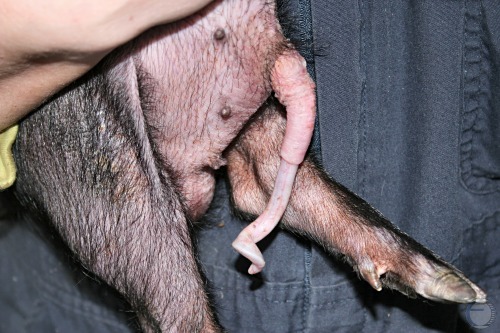
Paraphimosis in a Piglet.
This 5 week old piglet has been unable to withdraw his penis into the prepuce. No history of trauma.
Smith MC (2011)
Chronic Paraphimosis in a Piglet.
This 4.5 kg Torra piglet has been unable to withdraw his penis into the prepuce for 5 weeks.
Smith MC (2011)
Close up of Chronic Paraphimosis.
This 4.5 kg Torra piglet has been unable to withdraw his penis into the prepuce for 5 weeks. No signs of trauma nor inflammation.
Smith MC (2011)
Partial Paraphimosis.
Incomplete retraction of the penis into the prepuce.
Smith MC (2011)
Complete Preputial Prolapse.
The preputial lining including the dorsal diverticulum have prolapsed.
Shipley C (2006)
Chronic Preputial Prolapse.
Chronic preputial prolapse. The exposed lining of the prepuce has been traumatized and become indurated and infected.
Shipley C (2006)
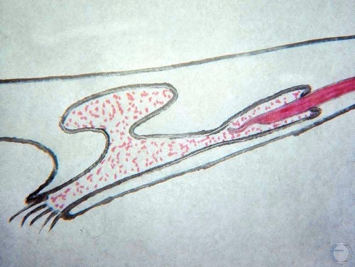
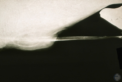
Diagram of the Preputial Cavity.
Preputial cavity in relationship to the retracted penis. Note the bi-lobed, dorsal diverticulum and the narrowing of the prepuce deeper into the preputial cavity.
Evans LE (2009)
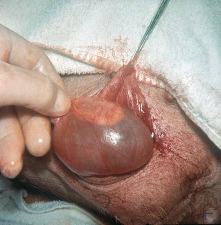
Preputial Diverticulum.
Direct surgical exposure of one lobe of the fluid filled diverticulum prior to removal. The diverticulum was filled through the sheath orifice in preparation for removal.
Evans LE (2009)
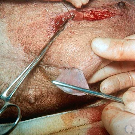
Preputial Diverticulum Removal.
Direct surgical exposure of the fluid filled diverticulum prior to removal.
Evans LE (2009)
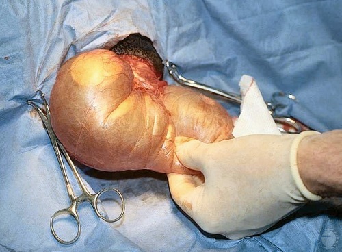
Preputial Diverticulum Excision.
Diverticula filled with fluid and dissected free in preparation for removal. Both lobes have been exposed and the delineation between them can be seen.
Evans LE (2009)
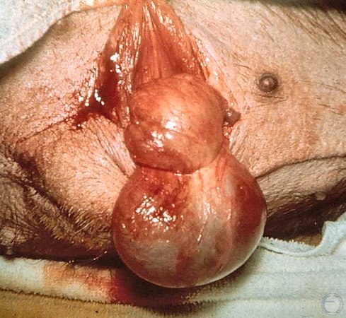
Preputial Diverticulum Excision.
Everted lobe of one diverticulum after a purse string suture has been placed around its common base with the other diverticulum lobe. Both are emptied and everted, after which the purse string suture is pulled tight and tied.
Evans LE (2009)
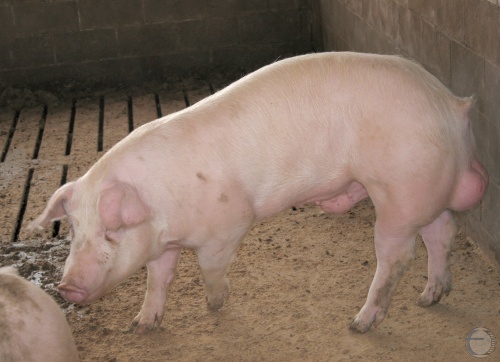
Preputial Swelling.
Boar with preputial swelling, possibly due to fluid accumulation in the preputial diverticulum.
Smith MC (2010)