The Visual Guide to
Porcine Reproduction
Obstetrics: Cesarean Section
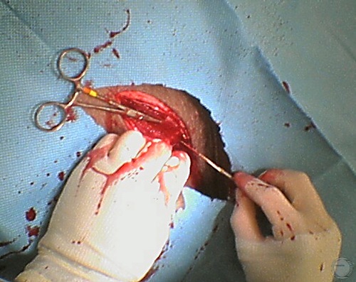
Controlling Bleeders.
Hemorrhage is controlled by ligating the major blood vessels and temporarily clamping the smaller ones.
Prado TM (2007)
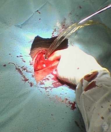
Peritoneal Fluid.
As the peritoneum is incised, it is common in pregnant sows that peritoneal fluid is released.
Prado TM (2007)
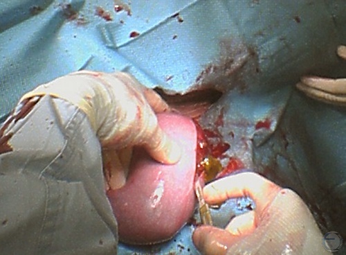
Uterine Incision.
The uterine wall including the diffuse placenta are gently incised over one of the fetuses.
Prado TM (2007)
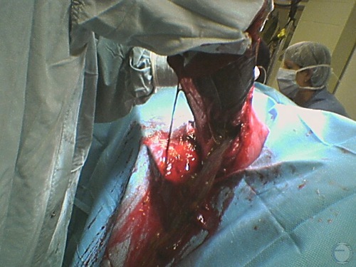
Extracting a Fetus.
The surgeon reaches into the lumen of the uterus to locate a fetus.
Prado TM (2007)
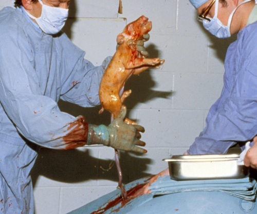
Delivering a Fetus.
The surgeon lifts a fetus out of the incision during a Cesarean Section. The umbilical cord is still attached.
Evans LE (2009)
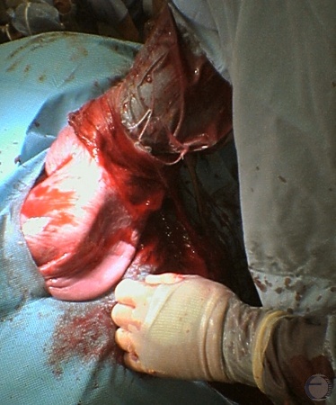
Reaching for the Next Fetus.
The next fetus is delivered via the same uterine incision. Generally, one uterine incision is required per horn.
Prado TM (2007)
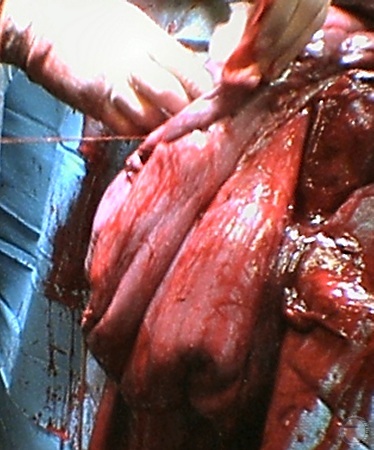
Uterine Closure.
The uterine incision is closed with a modified Cushing's pattern (the Utrecht pattern).
Prado TM (2007)
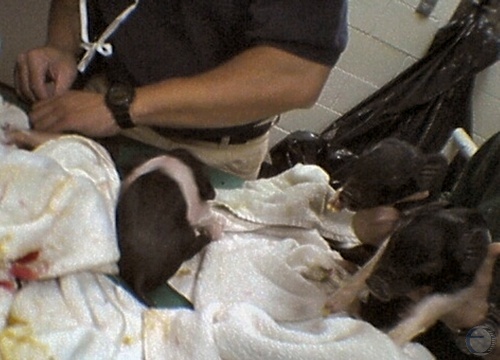
Live Neonates.
The live neonates are dried off and placed in a draft free environment.
Prado TM (2007)
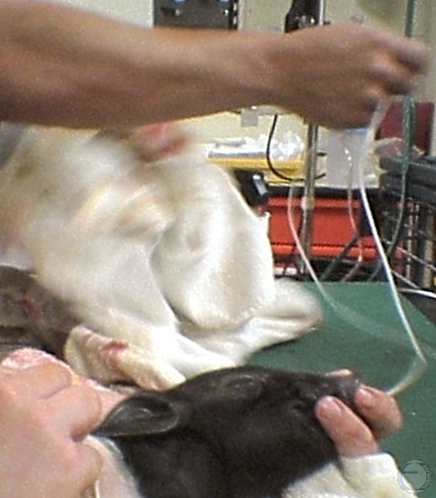
Oxygen Administration.
Neonates with a heartbeat but poor respiratory efforts are given supplemental oxygen via the nares.
Prado TM (2007)
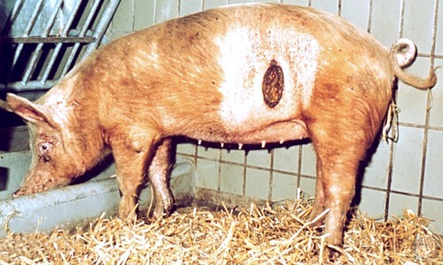
Dehiscence after C-section.
Dehiscense of the skin incision is uncommon after cesarean section in pigs. The wound is granulating in well, but must be kept clean.
Utrecht (1976)
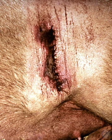
Infected Incision.
Early necrosis and dehiscence of an infected skin incision after cesarean section.
Utrecht (1976)
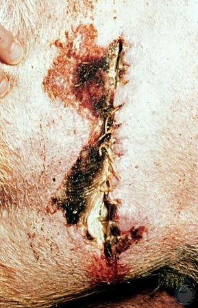
Necrotic Incision.
Tissue necrosis may be due to the excessive use of lidocaine containing epinephrine. Left flank incision for cesarean section.
Utrecht (1976)
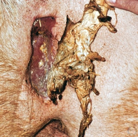
Sloughing Tissues.
The necrotic superficial tissues are sloughing off, after infection of the skin incision in the flank.
Utrecht (1976)
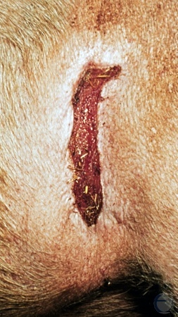
Granulation Tissue.
Partial dehiscence of a left flank incision. The wound is granulating in well but must be kept clean.
Utrecht (1976)
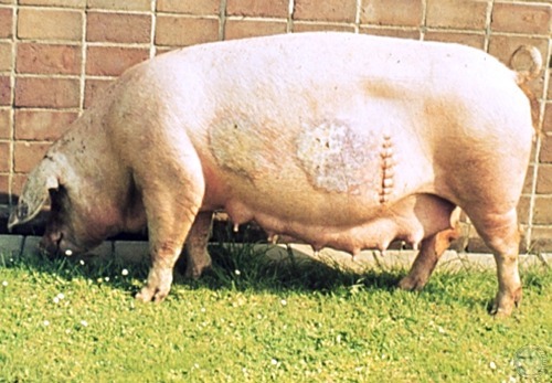
Burn Scars.
Sow is recovering from a cesarean section and from burns from a heat lamp which was placed too close to the body (less than 50 cm).
Utrecht (1976)
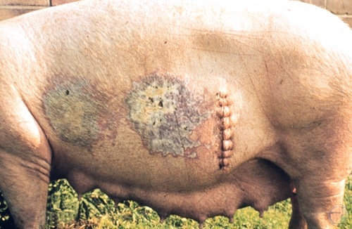
Burn Scars - Close-up.
Sow is recovering from a cesarean section and from burns from a heat lamp which was placed too close to the body (less than 50 cm).
Utrecht (1976)
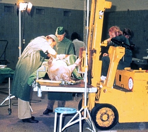
Mobile Surgery Table.
Fork lifts with a platform can be used as a mobile, adjustable, and expedient surgery table.
Utrecht (1976)
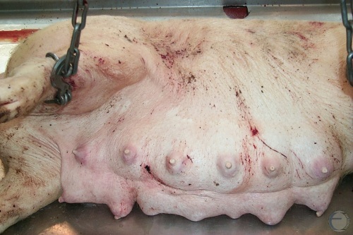
C-section - Dead Sow.
Positioning of a dead sow at term for a Cesarean section on a necropsy table.
Smith MC (2010)
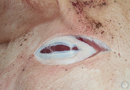
C-section - Skin Incision.
Cesarean section on a dead sow at term. Skin incision.
Smith MC (2011)
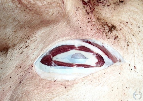
C-section - Peritoneal Incision.
Cesarean section on a dead sow at term. No hemorrhage. Grey uterus shows through the initial peritoneal incision.
Smith MC (2011)
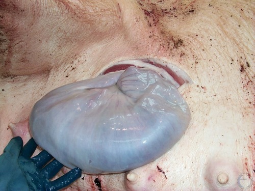
C-section - Exposure of Uterus.
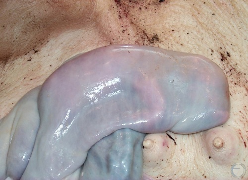
Cesarean section on a dead sow at term. No hemorrhage. Hemolytic uterus exteriorized for removal of the fetuses.
Smith MC (2011)
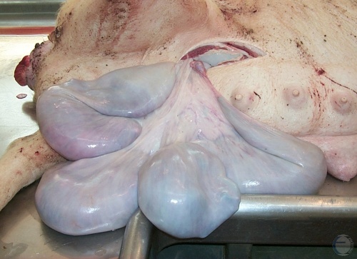
C-section - One Uterine Horn.
Most of the hemolytic uterus has been exteriorized.
Smith MC (2011)
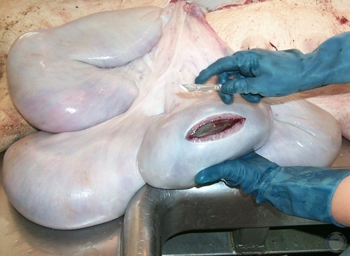
C-section - Uterine Incision.
Incision along the greater curvature over one of the fetuses.
Smith MC (2011)

