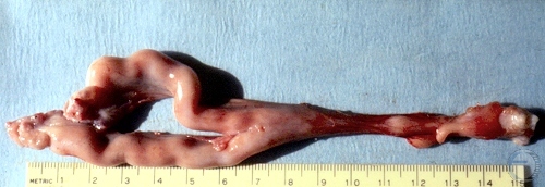
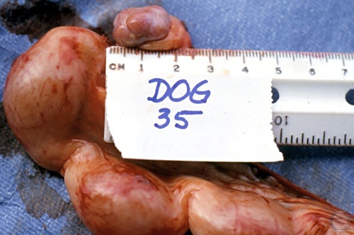
Normal Uterus.
Normal uterus and ovaries with recent corpora lutea, after ovariohysterectomy.
Shille VM (1980)
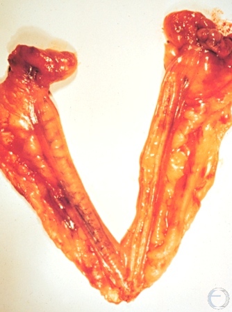
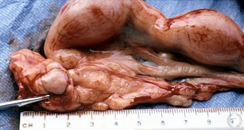
Uterus.
Dorsal view of the bicornuate uterus. Hemorrhagic ovaries.
Shille VM (1973)
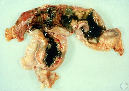
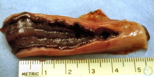
Postpartum Uterus.
Early postpartum uterus. Remnants of the zonary placentation are still present and covered with lochia. Normal very dark green discoloration.
Shille VM (1973)
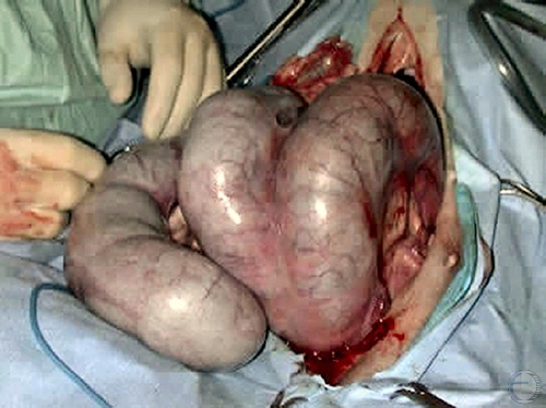
Gravid Uterus.
Intact gravid reproductive tract with ~12 conceptuses.
Shille VM (1981)
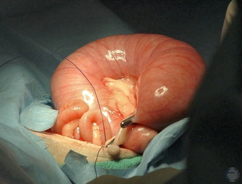
Gravid Uterine Horn.
Tip of the pregnant left horn. Large corpus luteum.
Shille VM (1981)
Pospartum Metritis.
Thickened uterus wall with scarring at the sites of the zonary placenta.
Utrecht (1976)
Hemorrhagic Endometritis.
Diffuse discolored swelling of the endometrium. This is an uncommon occurrence in postpartum bitches.
Shille VM (1973)
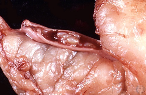
Cystic Endometrial Hyperplasia.
Hyperplastic cysts in the endometrium. Blood has accumulated in the lumen of the uterus.
Shille VM (1982)
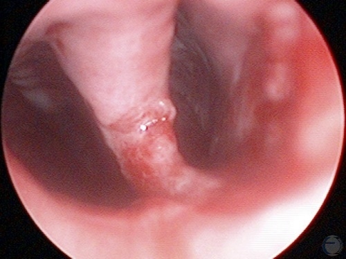
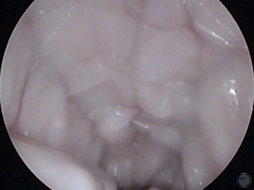
Intrauterine Bifurcation.
Endoscopic view of the bifurcation of the uterine horns. Presence of traumatic lesions on the septum and right uterine horn.
Verstegen J (2009)
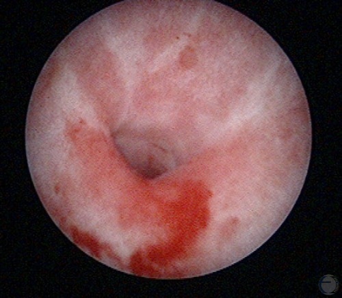
Uterine Lumen.
Laparoscopic view of the lumen of the vagina. Note the pale color and "dehydrated" appearance of the vaginal mucosal folds (called crenulations). These changes are indicative of estrus.
Verstegen J (2009)
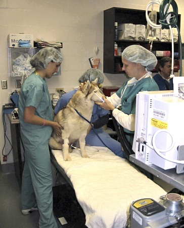
Induction of Anesthesia.
It is recommended to give a female dog exposure to pure oxygen for up to 20 minutes prior to induction for cesarean section.
Verstegen J (2014)
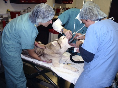
Tracheal Intubation.
Introduction of the endotracheal tube with direct visualization of the rima glottis.
Verstegen J (2009)
Postpartum Uterus.
Uterus 3 days post partum. The endometrial attachments of the zonary placenta are still clearly discernable.
Shille VM (1973)
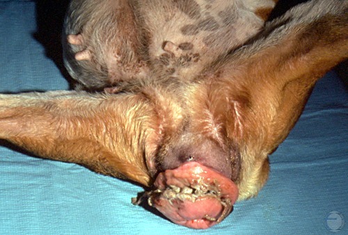
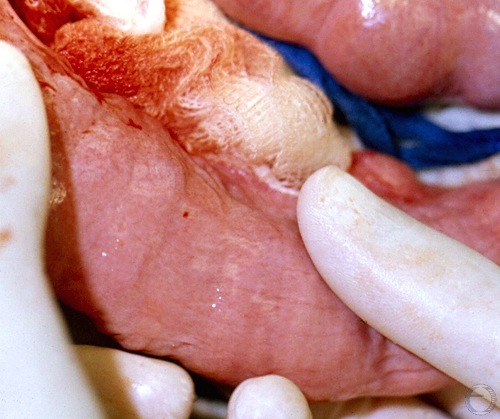
Cervicovaginal Prolapse.
A cervicovaginal prolapse. This condition is estrogen-responsive and often regresses with loss of estrogen. The mucous tissue may become excoriated from prolonged exposure.
Shille VM (1973)
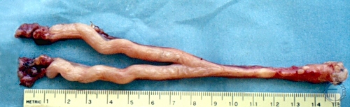
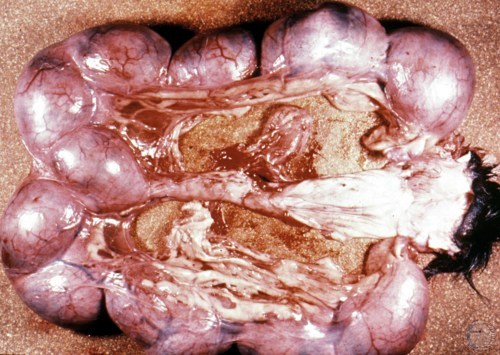
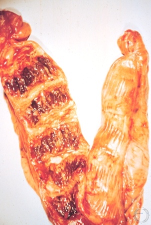
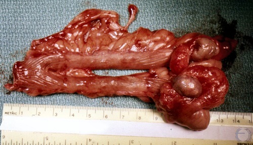
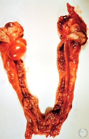
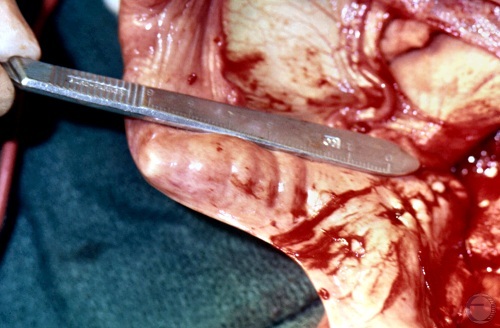
Subinvolution of Placental Sites.
Subinvolution of placental sites. Twelve sites can be identified.
McEntee K (1972)
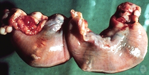
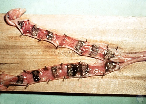
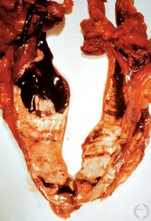
Subinvolution of Placental Sites.
Ten weeks post partum. Subinvolution of placental sites. There is evidence of bleeding at several of the sites.
McEntee K (1972)
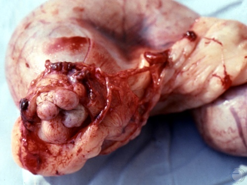
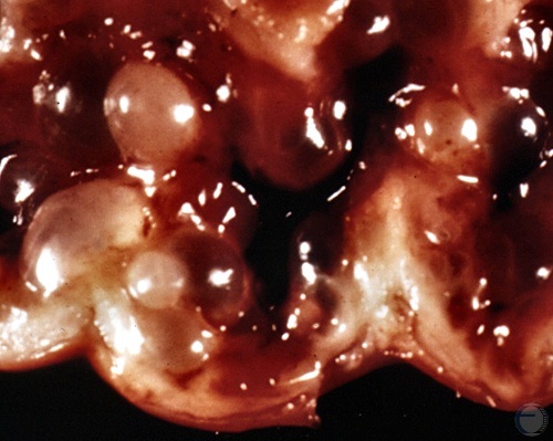
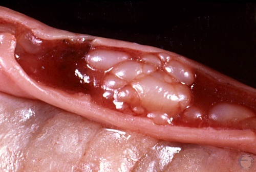
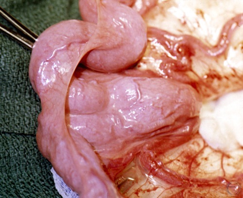
Cystic Endometrial Hyperplasia.
Cystic endometrial hyperplasia (CEH), a life threatening endocrine disorder.
Shille VM (1986)
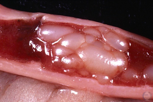
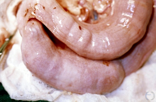
Cystic Endometrial Hyperplasia.
Cystic endometrial hyperplasia (CEH).
Shille VM (1986)
Cystic Endometrial Hyperplasia.
Cystic endometrial hyperplasia (CEH), a life threatening endocrine disorder in the bitch.
Shille VM (1986)
Cystic Endometrial Hyperplasia.
Cystic endometrial hyperplasia (CEH) which occurs in nulliparous animals after prolonged priming of the endometrium by estrogens.
Shille VM (1986)
Uterus and Ovaries.
Uterus and ovaries from an obese dog. Notice excessive fat in the broad ligament. The ovarian bursae have been opened to expose the ovaries.
Shille VM (1973)
Cystic Mesonephric Duct.
Regressing CL. Cystic mesonephric duct (Wolffian duct).
McEntee K (1973)
Early Cystic Hyperplasia.
This uterus is from a bitch that received promone (medroxyprogesterone acetate) 18 days earlier.
McEntee K (1973)
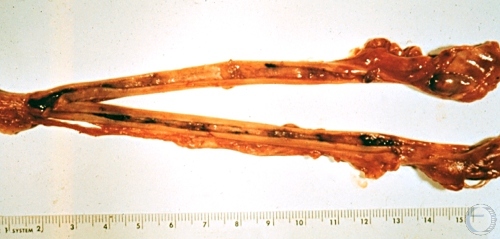
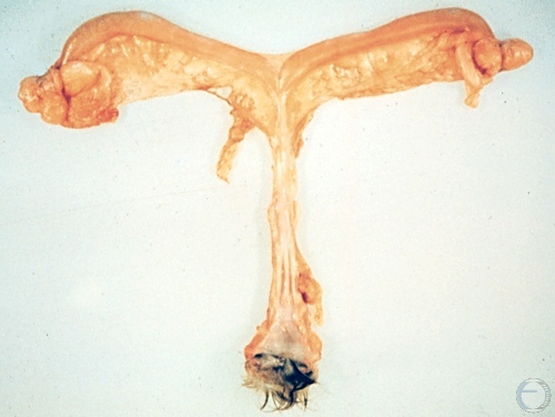
Segmental Aplasia.
Congenital malformation of the uterus. Segmental aplasia of the uterus. Both horns are dilated with trapped fluid. Cervix is closed. Bladder in the lower left corner of the image.
McEntee K (1972)
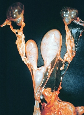
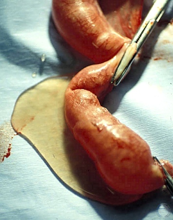
Hydrometra.
Hydrometra with limited amount of fluid. Ovarian Cysts. Atresia of the cervix.
McEntee K (1972)
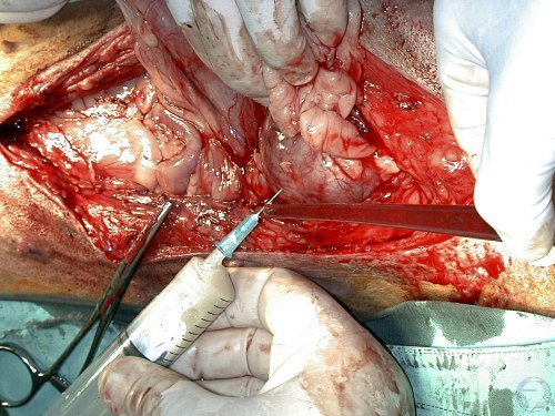
Pyometra - Aspiration of Pus.
Aspiration of pus from a distended uterine horn to diminish the amount of pus and to relieve tension on the uterine prior to hysterectomy.
Verstegen J (2009)
Pyometra Surgery.
Locating the dark, congested and enlarged uterus with pyometra.
Verstegen J (2009)
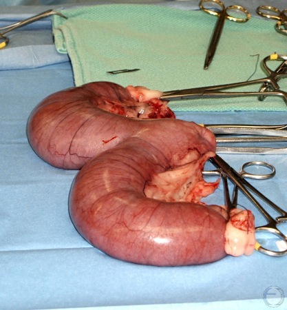
Hysterectomy.
The uterine body is clamped prior to removal of the pus filled uterus.
Verstegen J (2009)
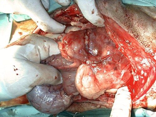
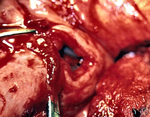
Pyometra.
Pyometra. Uterus exposed prior to hysterectomy. Compromised circulation.
Verstegen J (2009)
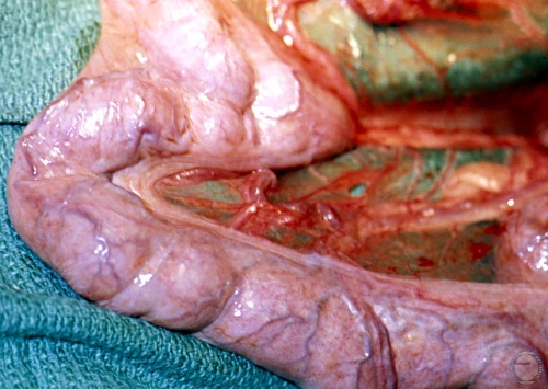
Pyometra Surgery.
Exposure of the ventral aspect of the uterine body at the posterior aspect of the abdominal incision.
Shille VM (1973)
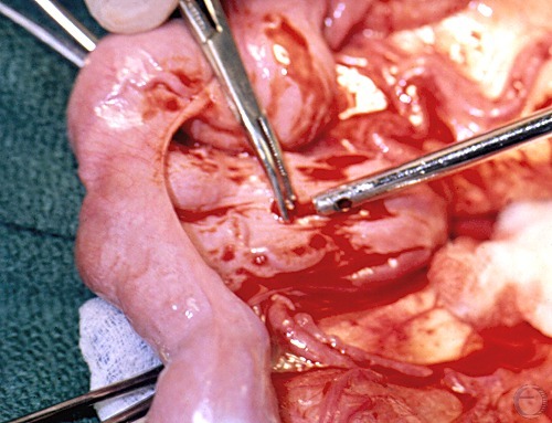
Pyometra Surgery.
Placement of a suction catheter into the body of the uterus.
Shille VM (1973)
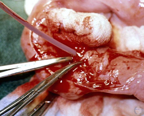
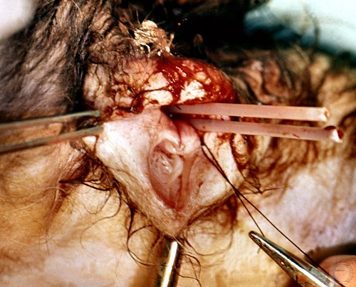
Pyometra Surgery.
After aspiration of pus from the uterine lumen a flexible catheter is inserted into the internal os of the cervix.
Shille VM (1973)
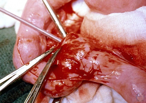
Pyometra Surgery.
Advancement of the flexible catheter through the cervix. The stainless steel catheter aspirates surrounding pus. Hemostats control hemorrhage.
Shille VM (1973)
Pyometra Surgery.
Expressing pus from the uterus via the stainless steel catheter.
Shille VM (1973)
Pyometra Surgery.
Uterine end of the drainage catheter through the cervix.
Shille VM (1973)
Pyometra Surgery.
Cranial end of the drainage catheter extends 6 cm from the body into the uterine horn.
Shille VM (1973)
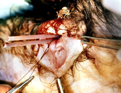
Pyometra Surgery.
Two uterine drainage catheters that were surgically inserted via the internal os of the cervix and then emerge here from the vagina.
Shille VM (1973)
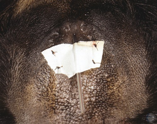
Pyometra Surgery.
The two uterine drainage catheters are anchored at the vulva.
Shille VM (1973)
Intrauterine Catheter.
Indwelling uterine catheter secured to the vulva.
Shille VM (1989)
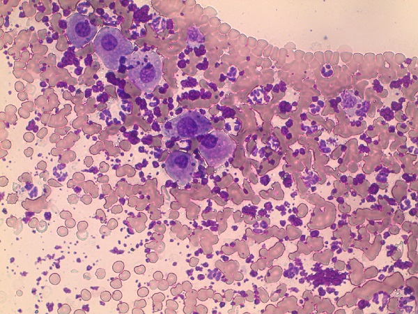
Vaginal Smear-Pyometra.
Vaginal cytology indicative of pyometra. Note the numerous, predominantly degenerate, neutrophils and red blood cells. Epithelial cells are of the intermediate type.
Khan FA (2021)