The Visual Guide to
Canine Reproduction
Estrus / Heat: Estrus Determination Aids
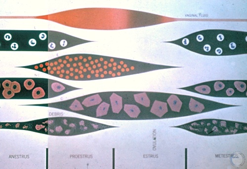
Vaginal Smear - Anestrus.
Cell types in Column 1 (of 4). Anestrus. From top to bottom shows the incidence of: lymphocytes, non-cornified epithelial cells, and debris.
Drost M (1973)
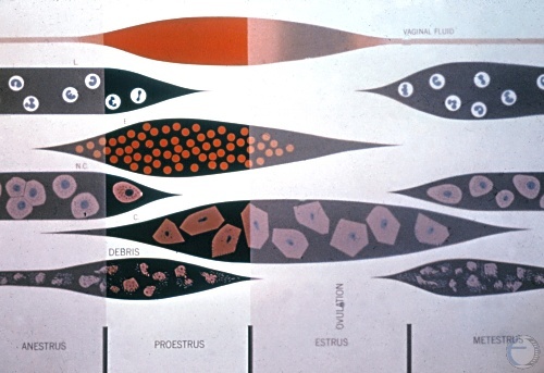
Vaginal Smear - Proestrus.
Cell types in Column 2 (of 4). Proestrus. From top to bottom shows the incidence of: vaginal fluid, erythrocytes, occasional non-cornified epithelial cell, cornified epithelial cells, debris. Erythrocytes dominate.
Drost M (1973)
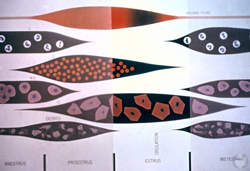
Vaginal Smear - Estrus.
Cell types in Column 3 (of 4). Estrus. From top to bottom shows the incidence of: vaginal fluid, some erythrocytes, cornified epithelial cells. Cornified epithelial cells dominate.
Drost M (1973)
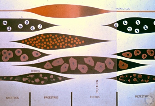
Vaginal Smear - Metestrus.
Cell types in Column 4 (of 4). Metestrus. From top to bottom shows the incidence of: leucocytes, non-cornified epithelial cells, occasional cornified epithelial cell, debris. Similar to anestrus.
Drost M (1973)
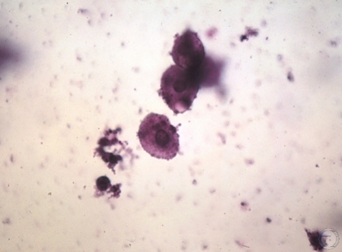
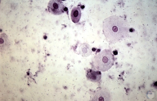
Cytology - Anestrus.
Vaginal smear during anestrus showing non-cornified epithelial cells and some cellular debris.
Roberts SJ (1973)
Cytology - Late Anestrus.
Vaginal smear during late anestrus showing predominantly non-cornified epithelial cells and some debris.
Roberts SJ (1973)
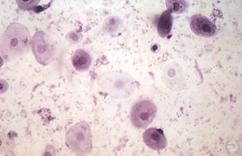
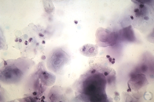
Cytology - Proestrus.
Vaginal smear during proestrus showing some erythrocytes, non-cornified epithelial cells, and debris.
Roberts SJ (1973)
Cytology - Early Estrus.
Vaginal smear during early estrus showing erythrocytes, cornified epithelial cells, and one non-cornified epithelial cell.
Roberts SJ (1973)
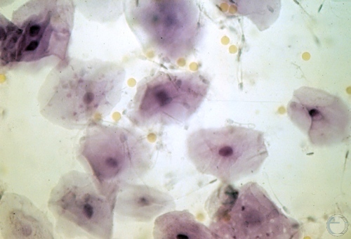
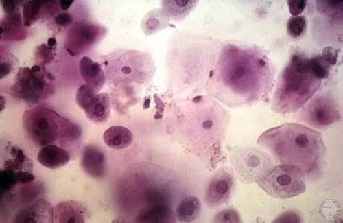
Cytology - Estrus.
Vaginal smear during estrus showing cornified epithelial cells and some erythrocytes.
Roberts SJ (1973)
Cytology - Metestrus.
Vaginal smear during metestrus showing some cornified epithelial cells, erythrocytes and debris.
Roberts SJ (1973)
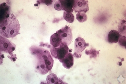
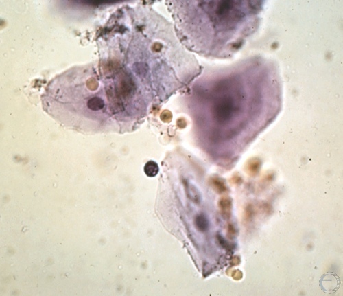
Cytology - Mid Metestrus.
Vaginal smear during mid-metestrus showing lymphocytes, non-cornified epithelial cells, occasional cornified epithelial cells, and debris.
Roberts SJ (1973)
Cytology - Late Metestrus.
Vaginal smear during late metestrus showing lymphocytes, non-cornified epithelial cells, and debris. Similar to anestrus.
Roberts SJ (1973)