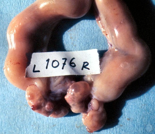
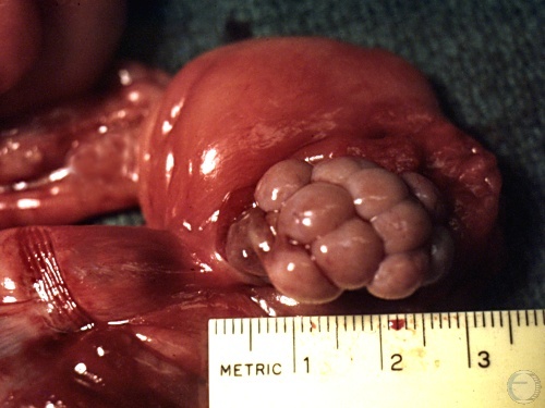
Ovulatory Follicle.
Ovulation in the dog may occur over a period of 12 to 24 hours, thus follicles and corpora hemorrhagica may be present at the same time. The ovary on the left shows a small protruding corpus hemorrhagicum and a medium size follicle, while the ovary on the right shows an ovulatory follicle. This occurs in other polytocous species (e.g. cats, pigs) and even in mares, where "asynchronous" ovulations (ovulations 12-24 hours apart)may occur.
Shille VM (1975)
Ovulatory Follicles.
Mature, ovulatory follicles. Ovulation occurs two days after the onset of the LH surge (Concannon), which may or may not coincide with the onset of estrus. Ovulation generally occurs when serum progesterone concentrations reach approximately 5ng / ml.
McEntee K (1972)
Corpora Hemorrhagica.
These ovaries show several corpora hemorrhagica which is compatible with mid- to late estrus. The pale structures are fat contained within the ovarian pedicle.
Shille VM (1975)
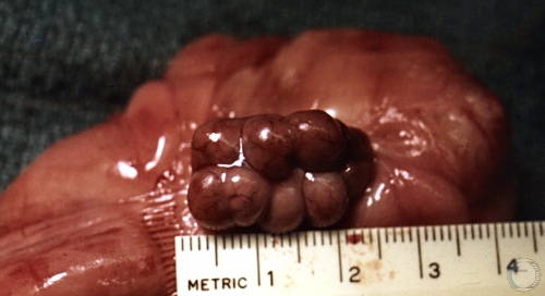
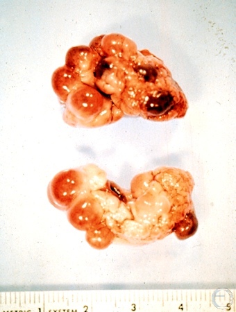
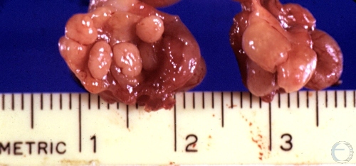
Corpora Lutea.
There are several corpora lutea. They are generally uniform in size which ranges from 5 to 8 mm. The ovaries are sometimes simply referred to as "mulberry ovaries".
Shille VM (1973)
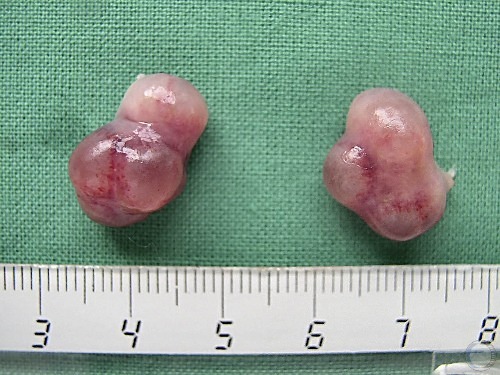
Corpora Lutea.
There are several corpora lutea. They are generally uniform in size which ranges from 5 to 8 mm.
Shille VM (1973)
CLs and Follicles.
Cross section of the ovary showing corpora lutea. Cavitated corpora lutea are functionally normal.
Shille VM (1973)
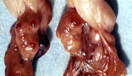
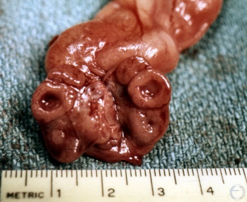
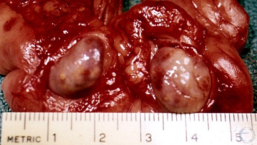
Cross-section of Ovary.
Three corpora lutea are present on this sectioned ovary. The two halves on the left section contain cavitated areas centrally.
Shille VM (1973)
Cross-section of Ovary.
Cross section of an ovary showing corpora hemorrhagica at different stages of development illustrating that ovulation occurs over a period of time (1-2 days after the LH peak).
Shille VM (1973)
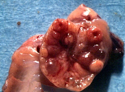
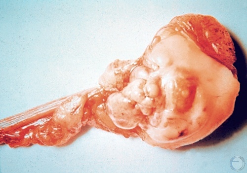
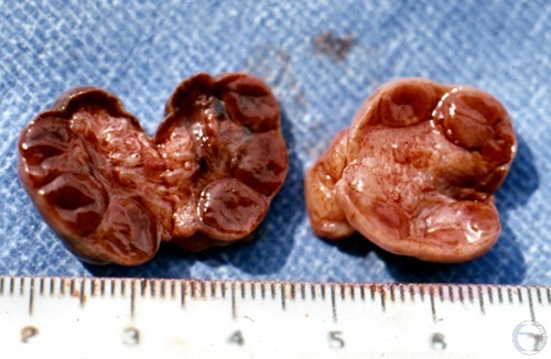
Granulosa Cell Tumor.
These neoplasms may have a smooth, nodular, or bosselated surface. The tumor may be accompanied by pyometra, or cystic endometrial hyperplasia.
McEntee K (1972)
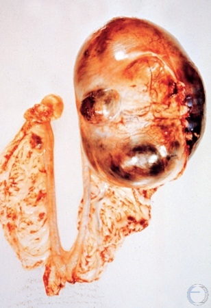
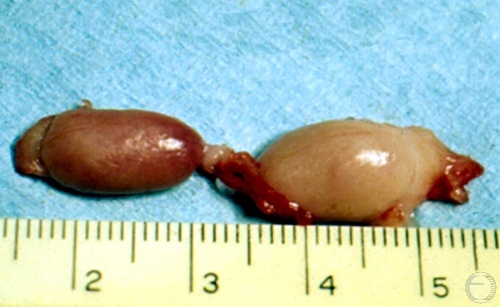
Cystic Granulosa Cell Tumor.
Prolonged proestrus with a bloody discharge is suggestive of a granulosa cell tumor. These tumors are uncommon and generally occur in older dogs.
McEntee K (1972)
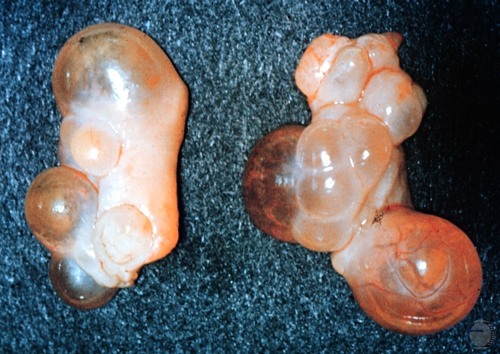
Ovarian Cystadenoma.
Cystadenomas are rare in domestic animals. They appear to arise from the epoopheron and / or rete ovarii. The neoplasms consist of multiple cysts. They occur predominantly in aged animals.
Shille VM (1980)
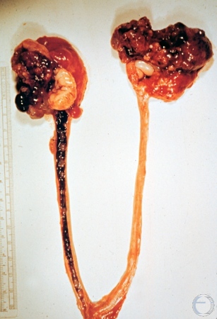
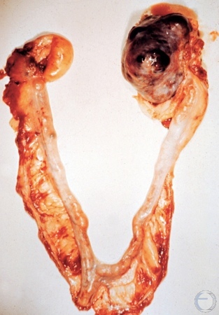
Cystic Adenocarcinoma.
Bilateral ovarian tumors. Proliferation of cauliflower-like tissue. Large ovarian cyst on the left ovary and a small cyst on the right ovary. These cysts metastasize around the abdominal cavity. There is a poor prognosis.
McEntee K (1972)
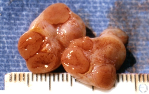
Ovaries.
Three luteal structures are evident in the ovary on the left and two are present on the ovary on the right.
Shille VM (1973)
Follicular Cysts.
Nonneoplastic follicular cysts do not occur as frequently in the bitch as other types of pathological ovarian cysts. They can be associated with various signs of hyperestrogenism.
McEntee K (1972)
Anestrous Ovaries.
Ovaries of a Greyhound racing bitch under treatment with testosterone. There is no evidence of any follicular activity.
Shille VM (1986)
CLs and Follicles.
Normal functioning ovaries with corpora lutea and one follicle on the right ovary.
Shille VM (1973)
Corpora Lutea.
Corpora lutea are evident on the surface of the ovaries.
Shille VM (1973)