The Visual Guide to
Bubaline Reproduction
Reproductive Technology: Ultrasonography
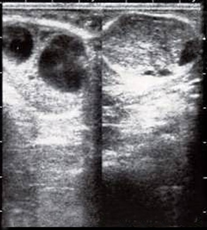
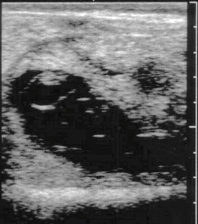
Dominant Follicle and CL.
Left ovary: dominant follicle and a co-dominant follicle which will undergo atresia. Right ovary: corpus luteum and a small follicle. About day 15 of the estrous cycle at the time of follicular deviation when the dominant follicle is selected (diameter 7.2 to 7.5 mm).
Zambrano-Varon J (2012)
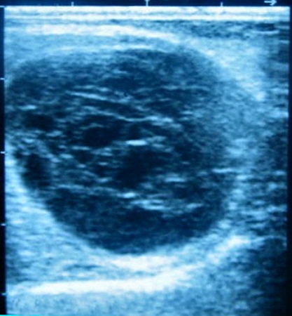
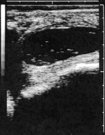
Anovulatory Follicle.
Single cyst like structure, reminiscent of an hemorrhagic corpus luteum commonly seen in mares but not in cows. Some of these anovulatory structures resolve spontaneously after a couple of cycles, others are treated with an ovsynch protocol with positive results.
Zambrano-Varon J (2012)
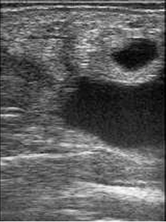
28-day Pregnancy.
Cross section of the pregnant horn distended by embryonic fluids. The large dark area below is the bladder.
Zambrano-Varon J (2012)
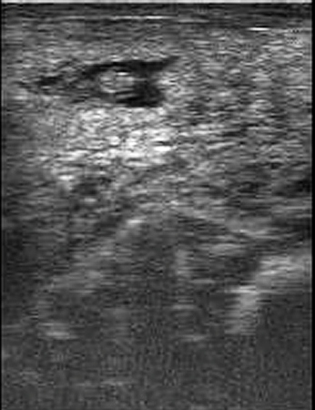
32-day Pregnancy.
The early embryo can be identified surrounded by fluids.
Zambrano-Varon J (2012)
Embryonic Death at 35 Days.
The 35-day embryo is disintegrating. Some flocculent material is present in the uterine fluid.
Zambrano-Varon J (2012)
Embryonic Death at 38 Days.
Flocculation at 38 days of gestation embryonic death. No evidence of an intact embryo.
Zambrano-Varon J (2012)
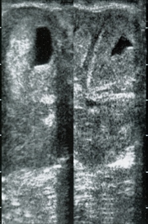
Flushing Medium in the Lumen.
Bilateral presence of flushing medium in the horns of a donor cow.
Zambrano-Varon J (2012)
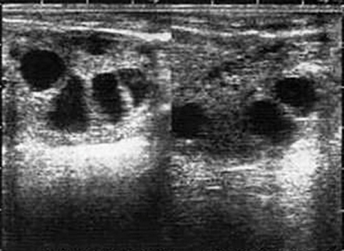
Superovulated Ovaries.
Follicular response to gonadotrophin treatment in a donor cow. There are seven follicles and there is no corpus luteum.
Zambrano-Varon J (2012)
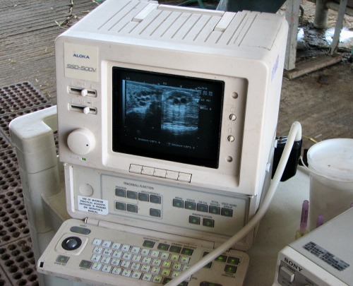
Ultrasound Unit.
Ultrasound unit showing superovulated ovaries on the screen.
Zambrano-Varon J (2012)
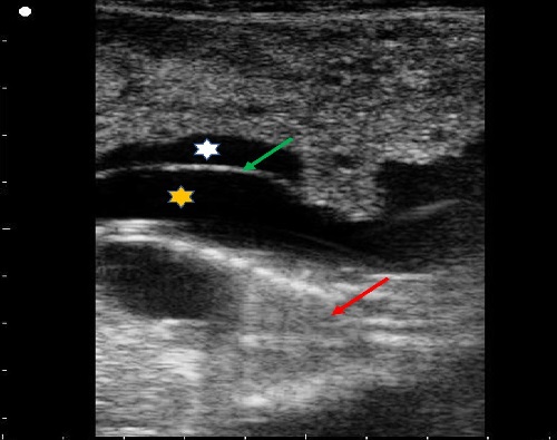
Fetus and fetal membranes.
This is an ultrasound image of the uterus in a Murrah buffalo at about 4 to 4.5 months of gestation. The fetus (red arrow), amnion (green arrow), amniotic fluid (yellow asterisk) and allantoic fluid (white asterisk) can be seen in the image.
Khan FA (2019)
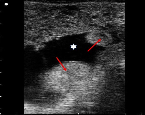
Placentomes.
This is an ultrasound image of the uterus in a Murrah buffalo at about 4 to 4.5 months of gestation. Placentomes (red arrows) and allantoic fluid (white asterisk) can be seen in the image.
Khan FA (2019)
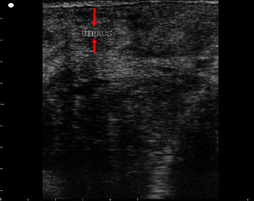
Non-pregnant uterus.
This is a longitudinal view ultrasound image of the uterus (red arrows) in a non-pregnant Murrah buffalo.
Khan FA (2019)
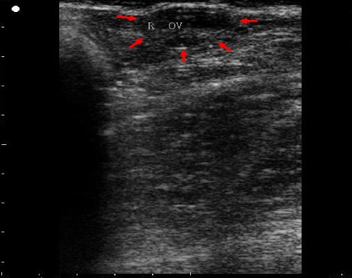
Ovary with small follicles.
This is an ultrasound image of the right ovary (periphery marked with red arrows) containing multiple small follicles (small anechoic areas within the ovary) in a non-pregnant Murrah buffalo.
Khan FA (2019)
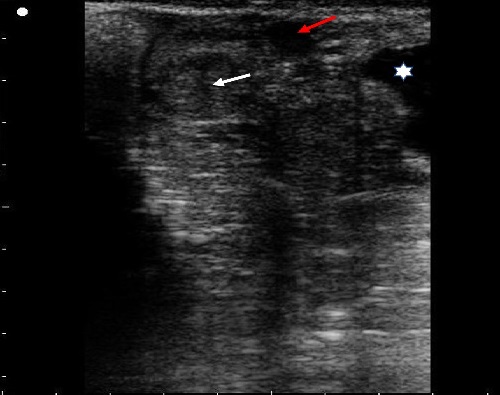
Ovary with a large follicle and a corpus luteum.
This is an ultrasound image of the left ovary containing a large follicle (red arrow) and a corpus luteum (white arrow) in a non-pregnant Murrah buffalo. The bladder can be seen on the right side of the image (white asterisk).
Khan FA (2019)
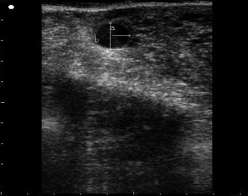
Ovary with a pre-ovulatory follicle.
This is an ultrasound image of a preovulatory size follicle of 12.7 mm.
Khan FA (2019)